2WGR
 
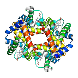
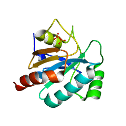 | | Combining crystallography and molecular dynamics: The case of Schistosoma mansoni phospholipid glutathione peroxidase | | 分子名称: | GLUTATHIONE PEROXIDASE, PYROPHOSPHATE 2- | | 著者 | Dimastrogiovanni, D, Anselmi, M, Miele, A.E, Boumis, G, Angelucci, F, Di Nola, A, Brunori, M, Bellelli, A. | | 登録日 | 2009-04-24 | | 公開日 | 2009-09-08 | | 最終更新日 | 2023-12-13 | | 実験手法 | X-RAY DIFFRACTION (1.7 Å) | | 主引用文献 | Combining Crystallography and Molecular Dynamics: The Case of Schistosoma Mansoni Phospholipid Glutathione Peroxidase.
Proteins, 78, 2010
|
|
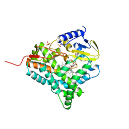
2V1M
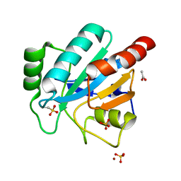 | | Crystal structure of Schistosoma mansoni glutathione peroxidase | | 分子名称: | ACETATE ION, GLUTATHIONE PEROXIDASE, LITHIUM ION, ... | | 著者 | Dimastrogiovanni, D, Miele, A.E, Angelucci, F, Boumis, G, Bellelli, A, Brunori, M. | | 登録日 | 2008-09-16 | | 公開日 | 2009-09-08 | | 最終更新日 | 2023-12-13 | | 実験手法 | X-RAY DIFFRACTION (1 Å) | | 主引用文献 | Combining Crystallography and Molecular Dynamics: The Case of Schistosoma Mansoni Phospholipid Glutathione Peroxidase.
Proteins, 78, 2010
|
|
2XC2
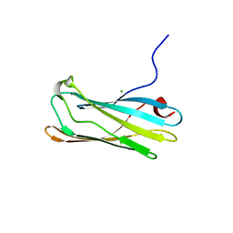
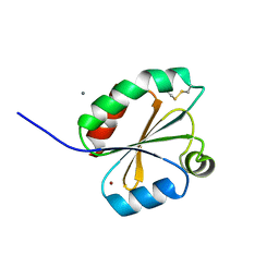 | | Crystal structure of oxidized Schistosoma mansoni Thioredoxin pre- protein at 1.6 Angstrom | | 分子名称: | CALCIUM ION, THIOREDOXINN, ZINC ION | | 著者 | Boumis, G, Miele, A.E, Dimastrogiovanni, D, Angelucci, F, Bellelli, A. | | 登録日 | 2010-04-15 | | 公開日 | 2010-08-11 | | 最終更新日 | 2023-12-20 | | 実験手法 | X-RAY DIFFRACTION (1.56 Å) | | 主引用文献 | Structural and Functional Characterization of Schistosoma Mansoni Thioredoxin.
Protein Sci., 20, 2011
|
|
3H4K
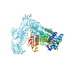 | | Crystal structure of the wild type Thioredoxin glutatione reductase from Schistosoma mansoni in complex with auranofin | | 分子名称: | FLAVIN-ADENINE DINUCLEOTIDE, GLUTATHIONE, GOLD ION, ... | | 著者 | Angelucci, F, Dimastrogiovanni, D, Miele, A.E, Boumis, G, Brunori, M, Bellelli, A. | | 登録日 | 2009-04-20 | | 公開日 | 2009-08-25 | | 最終更新日 | 2023-11-01 | | 実験手法 | X-RAY DIFFRACTION (2.55 Å) | | 主引用文献 | Inhibition of Schistosoma mansoni thioredoxin-glutathione reductase by auranofin: structural and kinetic aspects.
J.Biol.Chem., 284, 2009
|
|
1LQ9
 | | Crystal Structure of a Monooxygenase from the Gene ActVA-Orf6 of Streptomyces coelicolor Strain A3(2) | | 分子名称: | ACTVA-ORF6 MONOOXYGENASE, TETRAETHYLENE GLYCOL | | 著者 | Sciara, G, Kendrew, S.G, Miele, A.E, Marsh, N.G, Federici, L, Malatesta, F, Schimperna, G, Savino, C, Vallone, B. | | 登録日 | 2002-05-09 | | 公開日 | 2003-01-14 | | 最終更新日 | 2024-02-14 | | 実験手法 | X-RAY DIFFRACTION (1.3 Å) | | 主引用文献 | The structure of ActVA-Orf6, a novel type of monooxygenase involved in actinorhodin biosynthesis
EMBO J., 22, 2003
|
|
1GLI
 | |
1GYW
 | | Gamma-adaptin appendage domain from clathrin adaptor AP1 A753D mutant | | 分子名称: | ADAPTER-RELATED PROTEIN COMPLEX 1 GAMMA 1 SUBUNIT, CHLORIDE ION | | 著者 | Evans, P.R, Miele, A.E, Owen, D.J, McMahon, H.M, Kent, H.M. | | 登録日 | 2002-04-30 | | 公開日 | 2002-08-22 | | 最終更新日 | 2023-12-13 | | 実験手法 | X-RAY DIFFRACTION (2.4 Å) | | 主引用文献 | Gamma-adaptin appendage domain: structure and binding site for Eps15 and gamma-synergin.
Structure, 10, 2002
|
|
2JJP
 | | Structure of cytochrome P450 EryK in complex with inhibitor ketoconazole (KC) | | 分子名称: | 1-ACETYL-4-(4-{[(2S,4R)-2-(2,4-DICHLOROPHENYL)-2-(1H-IMIDAZOL-1-YLMETHYL)-1,3-DIOXOLAN-4-YL]METHOXY}PHENYL)PIPERAZINE, CYTOCHROME P450 113A1, PROTOPORPHYRIN IX CONTAINING FE, ... | | 著者 | Savino, C, Sciara, G, Miele, A.E, Kendrew, S.G, Vallone, B. | | 登録日 | 2008-04-15 | | 公開日 | 2009-07-14 | | 最終更新日 | 2023-12-13 | | 実験手法 | X-RAY DIFFRACTION (2.1 Å) | | 主引用文献 | Azole Drugs Trap Cytochrome P450 Eryk in Alternative Conformational States.
Biochemistry, 49, 2010
|
|


