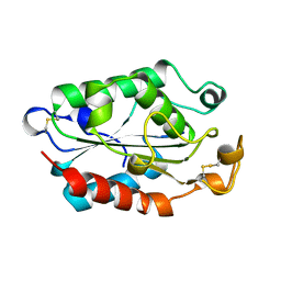
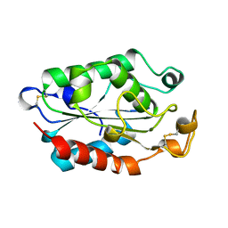
2FJN
 
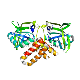
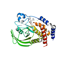 | | The structure of phosphotyrosine phosphatase 1B in complex with compound 2 | | 分子名称: | (4-{(2S,4E)-2-(1H-1,2,3-BENZOTRIAZOL-1-YL)-2-[4-(METHOXYCARBONYL)PHENYL]-5-PHENYLPENT-4-ENYL}PHENYL)(DIFLUORO)METHYLPHOSPHONIC ACID, CHLORIDE ION, Tyrosine-protein phosphatase, ... | | 著者 | Asante-Appiah, E, Patel, S, Desponts, C, Taylor, J.M, Lau, C, Dufresne, C, Therien, M, Friesen, R, Becker, J.W, Leblanc, Y, Kennedy, B.P, Scapin, G. | | 登録日 | 2006-01-03 | | 公開日 | 2006-01-17 | | 最終更新日 | 2023-08-30 | | 実験手法 | X-RAY DIFFRACTION (2.2 Å) | | 主引用文献 | Conformation-assisted inhibition of protein-tyrosine phosphatase-1B elicits inhibitor selectivity over T-cell protein-tyrosine phosphatase.
J.Biol.Chem., 281, 2006
|
|
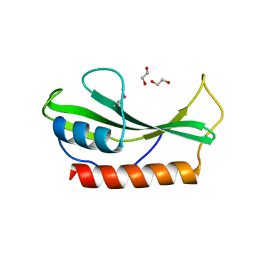
3KG4
 | | Crystal structure of an uncharacterized protein from Mannheimia succiniciproducens | | 分子名称: | Uncharacterized protein | | 著者 | Bonanno, J.B, Dickey, M, Bain, K.T, Lau, C, Romero, R, Wasserman, S, Sauder, J.M, Burley, S.K, Almo, S.C, New York SGX Research Center for Structural Genomics (NYSGXRC) | | 登録日 | 2009-10-28 | | 公開日 | 2009-11-24 | | 最終更新日 | 2024-02-21 | | 実験手法 | X-RAY DIFFRACTION (1.95 Å) | | 主引用文献 | Crystal structure of an uncharacterized protein from Mannheimia succiniciproducens
To be Published
|
|
2QGY
 | | Crystal structure of an enolase from the environmental genome shotgun sequencing of the Sargasso Sea | | 分子名称: | Enolase from the environmental genome shotgun sequencing of the Sargasso Sea, MAGNESIUM ION | | 著者 | Bonanno, J.B, Gilmore, M, Bain, K.T, Lau, C, Romero, R, Smith, D, Wasserman, S, Sauder, J.M, Burley, S.K, Almo, S.C, New York SGX Research Center for Structural Genomics (NYSGXRC) | | 登録日 | 2007-06-29 | | 公開日 | 2007-07-24 | | 最終更新日 | 2024-02-21 | | 実験手法 | X-RAY DIFFRACTION (1.8 Å) | | 主引用文献 | Crystal structure of an enolase from the environmental genome shotgun sequencing of the Sargasso Sea.
To be Published
|
|
2R1F
 | | Crystal structure of predicted aminodeoxychorismate lyase from Escherichia coli | | 分子名称: | CADMIUM ION, GLYCEROL, Predicted aminodeoxychorismate lyase, ... | | 著者 | Patskovsky, Y, Ramagopal, U.A, Toro, R, Meyer, A.J, Rutter, M, Lau, C, Maletic, M, Smith, D, Gheyi, T, Wasserman, S, Sauder, J.M, Burley, S.K, Almo, S.C, New York SGX Research Center for Structural Genomics (NYSGXRC) | | 登録日 | 2007-08-22 | | 公開日 | 2007-09-04 | | 最終更新日 | 2021-10-20 | | 実験手法 | X-RAY DIFFRACTION (2.21 Å) | | 主引用文献 | Crystal Structure of Predicted Aminodeoxychorismate Lyase from Escherichia coli.
To be Published
|
|
2RDX
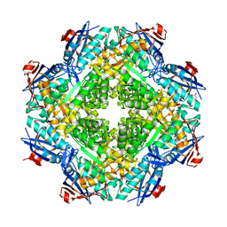 | | Crystal structure of mandelate racemase/muconate lactonizing enzyme from Roseovarius nubinhibens ISM | | 分子名称: | GLYCEROL, MAGNESIUM ION, Mandelate racemase/muconate lactonizing enzyme, ... | | 著者 | Patskovsky, Y, Bonanno, J, Sauder, J.M, Ozyurt, S, Gilmore, M, Lau, C, Maletic, M, Gheyi, T, Wasserman, S.R, Koss, J, Gerlt, J.A, Burley, S.K, Almo, S.C, New York SGX Research Center for Structural Genomics (NYSGXRC) | | 登録日 | 2007-09-25 | | 公開日 | 2007-10-09 | | 最終更新日 | 2024-02-21 | | 実験手法 | X-RAY DIFFRACTION (2 Å) | | 主引用文献 | Crystal structure of mandelate racemase/muconate lactonizing enzyme from Roseovarius nubinhibens ISM.
To be Published
|
|
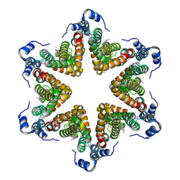
3FV9
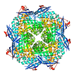 | | Crystal structure of putative mandelate racemase/muconatelactonizing enzyme from ROSEOVARIUS NUBINHIBENS ISM complexed with magnesium | | 分子名称: | MAGNESIUM ION, Mandelate racemase/muconate lactonizing enzyme | | 著者 | Malashkevich, V.N, Rutter, M, Bain, K.T, Lau, C, Ozyurt, S, Smith, D, Wasserman, S, Sauder, J.M, Burley, S.K, Almo, S.C, New York SGX Research Center for Structural Genomics (NYSGXRC) | | 登録日 | 2009-01-15 | | 公開日 | 2009-01-27 | | 最終更新日 | 2024-02-21 | | 実験手法 | X-RAY DIFFRACTION (1.9 Å) | | 主引用文献 | Crystal structure of putative mandelate racemase/muconatelactonizing enzyme from ROSEOVARIUS NUBINHIBENS ISM complexed with magnesium
to be published
|
|
3FVD
 | | Crystal structure of a member of enolase superfamily from ROSEOVARIUS NUBINHIBENS ISM complexed with magnesium | | 分子名称: | MAGNESIUM ION, Mandelate racemase/muconate lactonizing enzyme | | 著者 | Malashkevich, V.N, Rutter, M, Bain, K.T, Lau, C, Ozyurt, S, Smith, D, Wasserman, S, Sauder, J.M, Burley, S.K, Almo, S.C, New York SGX Research Center for Structural Genomics (NYSGXRC) | | 登録日 | 2009-01-15 | | 公開日 | 2009-01-27 | | 最終更新日 | 2024-02-21 | | 実験手法 | X-RAY DIFFRACTION (2.3 Å) | | 主引用文献 | Crystal structure of a member of enolase superfamily from ROSEOVARIUS NUBINHIBENS ISM complexed with magnesium
to be published
|
|
2OX7
 | | Crystal structure of protein EF1440 from Enterococcus faecalis | | 分子名称: | Hypothetical protein | | 著者 | Malashkevich, V.N, Toro, R, Sauder, J.M, Schwinn, K.D, Thompson, D.A, Bain, K.T, Adams, J.M, Reyes, C, Lau, C, Gilmore, J, Rooney, I, Gheyi, T, Wasserman, S.R, Emtage, S, Burley, S.K, Almo, S.C, New York SGX Research Center for Structural Genomics (NYSGXRC) | | 登録日 | 2007-02-19 | | 公開日 | 2007-03-06 | | 最終更新日 | 2021-02-03 | | 実験手法 | X-RAY DIFFRACTION (1.777 Å) | | 主引用文献 | Crystal structure of the hypothetical protein from Enterococcus faecalis
To be Published
|
|
2P84
 | | Crystal structure of ORF041 from Bacteriophage 37 | | 分子名称: | ORF041 | | 著者 | Malashkevich, V.N, Toro, R, Sauder, J.M, Schwinn, K.D, Thompson, D.A, Bain, K.T, Adams, J.M, Reyes, C, Lau, C, Gilmore, J, Rooney, I, Wasserman, T, Gheyi, S.R, Emtage, S, Burley, S.K, Almo, S.C, New York SGX Research Center for Structural Genomics (NYSGXRC) | | 登録日 | 2007-03-21 | | 公開日 | 2007-04-03 | | 最終更新日 | 2024-02-21 | | 実験手法 | X-RAY DIFFRACTION (1.8 Å) | | 主引用文献 | Crystal structure of the hypothetical protein from Staphylococcus phage 37
To be Published
|
|
2NRH
 | | Crystal structure of conserved putative Baf family transcriptional activator from Campylobacter jejuni | | 分子名称: | SULFATE ION, Transcriptional activator, putative, ... | | 著者 | Bonanno, J.B, Dickey, M, Bain, K.T, Lau, C, Wasserman, S, Smith, D, Sauder, J.M, Burley, S.K, Almo, S.C, New York SGX Research Center for Structural Genomics (NYSGXRC) | | 登録日 | 2006-11-02 | | 公開日 | 2006-11-14 | | 最終更新日 | 2023-12-27 | | 実験手法 | X-RAY DIFFRACTION (2.3 Å) | | 主引用文献 | Crystal structure of conserved putative Baf family transcriptional activator from Campylobacter jejuni
To be Published
|
|
2NR4
 | | Crystal structure of FMN-bound protein MM1853 from Methanosarcina mazei, Pfam DUF447 | | 分子名称: | Conserved hypothetical protein, FLAVIN MONONUCLEOTIDE | | 著者 | Bonanno, J.B, Gilmore, J, Bain, K.T, Lau, C, Wasserman, S, Smith, D, Sauder, J.M, Burley, S.K, Almo, S.C, New York SGX Research Center for Structural Genomics (NYSGXRC) | | 登録日 | 2006-11-01 | | 公開日 | 2006-11-07 | | 最終更新日 | 2023-12-27 | | 実験手法 | X-RAY DIFFRACTION (1.85 Å) | | 主引用文献 | Crystal structure of conserved FMN bound hypothetical protein from Methanosarcina mazei
To be Published
|
|
2PWW
 | | Crystal structure of ABC2387 from Bacillus clausii | | 分子名称: | 1,2-ETHANEDIOL, Uncharacterized protein | | 著者 | Ramagopal, U.A, Freeman, J, Lau, C, Toro, R, Bain, K, Rodgers, L, Sauder, J.M, Burley, S.K, Almo, S.C, New York SGX Research Center for Structural Genomics (NYSGXRC) | | 登録日 | 2007-05-14 | | 公開日 | 2007-05-22 | | 最終更新日 | 2021-02-03 | | 実験手法 | X-RAY DIFFRACTION (1.82 Å) | | 主引用文献 | Crystal structure of ABC2387 from Bacillus clausii.
To be Published
|
|
2OYN
 | | Crystal structure of CDP-bound protein MJ0056 from Methanococcus jannaschii, Pfam DUF120 | | 分子名称: | CYTIDINE-5'-DIPHOSPHATE, Hypothetical protein MJ0056, SODIUM ION | | 著者 | Bonanno, J.B, Dickey, M, Bain, K.T, Lau, C, Romero, R, Smith, D, Wasserman, S, Sauder, J.M, Burley, S.K, Almo, S.C, New York SGX Research Center for Structural Genomics (NYSGXRC) | | 登録日 | 2007-02-22 | | 公開日 | 2007-03-06 | | 最終更新日 | 2024-02-21 | | 実験手法 | X-RAY DIFFRACTION (1.85 Å) | | 主引用文献 | Crystal structure of hypothetical protein from Methanococcus jannaschii bound to CDP
TO BE PUBLISHED
|
|
2PCE
 | | Crystal structure of putative mandelate racemase/muconate lactonizing enzyme from Roseovarius nubinhibens ISM | | 分子名称: | PHOSPHATE ION, putative mandelate racemase/muconate lactonizing enzyme | | 著者 | Bonanno, J.B, Rutter, M, Bain, K.T, Lau, C, Ozyurt, S, Smith, D, Wasserman, S, Sauder, J.M, Burley, S.K, Almo, S.C, New York SGX Research Center for Structural Genomics (NYSGXRC) | | 登録日 | 2007-03-29 | | 公開日 | 2007-04-10 | | 最終更新日 | 2024-02-21 | | 実験手法 | X-RAY DIFFRACTION (2 Å) | | 主引用文献 | Crystal structure of putative mandelate racemase/muconate lactonizing enzyme from Roseovarius nubinhibens ISM
To be Published
|
|
4YO5
 | | EAEC T6SS TssA-Cterminus | | 分子名称: | TssA | | 著者 | Durand, E, Zoued, A, Spinelli, S, Douzi, B, Brunet, Y.R, Bebeacua, C, Legrand, P, Journet, L, Mignot, T, Cambillau, C, Cascales, E. | | 登録日 | 2015-03-11 | | 公開日 | 2016-02-17 | | 最終更新日 | 2017-06-14 | | 実験手法 | X-RAY DIFFRACTION (3.35 Å) | | 主引用文献 | Priming and polymerization of a bacterial contractile tail structure.
Nature, 531, 2016
|
|
4V5I
 | | Structure of the Phage P2 Baseplate in its Activated Conformation with Ca | | 分子名称: | CALCIUM ION, ORF15, ORF16, ... | | 著者 | Sciara, G, Bebeacua, C, Bron, P, Tremblay, D, Ortiz-Lombardia, M, Lichiere, J, van Heel, M, Campanacci, V, Moineau, S, Cambillau, C. | | 登録日 | 2010-02-05 | | 公開日 | 2014-07-09 | | 最終更新日 | 2024-01-10 | | 実験手法 | X-RAY DIFFRACTION (5.464 Å) | | 主引用文献 | Structure of Lactococcal Phage P2 Baseplate and its Mechanism of Activation.
Proc.Natl.Acad.Sci.USA, 107, 2010
|
|
3D75
 | | Crystal structure of a pheromone binding protein mutant D35N, from Apis mellifera, at pH 5.5 | | 分子名称: | N-BUTYL-BENZENESULFONAMIDE, Pheromone-binding protein ASP1 | | 著者 | Pesenti, M.E, Spinelli, S, Bezirard, V, Briand, L, Pernollet, J.C, Tegoni, M, Cambillau, C. | | 登録日 | 2008-05-20 | | 公開日 | 2009-05-26 | | 最終更新日 | 2023-11-01 | | 実験手法 | X-RAY DIFFRACTION (2.3 Å) | | 主引用文献 | Queen bee pheromone binding protein pH-induced domain swapping favors pheromone release
J.Mol.Biol., 390, 2009
|
|
3D77
 | | Crystal structure of a pheromone binding protein mutant D35N, from Apis mellifera, soaked at pH 4.0 | | 分子名称: | 1,2-ETHANEDIOL, N-BUTYL-BENZENESULFONAMIDE, Pheromone-binding protein ASP1, ... | | 著者 | Pesenti, M.E, Spinelli, S, Bezirard, V, Briand, L, Pernollet, J.C, Tegoni, M, Cambillau, C. | | 登録日 | 2008-05-20 | | 公開日 | 2009-05-26 | | 最終更新日 | 2023-11-01 | | 実験手法 | X-RAY DIFFRACTION (1.7 Å) | | 主引用文献 | Queen bee pheromone binding protein pH-induced domain swapping favors pheromone release
J.Mol.Biol., 390, 2009
|
|
3UH8
 | | N-terminal domain of phage TP901-1 ORF48 | | 分子名称: | ORF48 | | 著者 | Veesler, D, Spinelli, S, Mahony, J, Lichiere, J, Blangy, S, Bricogne, G, Legrand, P, Ortiz-Lombardia, M, Campanacci, V.I, van Sinderen, D, Cambillau, C. | | 登録日 | 2011-11-03 | | 公開日 | 2012-05-30 | | 最終更新日 | 2024-02-28 | | 実験手法 | X-RAY DIFFRACTION (2.3 Å) | | 主引用文献 | Structure of the phage TP901-1 1.8 MDa baseplate suggests an alternative host adhesion mechanism.
Proc.Natl.Acad.Sci.USA, 109, 2012
|
|
1CUJ
 | | CUTINASE, S120C MUTANT | | 分子名称: | CUTINASE | | 著者 | Martinez, C, Cambillau, C. | | 登録日 | 1995-11-16 | | 公開日 | 1996-07-11 | | 最終更新日 | 2021-11-03 | | 実験手法 | X-RAY DIFFRACTION (1.6 Å) | | 主引用文献 | Dynamics of Fusarium solani cutinase investigated through structural comparison among different crystal forms of its variants.
Proteins, 26, 1996
|
|
1CUY
 | | CUTINASE, L189F MUTANT | | 分子名称: | CUTINASE | | 著者 | Longhi, S, Cambillau, C. | | 登録日 | 1995-11-16 | | 公開日 | 1996-07-11 | | 最終更新日 | 2021-11-03 | | 実験手法 | X-RAY DIFFRACTION (1.69 Å) | | 主引用文献 | Dynamics of Fusarium solani cutinase investigated through structural comparison among different crystal forms of its variants.
Proteins, 26, 1996
|
|
1CUW
 | | CUTINASE, G82A, A85F, V184I, A185L, L189F MUTANT | | 分子名称: | CUTINASE | | 著者 | Longhi, S, Cambillau, C. | | 登録日 | 1995-11-16 | | 公開日 | 1996-07-11 | | 最終更新日 | 2021-11-03 | | 実験手法 | X-RAY DIFFRACTION (2.7 Å) | | 主引用文献 | Dynamics of Fusarium solani cutinase investigated through structural comparison among different crystal forms of its variants.
Proteins, 26, 1996
|
|
1CUE
 | | CUTINASE, Q121L MUTANT | | 分子名称: | CUTINASE | | 著者 | Martinez, C, Cambillau, C. | | 登録日 | 1995-11-16 | | 公開日 | 1996-07-11 | | 最終更新日 | 2021-11-03 | | 実験手法 | X-RAY DIFFRACTION (2.1 Å) | | 主引用文献 | Dynamics of Fusarium solani cutinase investigated through structural comparison among different crystal forms of its variants.
Proteins, 26, 1996
|
|
1CUC
 | |
1CUB
 | |


