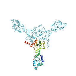
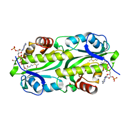
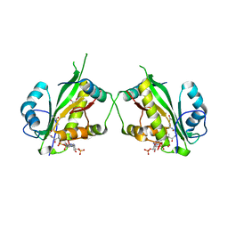
7ZG5
 
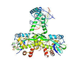 | | The crystal structure of Salmonella TacAT3-DNA complex | | 分子名称: | (4S)-2-METHYL-2,4-PENTANEDIOL, Acetyltransferase, BARIUM ION, ... | | 著者 | Grabe, G.J, Morgan, R.M.L, Helaine, S. | | 登録日 | 2022-04-01 | | 公開日 | 2023-10-11 | | 最終更新日 | 2024-04-10 | | 実験手法 | X-RAY DIFFRACTION (2 Å) | | 主引用文献 | Molecular stripping underpins derepression of a toxin-antitoxin system.
Nat.Struct.Mol.Biol., 2024
|
|
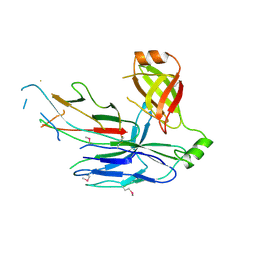
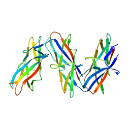
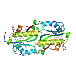
6FJY
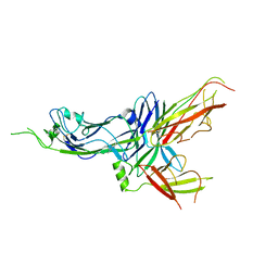 | | Crystal structure of CsuC-CsuE chaperone-tip adhesion subunit pre-assembly complex from archaic chaperone-usher Csu pili of Acinetobacter baumannii | | 分子名称: | CsuC, Protein CsuE | | 著者 | Pakharukova, N.A, Tuitilla, M, Paavilainen, S, Zavialov, A.V. | | 登録日 | 2018-01-23 | | 公開日 | 2018-05-16 | | 最終更新日 | 2018-09-19 | | 実験手法 | X-RAY DIFFRACTION (2.31 Å) | | 主引用文献 | Structural basis forAcinetobacter baumanniibiofilm formation.
Proc. Natl. Acad. Sci. U.S.A., 115, 2018
|
|
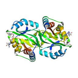
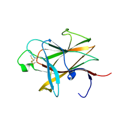
6FM5
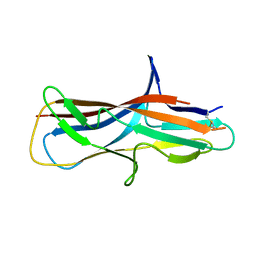 | | Crystal structure of self-complemented CsuA/B major subunit from archaic chaperone-usher Csu pili of Acinetobacter baumannii | | 分子名称: | CsuA/B,CsuA/B,CsuA/B,CsuA/B | | 著者 | Pakharukova, N.A, Tuitilla, M, Paavilainen, S, Zavialov, A.V. | | 登録日 | 2018-01-30 | | 公開日 | 2018-09-26 | | 最終更新日 | 2018-11-14 | | 実験手法 | X-RAY DIFFRACTION (1.47 Å) | | 主引用文献 | Archaic and alternative chaperones preserve pilin folding energy by providing incomplete structural information.
J. Biol. Chem., 293, 2018
|
|
6FQ0
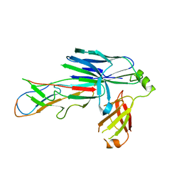 | | Crystal structure of the CsuC-CsuA/B chaperone-subunit preassembly complex of the archaic chaperone-usher Csu pili of Acinetobacter baumannii | | 分子名称: | CsuA/B,CsuA/B, CsuC | | 著者 | Pakharukova, N.A, Tuitilla, M, Paavilainen, S, Zavialov, A.V. | | 登録日 | 2018-02-12 | | 公開日 | 2018-09-26 | | 最終更新日 | 2018-11-14 | | 実験手法 | X-RAY DIFFRACTION (2.5 Å) | | 主引用文献 | Archaic and alternative chaperones preserve pilin folding energy by providing incomplete structural information.
J. Biol. Chem., 293, 2018
|
|
6FQA
 | | Crystal structure of the CsuC-CsuA/B chaperone-subunit preassembly complex of the archaic chaperone-usher Csu pili of Acinetobacter baumannii | | 分子名称: | CsuA/B,CsuA/B, CsuC | | 著者 | Parilova, O, Pakharukova, N.A, Malmi, H, Tuitilla, M, Paavilainen, S, Zavialov, A.V. | | 登録日 | 2018-02-13 | | 公開日 | 2018-09-26 | | 最終更新日 | 2024-01-17 | | 実験手法 | X-RAY DIFFRACTION (2.85 Å) | | 主引用文献 | Archaic and alternative chaperones preserve pilin folding energy by providing incomplete structural information.
J. Biol. Chem., 293, 2018
|
|
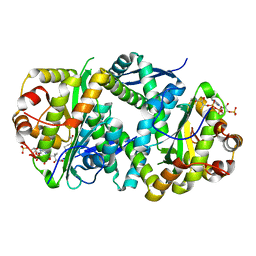
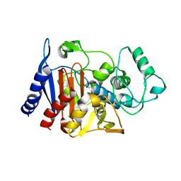
1KO7
 | | X-ray structure of the HPr kinase/phosphatase from Staphylococcus xylosus at 1.95 A resolution | | 分子名称: | Hpr kinase/phosphatase, PHOSPHATE ION | | 著者 | Marquez, J.A, Hasenbein, S, Koch, B, Fieulaine, S, Nessler, S, Hengstenberg, W, Scheffzek, K. | | 登録日 | 2001-12-20 | | 公開日 | 2002-04-03 | | 最終更新日 | 2023-08-16 | | 実験手法 | X-RAY DIFFRACTION (1.95 Å) | | 主引用文献 | Structure of the full-length HPr kinase/phosphatase from Staphylococcus xylosus at 1.95 A resolution: Mimicking the product/substrate of the phospho transfer reactions.
Proc.Natl.Acad.Sci.USA, 99, 2002
|
|
5D6H
 | | Crystal structure of CsuC-CsuA/B chaperone-major subunit pre-assembly complex from Csu biofilm-mediating pili of Acinetobacter baumannii | | 分子名称: | CsuA/B, CsuC | | 著者 | Pakharukova, N.A, Tuitilla, M, Paavilainen, S, Zavialov, A. | | 登録日 | 2015-08-12 | | 公開日 | 2015-11-04 | | 最終更新日 | 2015-12-02 | | 実験手法 | X-RAY DIFFRACTION (2.4 Å) | | 主引用文献 | Structural Insight into Archaic and Alternative Chaperone-Usher Pathways Reveals a Novel Mechanism of Pilus Biogenesis.
Plos Pathog., 11, 2015
|
|
2KN8
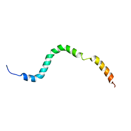 | | NMR structure of the C-terminal domain of pUL89 | | 分子名称: | DNA cleavage and packaging protein large subunit, UL89 | | 著者 | Couvreux, A, Hantz, S, Marquant, R, Champier, G, Alain, S, Morellet, N, Bouaziz, S. | | 登録日 | 2009-08-18 | | 公開日 | 2010-06-09 | | 最終更新日 | 2024-05-01 | | 実験手法 | SOLUTION NMR | | 主引用文献 | Insight into the structure of the pUL89 C-terminal domain of the human cytomegalovirus terminase complex.
Proteins, 78, 2010
|
|
6G96
 | | Crystal structure of TacT3 (tRNA acetylating toxin) from Salmonella | | 分子名称: | ACETYL COENZYME *A, Acetyltransferase, BICINE, ... | | 著者 | Grabe, G.J, Rycroft, J.A, Gollan, B, Hall, A, Cheverton, A.M, Larrouy-Maumus, G, Hare, S.A, Helaine, S. | | 登録日 | 2018-04-10 | | 公開日 | 2018-05-16 | | 最終更新日 | 2024-01-17 | | 実験手法 | X-RAY DIFFRACTION (1.4766078 Å) | | 主引用文献 | Activity of acetyltransferase toxins involved in Salmonella persister formation during macrophage infection.
Nat Commun, 9, 2018
|
|
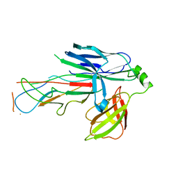
7ZL4
 | | Cryo-EM structure of archaic chaperone-usher Csu pilus of Acinetobacter baumannii | | 分子名称: | CsuA/B | | 著者 | Pakharukova, N, Malmi, H, Tuittila, M, Paavilainen, S, Ghosal, D, Chang, Y.W, Jensen, G.J, Zavialov, A.V. | | 登録日 | 2022-04-13 | | 公開日 | 2022-08-03 | | 最終更新日 | 2022-09-21 | | 実験手法 | ELECTRON MICROSCOPY (3.45 Å) | | 主引用文献 | Archaic chaperone-usher pili self-secrete into superelastic zigzag springs.
Nature, 609, 2022
|
|
7AK9
 | | Structure of Salmonella TacT3 toxin bound to TacA3 antitoxin C-terminal peptide | | 分子名称: | ABC transporter, Acetyltransferase, DEPHOSPHO COENZYME A | | 著者 | Grabe, G.J, Morgan, R.M.L, Hare, S.A, Helaine, S. | | 登録日 | 2020-09-30 | | 公開日 | 2021-08-18 | | 最終更新日 | 2024-01-31 | | 実験手法 | X-RAY DIFFRACTION (2.55 Å) | | 主引用文献 | Auxiliary interfaces support the evolution of specific toxin-antitoxin pairing.
Nat.Chem.Biol., 17, 2021
|
|
7AK7
 | | Structure of Salmonella TacT2 toxin bound to TacA2 antitoxin | | 分子名称: | ACETYL COENZYME *A, Acetyltransferase, CHLORIDE ION, ... | | 著者 | Grabe, G.J, Morgan, R.M.L, Hare, S.A, Helaine, S. | | 登録日 | 2020-09-30 | | 公開日 | 2021-08-18 | | 最終更新日 | 2024-01-31 | | 実験手法 | X-RAY DIFFRACTION (2.14 Å) | | 主引用文献 | Auxiliary interfaces support the evolution of specific toxin-antitoxin pairing.
Nat.Chem.Biol., 17, 2021
|
|
7AK8
 | | Structure of Salmonella TacT1 toxin bound to TacA1 antitoxin C-terminal peptide | | 分子名称: | ACETYL COENZYME *A, GCN5 family acetyltransferase, GLYCEROL, ... | | 著者 | Grabe, G.J, Morgan, R.M.L, Helaine, S, Hare, S.A. | | 登録日 | 2020-09-30 | | 公開日 | 2021-08-18 | | 最終更新日 | 2024-01-31 | | 実験手法 | X-RAY DIFFRACTION (2.5 Å) | | 主引用文献 | Auxiliary interfaces support the evolution of specific toxin-antitoxin pairing.
Nat.Chem.Biol., 17, 2021
|
|
5FVJ
 | | Crystal structure of TacT (tRNA acetylating toxin) from Salmonella | | 分子名称: | ACETYL COENZYME *A, PUTATIVE ACETYLTRANSFERASE | | 著者 | Przydacz, M, Wong, C.T, Cheverton, A.M, Gollan, B, Mylona, A, Helaine, S, Hare, S.A. | | 登録日 | 2016-02-08 | | 公開日 | 2016-06-01 | | 最終更新日 | 2024-05-08 | | 実験手法 | X-RAY DIFFRACTION (1.7 Å) | | 主引用文献 | A Salmonella Toxin Promotes Persister Formation Through Acetylation of tRNA.
Mol.Cell, 63, 2016
|
|
3ETP
 | | The crystal structure of the ligand-binding domain of the EphB2 receptor at 2.0 A resolution | | 分子名称: | Ephrin type-B receptor 2 | | 著者 | Goldgur, Y, Paavilainen, S, Nikolov, D.B, Himanen, J.P. | | 登録日 | 2008-10-08 | | 公開日 | 2008-10-21 | | 最終更新日 | 2023-11-29 | | 実験手法 | X-RAY DIFFRACTION (2 Å) | | 主引用文献 | Structure of the ligand-binding domain of the EphB2 receptor at 2 A resolution.
Acta Crystallogr.,Sect.F, 65, 2009
|
|
1ZC2
 | | Crystal Structure of plasmid-encoded class C beta-lactamase CMY-2 complexed with citrate molecule | | 分子名称: | CITRIC ACID, beta-lactamase class C | | 著者 | Bauvois, C, Jacquamet, L, Fieulaine, S, Frere, J.-M, Galleni, M, Ferrer, J.-L. | | 登録日 | 2005-04-10 | | 公開日 | 2006-04-25 | | 最終更新日 | 2024-03-13 | | 実験手法 | X-RAY DIFFRACTION (2.09 Å) | | 主引用文献 | Crystallographic structure of plasmid-encoded CMY-2 beta-lactamase revealed citrate molecule in the active site.
To be Published
|
|
3EKK
 | | Insulin receptor kinase complexed with an inhibitor | | 分子名称: | 2-[(2-{[1-(N,N-dimethylglycyl)-5-methoxy-1H-indol-6-yl]amino}-7H-pyrrolo[2,3-d]pyrimidin-4-yl)amino]-6-fluoro-N-methylbenzamide, Insulin receptor | | 著者 | Chamberlain, S, Atkins, C, Deanda, F, Dumble, M, Gerding, R, Groy, A, Korenchuk, S, Kumar, R, Lei, H, Mook, R, Moorthy, G, Redman, A, Rowland, J, Sabbatini, P, Shewchuk, L. | | 登録日 | 2008-09-19 | | 公開日 | 2008-12-23 | | 最終更新日 | 2023-08-30 | | 実験手法 | X-RAY DIFFRACTION (2.1 Å) | | 主引用文献 | Discovery of 4,6-bis-anilino-1H-pyrrolo[2,3-d]pyrimidines: Potent inhibitors of the IGF-1R receptor tyrosine kinase.
Bioorg.Med.Chem.Lett., 19, 2009
|
|
3ELJ
 | | Jnk1 complexed with a bis-anilino-pyrrolopyrimidine inhibitor. | | 分子名称: | 2-fluoro-6-{[2-({2-methoxy-4-[(methylsulfonyl)methyl]phenyl}amino)-7H-pyrrolo[2,3-d]pyrimidin-4-yl]amino}benzamide, Mitogen-activated protein kinase 8 | | 著者 | Chamberlain, S, Atkins, C, Deanda, F, Dumble, M, Gerding, R, Groy, A, Korenchuk, S, Kumar, R, Lei, H, Mook, R, Moorthy, G, Redman, A, Rowland, J, Shewchuk, L, Vicentini, G, Mosley, J. | | 登録日 | 2008-09-22 | | 公開日 | 2008-12-30 | | 最終更新日 | 2023-09-06 | | 実験手法 | X-RAY DIFFRACTION (1.8 Å) | | 主引用文献 | Optimization of 4,6-bis-anilino-1H-pyrrolo[2,3-d]pyrimidine IGF-1R tyrosine kinase inhibitors towards JNK selectivity.
Bioorg.Med.Chem.Lett., 19, 2009
|
|
3EKN
 | | Insulin receptor kinase complexed with an inhibitor | | 分子名称: | 2-fluoro-6-{[2-({2-methoxy-4-[4-(1-methylethyl)piperazin-1-yl]phenyl}amino)-7H-pyrrolo[2,3-d]pyrimidin-4-yl]amino}benzamide, Insulin receptor | | 著者 | Chamberlain, S, Atkins, C, Deanda, F, Dumble, M, Gerding, R, Groy, A, Korenchuk, S, Kumar, R, Lei, H, Mook, R, Moorthy, G, Redman, A, Rowland, J, Shewchuk, L. | | 登録日 | 2008-09-19 | | 公開日 | 2008-12-30 | | 最終更新日 | 2023-08-30 | | 実験手法 | X-RAY DIFFRACTION (2.2 Å) | | 主引用文献 | Optimization of 4,6-bis-anilino-1H-pyrrolo[2,3-d]pyrimidine IGF-1R tyrosine kinase inhibitors towards JNK selectivity.
Bioorg.Med.Chem.Lett., 19, 2009
|
|
6Z9I
 | | Escherichia coli D-2-deoxyribose-5-phosphate aldolase - N21K mutant complex with reaction products | | 分子名称: | 1,2-ETHANEDIOL, Deoxyribose-phosphate aldolase, GLYCERALDEHYDE-3-PHOSPHATE, ... | | 著者 | Paakkonen, J, Hakulinen, N, Rouvinen, J. | | 登録日 | 2020-06-04 | | 公開日 | 2020-11-18 | | 最終更新日 | 2024-01-24 | | 実験手法 | X-RAY DIFFRACTION (1.86 Å) | | 主引用文献 | Substrate specificity of 2-deoxy-D-ribose 5-phosphate aldolase (DERA) assessed by different protein engineering and machine learning methods.
Appl.Microbiol.Biotechnol., 104, 2020
|
|
6Z9H
 | | Escherichia coli D-2-deoxyribose-5-phosphate aldolase - C47V/G204A/S239D mutant | | 分子名称: | 1,2-ETHANEDIOL, Deoxyribose-phosphate aldolase, FORMIC ACID, ... | | 著者 | Paakkonen, J, Hakulinen, N, Rouvinen, J. | | 登録日 | 2020-06-04 | | 公開日 | 2020-11-18 | | 最終更新日 | 2024-01-24 | | 実験手法 | X-RAY DIFFRACTION (1.72 Å) | | 主引用文献 | Substrate specificity of 2-deoxy-D-ribose 5-phosphate aldolase (DERA) assessed by different protein engineering and machine learning methods.
Appl.Microbiol.Biotechnol., 104, 2020
|
|
6Z9J
 | |
4CHK
 | | Crystal Structure of the ARF5 oligomerization domain | | 分子名称: | AUXIN RESPONSE FACTOR 5 | | 著者 | Nanao, M.H, Mazzoleni, M, Thevenon, E, Brunoud, G, Vernoux, T, Parcy, F, Dumas, R. | | 登録日 | 2013-12-03 | | 公開日 | 2014-04-16 | | 最終更新日 | 2024-05-08 | | 実験手法 | X-RAY DIFFRACTION (2.85 Å) | | 主引用文献 | Structural Basis for Oligomerisation of Auxin Transcriptional Regulators
Nat.Commun., 5, 2014
|
|
3ETA
 | | Kinase domain of insulin receptor complexed with a pyrrolo pyridine inhibitor | | 分子名称: | 1-(3-{5-[4-(aminomethyl)phenyl]-1H-pyrrolo[2,3-b]pyridin-3-yl}phenyl)-3-(2-phenoxyphenyl)urea, insulin receptor, kinase domain | | 著者 | Patnaik, S, Stevens, K, Gerding, R, Deanda, F, Shotwell, B, Tang, J, Hamajima, T, Nakamura, H, Leesnitzer, A, Hassell, A, Shewchuk, L, Kumar, R, Lei, H, Chamberlain, S. | | 登録日 | 2008-10-07 | | 公開日 | 2009-05-26 | | 最終更新日 | 2023-09-06 | | 実験手法 | X-RAY DIFFRACTION (2.6 Å) | | 主引用文献 | Discovery of 3,5-disubstituted-1H-pyrrolo[2,3-b]pyridines as potent inhibitors of the insulin-like growth factor-1 receptor (IGF-1R) tyrosine kinase.
Bioorg.Med.Chem.Lett., 19, 2009
|
|
5LN8
 | | Crystal structure of self-complemented MyfA, the major subunit of Myf fimbriae from Yersinia enterocolitica, in complex with galactose | | 分子名称: | Fimbrial protein MyfA,Fimbrial protein MyfA, beta-D-galactopyranose | | 著者 | Pakharukova, N.A, Roy, S, Rahman, M.M, Tuitilla, M, Zavialov, A.V. | | 登録日 | 2016-08-03 | | 公開日 | 2016-08-24 | | 最終更新日 | 2024-01-10 | | 実験手法 | X-RAY DIFFRACTION (1.65 Å) | | 主引用文献 | Structural basis for Myf and Psa fimbriae-mediated tropism of pathogenic strains of Yersinia for host tissues.
Mol.Microbiol., 102, 2016
|
|


