7MPP
 
 | |
7MPL
 | |
7MPR
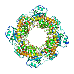
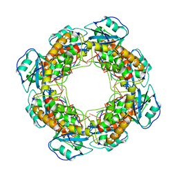
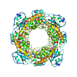
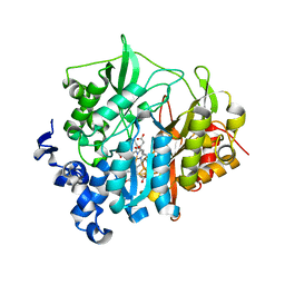
 | | Brucella melitensis NrnC | | 分子名称: | NanoRNase C, SULFATE ION | | 著者 | Lormand, J.D, Sondermann, H. | | 登録日 | 2021-05-04 | | 公開日 | 2021-09-15 | | 最終更新日 | 2023-10-18 | | 実験手法 | X-RAY DIFFRACTION (1.42 Å) | | 主引用文献 | Structural characterization of NrnC identifies unifying features of dinucleotidases.
Elife, 10, 2021
|
|
7MQC
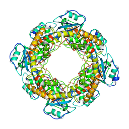
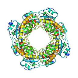
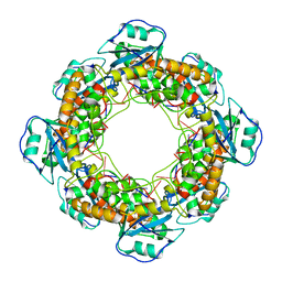
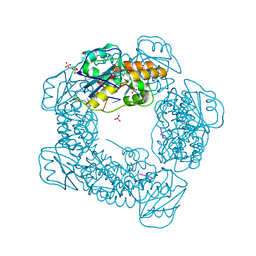
 | | Bartonella henselae NrnC bound to pGG. C1 reconstruction. | | 分子名称: | NanoRNase C, RNA (5'-R(P*GP*G)-3') | | 著者 | Lormand, J.D, Brownfield, B, Fromme, J.C, Sondermann, H. | | 登録日 | 2021-05-05 | | 公開日 | 2021-09-15 | | 最終更新日 | 2024-05-29 | | 実験手法 | ELECTRON MICROSCOPY (3.64 Å) | | 主引用文献 | Structural characterization of NrnC identifies unifying features of dinucleotidases.
Elife, 10, 2021
|
|
7MQG
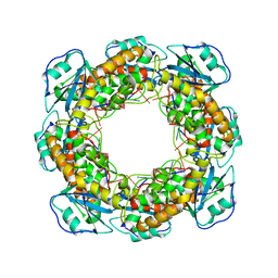
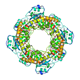
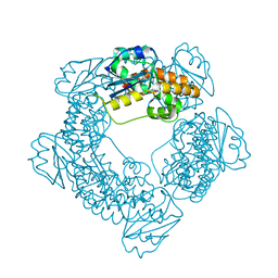
 | | Bartonella henselae NrnC complexed with pAAAGG. C1 reconstruction. | | 分子名称: | NanoRNase C, RNA (5'-R(P*AP*AP*AP*GP*G)-3') | | 著者 | Lormand, J.D, Brownfield, B, Fromme, J.C, Sondermann, H. | | 登録日 | 2021-05-05 | | 公開日 | 2021-09-15 | | 最終更新日 | 2024-05-29 | | 実験手法 | ELECTRON MICROSCOPY (3.25 Å) | | 主引用文献 | Structural characterization of NrnC identifies unifying features of dinucleotidases.
Elife, 10, 2021
|
|
7MPM
 | |
7MPO
 | |
7MPN
 | |
7MQI
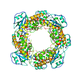
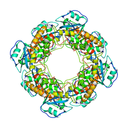 | | Bartonella henselae NrnC complexed with pAAAGG in the presence of Ca2+. C1 reconstruction. | | 分子名称: | NanoRNase C, RNA (5'-R(P*AP*AP*AP*GP*G)-3') | | 著者 | Lormand, J.D, Brownfield, B, Fromme, J.C, Sondermann, H. | | 登録日 | 2021-05-05 | | 公開日 | 2021-09-15 | | 最終更新日 | 2024-05-29 | | 実験手法 | ELECTRON MICROSCOPY (3.21 Å) | | 主引用文献 | Structural characterization of NrnC identifies unifying features of dinucleotidases.
Elife, 10, 2021
|
|
7MPQ
 | |
7MQH
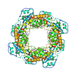
 | | Bartonella henselae NrnC complexed with pAAAGG in the presence of Ca2+. D4 Symmetry. | | 分子名称: | NanoRNase C, RNA (5'-R(P*AP*AP*AP*GP*G)-3') | | 著者 | Lormand, J.D, Brownfield, B, Fromme, J.C, Sondermann, H. | | 登録日 | 2021-05-05 | | 公開日 | 2021-09-15 | | 最終更新日 | 2024-05-29 | | 実験手法 | ELECTRON MICROSCOPY (3.1 Å) | | 主引用文献 | Structural characterization of NrnC identifies unifying features of dinucleotidases.
Elife, 10, 2021
|
|
7MPT
 | |
7MQF
 | | Bartonella henselae NrnC complexed with pAAAGG. D4 symmetry. | | 分子名称: | NanoRNase C, RNA (5'-R(P*AP*AP*AP*GP*G)-3') | | 著者 | Lormand, J.D, Brownfield, B, Fromme, J.C, Sondermann, H. | | 登録日 | 2021-05-05 | | 公開日 | 2021-09-15 | | 最終更新日 | 2024-05-29 | | 実験手法 | ELECTRON MICROSCOPY (2.88 Å) | | 主引用文献 | Structural characterization of NrnC identifies unifying features of dinucleotidases.
Elife, 10, 2021
|
|
7MPU
 | |
7MQE
 | | Bartonella henselae NrnC complexed with pAGG. C1 reconstruction. | | 分子名称: | NanoRNase C, RNA (5'-R(P*AP*GP*G)-3') | | 著者 | Lormand, J.D, Brownfield, B, Fromme, J.C, Sondermann, H. | | 登録日 | 2021-05-05 | | 公開日 | 2021-09-15 | | 最終更新日 | 2024-05-29 | | 実験手法 | ELECTRON MICROSCOPY (3.69 Å) | | 主引用文献 | Structural characterization of NrnC identifies unifying features of dinucleotidases.
Elife, 10, 2021
|
|
2AKJ
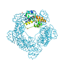
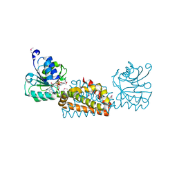
 | | Structure of spinach nitrite reductase | | 分子名称: | Ferredoxin--nitrite reductase, chloroplast, IRON/SULFUR CLUSTER, ... | | 著者 | Swamy, U, Wang, M, Tripathy, J.N, Kim, S.-K, Hirasawa, M, Knaff, D.B, Allen, J.P. | | 登録日 | 2005-08-03 | | 公開日 | 2006-01-24 | | 最終更新日 | 2023-12-27 | | 実験手法 | X-RAY DIFFRACTION (2.8 Å) | | 主引用文献 | Structure of Spinach Nitrite Reductase: Implications for Multi-electron Reactions by the Iron-Sulfur:Siroheme Cofactor
Biochemistry, 44, 2005
|
|
3B1F
 | | Crystal structure of prephenate dehydrogenase from Streptococcus mutans | | 分子名称: | NICOTINAMIDE-ADENINE-DINUCLEOTIDE, Putative prephenate dehydrogenase | | 著者 | Ku, H.K, Do, N.H, Song, J.S, Choi, S, Shin, M.H, Kim, K.J, Lee, S.J. | | 登録日 | 2011-07-02 | | 公開日 | 2011-10-26 | | 実験手法 | X-RAY DIFFRACTION (2.1 Å) | | 主引用文献 | Crystal structure of prephenate dehydrogenase from Streptococcus mutans.
Int.J.Biol.Macromol., 49, 2011
|
|
3QMX
 | |
1ZDN
 | | Ubiquitin-conjugating enzyme E2S | | 分子名称: | SODIUM ION, Ubiquitin-conjugating enzyme E2S | | 著者 | Walker, J.R, Avvakumov, G.V, Xue, S, Newman, E.M, Mackenzie, F, Sundstrom, M, Arrowsmith, C, Edwards, A, Bochkarev, A, Dhe-Paganon, S, Structural Genomics Consortium (SGC) | | 登録日 | 2005-04-14 | | 公開日 | 2005-05-03 | | 最終更新日 | 2023-08-23 | | 実験手法 | X-RAY DIFFRACTION (1.93 Å) | | 主引用文献 | A human ubiquitin conjugating enzyme (E2)-HECT E3 ligase structure-function screen.
Mol Cell Proteomics, 11, 2012
|
|
1YRV
 | | Novel Ubiquitin-Conjugating Enzyme | | 分子名称: | ubiquitin-conjugating ligase MGC351130 | | 著者 | Walker, J.R, Choe, J, Avvakumov, G.V, Newman, E.M, MacKenzie, F, Sundstrom, M, Arrowsmith, C, Edwards, A, Bochkarev, A, Dhe-Paganon, S, Structural Genomics Consortium (SGC) | | 登録日 | 2005-02-04 | | 公開日 | 2005-03-22 | | 最終更新日 | 2023-08-23 | | 実験手法 | X-RAY DIFFRACTION (2.18 Å) | | 主引用文献 | A human ubiquitin conjugating enzyme (E2)-HECT E3 ligase structure-function screen.
Mol Cell Proteomics, 11, 2012
|
|
1YH2
 | | Ubiquitin-Conjugating Enzyme HSPC150 | | 分子名称: | HSPC150 protein similar to ubiquitin-conjugating enzyme | | 著者 | Walker, J.R, Avvakumov, G.V, Newman, E.M, Mackenzie, F, Kozieradzki, I, Sundstrom, M, Arrowsmith, C, Edwards, A, Bochkarev, A, Dhe-paganon, S, Structural Genomics Consortium (SGC) | | 登録日 | 2005-01-06 | | 公開日 | 2005-02-15 | | 最終更新日 | 2023-08-23 | | 実験手法 | X-RAY DIFFRACTION (2 Å) | | 主引用文献 | A human ubiquitin conjugating enzyme (E2)-HECT E3 ligase structure-function screen.
Mol Cell Proteomics, 11, 2012
|
|
1ZUO
 | | Structure of Human Ubiquitin-Conjugating Enzyme (UBCi) Involved in Embryo Attachment and Implantation | | 分子名称: | BETA-MERCAPTOETHANOL, Hypothetical protein LOC92912 | | 著者 | Walker, J.R, Avvakumov, G.V, Cui, H, Newman, E.M, Mackenzie, F, Sundstrom, M, Arrowsmith, C, Edwards, A, Bochkarev, A, Dhe-Paganon, S, Structural Genomics Consortium (SGC) | | 登録日 | 2005-05-31 | | 公開日 | 2005-07-05 | | 最終更新日 | 2012-11-28 | | 実験手法 | X-RAY DIFFRACTION (1.8 Å) | | 主引用文献 | A human ubiquitin conjugating enzyme (E2)-HECT E3 ligase structure-function screen.
Mol Cell Proteomics, 11, 2012
|
|
1Y6L
 | | Human ubiquitin conjugating enzyme E2E2 | | 分子名称: | Ubiquitin-conjugating enzyme E2E2 | | 著者 | Walker, J.R, Avvakumov, G.V, Newman, E.M, Mackenzie, F, Kozieradzki, I, Bochkarev, A, Sundstrom, M, Arrowsmith, C, Edwards, A, Dhe-Paganon, S, Structural Genomics Consortium (SGC) | | 登録日 | 2004-12-06 | | 公開日 | 2005-01-11 | | 最終更新日 | 2023-08-23 | | 実験手法 | X-RAY DIFFRACTION (1.85 Å) | | 主引用文献 | A human ubiquitin conjugating enzyme (E2)-HECT E3 ligase structure-function screen.
Mol Cell Proteomics, 11, 2012
|
|
2A7L
 | | Structure of the human hypothetical ubiquitin-conjugating enzyme, LOC55284 | | 分子名称: | Hypothetical ubiquitin-conjugating enzyme LOC55284, SODIUM ION | | 著者 | Walker, J.R, Avvakumov, G.V, Xue, S, Newman, E.M, Mackenzie, F, Weigelt, J, Sundstrom, M, Arrowsmith, C, Edwards, A, Bochkarev, A, Dhe-Paganon, S, Structural Genomics Consortium (SGC) | | 登録日 | 2005-07-05 | | 公開日 | 2005-09-06 | | 最終更新日 | 2023-08-23 | | 実験手法 | X-RAY DIFFRACTION (1.82 Å) | | 主引用文献 | A human ubiquitin conjugating enzyme (E2)-HECT E3 ligase structure-function screen.
Mol Cell Proteomics, 11, 2012
|
|
2AWF
 | | Structure of human Ubiquitin-conjugating enzyme E2 G1 | | 分子名称: | Ubiquitin-conjugating enzyme E2 G1 | | 著者 | Walker, J.R, Avvakumov, G.V, Xue, S, Newman, E.M, Finerty, P, Mackenzie, F, Weigelt, J, Sundstrom, M, Arrowsmith, C, Edwards, A, Bochkarev, A, Dhe-Paganon, S, Structural Genomics Consortium (SGC) | | 登録日 | 2005-09-01 | | 公開日 | 2005-09-20 | | 最終更新日 | 2023-08-23 | | 実験手法 | X-RAY DIFFRACTION (2.1 Å) | | 主引用文献 | A human ubiquitin conjugating enzyme (E2)-HECT E3 ligase structure-function screen.
Mol Cell Proteomics, 11, 2012
|
|


