Loading
PDBj
メニューPDBj@FacebookPDBj@TwitterPDBj@YouTubewwPDB FoundationwwPDB
RCSB PDBPDBeBMRBAdv. SearchSearch help
Search by PDB author
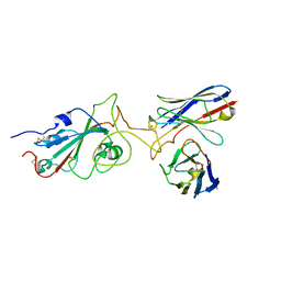
7Y6N
DownloadVisualize
BU of 7y6n by Molmil
The SARS-CoV-2 receptor binding domain bound with the Fab fragment of a human neutralizing antibody Ab803
分子名称: 2-acetamido-2-deoxy-beta-D-glucopyranose, Ab803 heavy chain, Ab803 light chain, ...
著者Uchikubo, T, Shirouzu, M.
登録日2022-06-21
公開日2023-06-21
最終更新日2023-09-13
実験手法ELECTRON MICROSCOPY (4.4 Å)
主引用文献Potent neutralizing broad-spectrum antibody against SARS-CoV-2 generated from dual-antigen-specific B cells from convalescents.
Iscience, 26, 2023
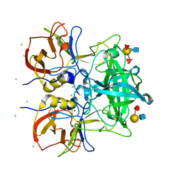
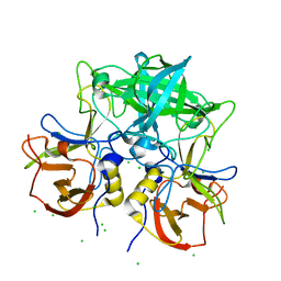
7VS9
DownloadVisualize
BU of 7vs9 by Molmil
Crystal structure of P domain from norovirus GI.9 capsid protein in complex with Lewis x antigen.
分子名称: CHLORIDE ION, MAGNESIUM ION, VP1, ...
著者Murayama, K, Kato-Murayama, M, Shirouzu, M.
登録日2021-10-26
公開日2022-08-31
最終更新日2023-11-29
実験手法X-RAY DIFFRACTION (2.26 Å)
主引用文献Lewis fucose is a key moiety for the recognition of histo-blood group antigens by GI.9 norovirus, as revealed by structural analysis.
Febs Open Bio, 12, 2022
7VS8
DownloadVisualize
BU of 7vs8 by Molmil
Crystal structure of P domain from norovirus GI.9 capsid protein in complex with Lewis b antigen.
分子名称: CHLORIDE ION, MAGNESIUM ION, VP1, ...
著者Murayama, K, Kato-Murayama, M, Shirouzu, M.
登録日2021-10-26
公開日2022-08-31
最終更新日2023-11-29
実験手法X-RAY DIFFRACTION (2.4 Å)
主引用文献Lewis fucose is a key moiety for the recognition of histo-blood group antigens by GI.9 norovirus, as revealed by structural analysis.
Febs Open Bio, 12, 2022
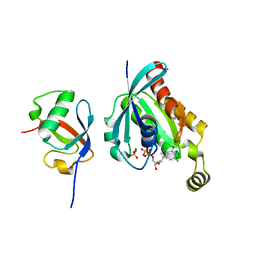
7Y4A
DownloadVisualize
BU of 7y4a by Molmil
Crystal structure of human ELMO1 RBD-RhoG complex
分子名称: Engulfment and cell motility protein 1, GUANOSINE-5'-DIPHOSPHATE, MAGNESIUM ION, ...
著者Tsuda, K, Kukimoto-Niino, M, Shirouzu, M.
登録日2022-06-14
公開日2023-03-15
最終更新日2023-11-29
実験手法X-RAY DIFFRACTION (1.6 Å)
主引用文献Targeting Ras-binding domain of ELMO1 by computational nanobody design.
Commun Biol, 6, 2023
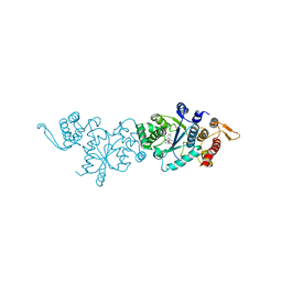
7YNW
DownloadVisualize
BU of 7ynw by Molmil
Crystal structure of O-(2-nitrobenzyl)-L-tyrosine-tRNA sythetase in complex with O-(2-nitrobenzyl)-L-tyrosine
分子名称: (2~{S})-2-azanyl-3-[4-[(2-nitrophenyl)methoxy]phenyl]propanoic acid, Tyrosine--tRNA ligase
著者Hosaka, T, Shirouzu, M.
登録日2022-08-01
公開日2022-09-21
最終更新日2023-11-29
実験手法X-RAY DIFFRACTION (2.79 Å)
主引用文献Crystal Structure of an Archaeal Tyrosyl-tRNA Synthetase Bound to Photocaged L-Tyrosine and Its Potential Application to Time-Resolved X-ray Crystallography.
Int J Mol Sci, 23, 2022
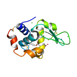
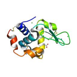
7YNU
DownloadVisualize
BU of 7ynu by Molmil
Crystal structure of Hen Egg white LYSOZYME introduced with O-(2-nitrobenzyl)-L-tyrosine
分子名称: ACETATE ION, CHLORIDE ION, Lysozyme C, ...
著者Hosaka, T, Shirouzu, M.
登録日2022-08-01
公開日2022-09-21
最終更新日2024-11-06
実験手法X-RAY DIFFRACTION (1.44 Å)
主引用文献Crystal Structure of an Archaeal Tyrosyl-tRNA Synthetase Bound to Photocaged L-Tyrosine and Its Potential Application to Time-Resolved X-ray Crystallography.
Int J Mol Sci, 23, 2022
7YNV
DownloadVisualize
BU of 7ynv by Molmil
Crystal structure of photolysed Hen Egg white LYSOZYME introduced with O-(2-nitrobenzyl)-L-tyrosine
分子名称: ACETATE ION, CHLORIDE ION, Lysozyme C, ...
著者Hosaka, T, Shirouzu, M.
登録日2022-08-01
公開日2022-09-21
最終更新日2023-11-29
実験手法X-RAY DIFFRACTION (1.39 Å)
主引用文献Crystal Structure of an Archaeal Tyrosyl-tRNA Synthetase Bound to Photocaged L-Tyrosine and Its Potential Application to Time-Resolved X-ray Crystallography.
Int J Mol Sci, 23, 2022
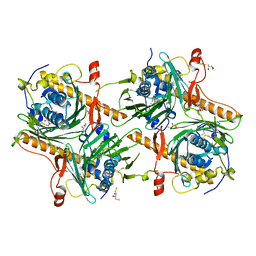
7V62
DownloadVisualize
BU of 7v62 by Molmil
Crystal structure of human OSBP ORD in complex with cholesterol
分子名称: 2,3-DIHYDROXY-1,4-DITHIOBUTANE, CHOLESTEROL, CITRIC ACID, ...
著者Kobayashi, J, Kato, R.
登録日2021-08-19
公開日2022-06-08
最終更新日2023-11-29
実験手法X-RAY DIFFRACTION (3.25 Å)
主引用文献Ligand Recognition by the Lipid Transfer Domain of Human OSBP Is Important for Enterovirus Replication.
Acs Infect Dis., 8, 2022
<123


227111

件を2024-11-06に公開中

PDB statisticsPDBj update infoContact PDBjnumon