3E9B
 
 | |
3E8Z
 | |
5SBG
 | |
1NVO
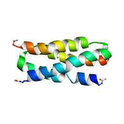
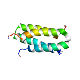 | | Solution structure of a four-helix bundle model, apo-DF1 | | 分子名称: | Homodimeric Alpha2 Four-Helix Bundle | | 著者 | Maglio, O, Nastri, F, Pavone, V, Lombardi, A, DeGrado, W.F. | | 登録日 | 2003-02-04 | | 公開日 | 2003-03-25 | | 最終更新日 | 2022-02-23 | | 実験手法 | SOLUTION NMR | | 主引用文献 | Preorganization of molecular binding sites in designed diiron proteins
Proc.Natl.Acad.Sci.USA, 100, 2003
|
|
2KIK
 | |
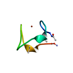
3BKD
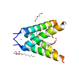 | | High resolution Crystal structure of Transmembrane domain of M2 protein | | 分子名称: | CHLORIDE ION, DI(HYDROXYETHYL)ETHER, Transmembrane Domain of Matrix protein M2, ... | | 著者 | Stouffer, A.L, Acharya, R, Salom, D. | | 登録日 | 2007-12-06 | | 公開日 | 2008-01-29 | | 最終更新日 | 2024-04-03 | | 実験手法 | X-RAY DIFFRACTION (2.05 Å) | | 主引用文献 | Structural basis for the function and inhibition of an influenza virus proton channel
Nature, 451, 2008
|
|
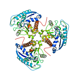
3SL1
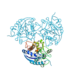 | | Crystal Structure of P. falciparum arginase complexed with 2-amino-6-borono-2-methylhexanoic acid | | 分子名称: | 6-(dihydroxyboranyl)-2-methyl-L-norleucine, Arginase, MANGANESE (II) ION | | 著者 | Dowling, D.P, Ilies, M, Christianson, D.W. | | 登録日 | 2011-06-23 | | 公開日 | 2011-07-20 | | 最終更新日 | 2023-09-13 | | 実験手法 | X-RAY DIFFRACTION (1.902 Å) | | 主引用文献 | Binding of alpha , alpha-disubstituted amino acids to arginase suggests new avenues for inhibitor design.
J.Med.Chem., 54, 2011
|
|
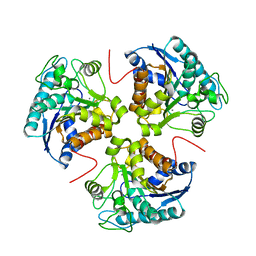
3SL0
 | | Crystal Structure of P. falciparum arginase complexed with 2-amino-6-borono-2-(difluoromethyl)hexanoic acid | | 分子名称: | 2-(difluoromethyl)-6-(dihydroxyboranyl)-L-norleucine, Arginase, MANGANESE (II) ION | | 著者 | Dowling, D.P, Ilies, M, Christianson, D.W. | | 登録日 | 2011-06-23 | | 公開日 | 2011-07-20 | | 最終更新日 | 2023-09-13 | | 実験手法 | X-RAY DIFFRACTION (1.997 Å) | | 主引用文献 | Binding of alpha , alpha-disubstituted amino acids to arginase suggests new avenues for inhibitor design.
J.Med.Chem., 54, 2011
|
|


