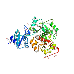
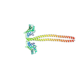
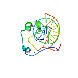
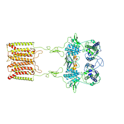
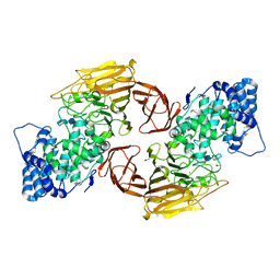
6KTW
 
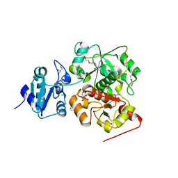 | | structure of EanB with hercynine | | 分子名称: | 1,2-ETHANEDIOL, CHLORIDE ION, GLYCEROL, ... | | 著者 | Wu, L, Liu, P.H, Zhou, J.H. | | 登録日 | 2019-08-29 | | 公開日 | 2020-08-26 | | 最終更新日 | 2023-11-22 | | 実験手法 | X-RAY DIFFRACTION (1.931 Å) | | 主引用文献 | Single-Step Replacement of an Unreactive C-H Bond by a C-S Bond Using Polysulfide as the Direct Sulfur Source in the Anaerobic Ergothioneine Biosynthesis
Acs Catalysis, 10, 2020
|
|
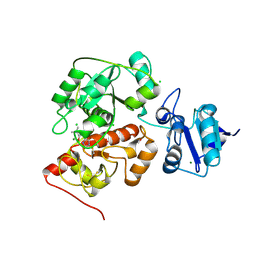
6KU2
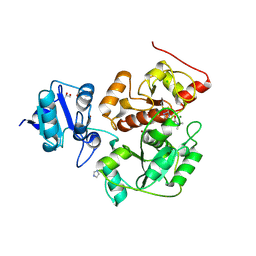 | | The structure of EanB/Y353A complex with ergothioneine covalent linked with persulfide Cys412 | | 分子名称: | 1,2-ETHANEDIOL, BROMIDE ION, CHLORIDE ION, ... | | 著者 | Wu, L, Liu, P.H, Zhou, J.H. | | 登録日 | 2019-08-30 | | 公開日 | 2020-08-26 | | 実験手法 | X-RAY DIFFRACTION (2.34 Å) | | 主引用文献 | Single-Step Replacement of an Unreactive C-H Bond by a C-S Bond Using Polysulfide as the Direct Sulfur Source in the Anaerobic Ergothioneine Biosynthesis
Acs Catalysis, 10, 2020
|
|
6KTZ
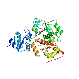 | | The complex structure of EanB/C412S with hercynine | | 分子名称: | 1,2-ETHANEDIOL, BROMIDE ION, CHLORIDE ION, ... | | 著者 | Wu, L, Liu, P.H, Zhou, J.H. | | 登録日 | 2019-08-29 | | 公開日 | 2020-08-26 | | 実験手法 | X-RAY DIFFRACTION (2 Å) | | 主引用文献 | Single-Step Replacement of an Unreactive C-H Bond by a C-S Bond Using Polysulfide as the Direct Sulfur Source in the Anaerobic Ergothioneine Biosynthesis
Acs Catalysis, 10, 2020
|
|
6KTV
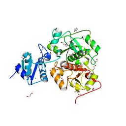 | | The structure of EanB complex with hercynine and persulfided Cys412 | | 分子名称: | 1,2-ETHANEDIOL, 1,3-PROPANDIOL, CHLORIDE ION, ... | | 著者 | Wu, L, Liu, P.H, Zhou, J.H. | | 登録日 | 2019-08-29 | | 公開日 | 2020-08-26 | | 最終更新日 | 2023-11-22 | | 実験手法 | X-RAY DIFFRACTION (2.2 Å) | | 主引用文献 | Single-Step Replacement of an Unreactive C-H Bond by a C-S Bond Using Polysulfide as the Direct Sulfur Source in the Anaerobic Ergothioneine Biosynthesis
Acs Catalysis, 10, 2020
|
|
6KTX
 | | The wildtype structure of EanB | | 分子名称: | 1,2-ETHANEDIOL, 3[N-MORPHOLINO]PROPANE SULFONIC ACID, CHLORIDE ION, ... | | 著者 | Wu, L, Liu, P.H, Zhou, J.H. | | 登録日 | 2019-08-29 | | 公開日 | 2020-08-26 | | 最終更新日 | 2023-11-22 | | 実験手法 | X-RAY DIFFRACTION (2.189 Å) | | 主引用文献 | Single-Step Replacement of an Unreactive C-H Bond by a C-S Bond Using Polysulfide as the Direct Sulfur Source in the Anaerobic Ergothioneine Biosynthesis
Acs Catalysis, 10, 2020
|
|
6KU1
 | | The structure of EanB/Y353A complex with ergothioneine | | 分子名称: | 1,2-ETHANEDIOL, CHLORIDE ION, MAGNESIUM ION, ... | | 著者 | Wu, L, Liu, P.H, Zhou, J.H. | | 登録日 | 2019-08-30 | | 公開日 | 2020-08-26 | | 最終更新日 | 2023-11-22 | | 実験手法 | X-RAY DIFFRACTION (2.25 Å) | | 主引用文献 | Single-Step Replacement of an Unreactive C-H Bond by a C-S Bond Using Polysulfide as the Direct Sulfur Source in the Anaerobic Ergothioneine Biosynthesis
Acs Catalysis, 10, 2020
|
|
6KG9
 | |
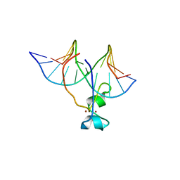
6KGF
 | | Crystal structure of CaDoc0917(R16D)-CaCohA2 complex at pH 8.2 | | 分子名称: | And cellulose-binding endoglucanase family 9 CelL ortholog dockerin domain, CALCIUM ION, Probably cellulosomal scaffolding protein, ... | | 著者 | Feng, Y, Yao, X. | | 登録日 | 2019-07-11 | | 公開日 | 2020-07-08 | | 最終更新日 | 2023-11-22 | | 実験手法 | X-RAY DIFFRACTION (2.3 Å) | | 主引用文献 | Discovery and mechanism of a pH-dependent dual-binding-site switch in the interaction of a pair of protein modules.
Sci Adv, 6, 2020
|
|
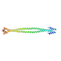
4XA1
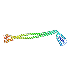 | | Crystal Structure of the coiled-coil surrounding Skip 1 of MYH7 | | 分子名称: | Gp7-MYH7(1173-1238)-EB1 chimera protein | | 著者 | Taylor, K.C, Buvoli, M, Korkmaz, E.N, Buvoli, A, Zheng, Y, Heinz, N.T, Qiang, C, Leinwand, L.A, Rayment, I. | | 登録日 | 2014-12-12 | | 公開日 | 2015-07-01 | | 最終更新日 | 2023-09-27 | | 実験手法 | X-RAY DIFFRACTION (3.2 Å) | | 主引用文献 | Skip residues modulate the structural properties of the myosin rod and guide thick filament assembly.
Proc.Natl.Acad.Sci.USA, 112, 2015
|
|
4XA6
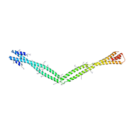 | | Crystal Structure of the coiled-coil surrounding Skip 4 of MYH7 | | 分子名称: | Gp7-MYH7(1777-1855)-EB1 chimera protein | | 著者 | Taylor, K.C, Buvoli, M, Korkmaz, E.N, Buvoli, A, Zheng, Y, Heinz, N.T, Qiang, C, Leinwand, L.A, Rayment, I. | | 登録日 | 2014-12-12 | | 公開日 | 2015-07-01 | | 最終更新日 | 2023-11-15 | | 実験手法 | X-RAY DIFFRACTION (3.42 Å) | | 主引用文献 | Skip residues modulate the structural properties of the myosin rod and guide thick filament assembly.
Proc.Natl.Acad.Sci.USA, 112, 2015
|
|
4XA4
 | | Crystal Structure of the coiled-coil surrounding Skip 3 of MYH7 | | 分子名称: | Xrcc4-MYH7(1551-1609) chimera protein | | 著者 | Taylor, K.C, Buvoli, M, Korkmaz, E.N, Buvoli, A, Zheng, Y, Heinz, N.T, Qiang, C, Leinwand, L.A, Rayment, I. | | 登録日 | 2014-12-12 | | 公開日 | 2015-07-01 | | 最終更新日 | 2023-09-27 | | 実験手法 | X-RAY DIFFRACTION (2.327 Å) | | 主引用文献 | Skip residues modulate the structural properties of the myosin rod and guide thick filament assembly.
Proc.Natl.Acad.Sci.USA, 112, 2015
|
|
4XA3
 | | Crystal structure of the coiled-coil surrounding Skip 2 of MYH7 | | 分子名称: | Gp7-MYH7(1361-1425)-Eb1 chimera protein | | 著者 | Taylor, K.C, Buvoli, M, Korkmaz, E.N, Buvoli, A, Zheng, Y, Heinz, N.T, Qiang, C, Leinwand, L.A, Rayment, I. | | 登録日 | 2014-12-12 | | 公開日 | 2015-07-01 | | 最終更新日 | 2023-09-27 | | 実験手法 | X-RAY DIFFRACTION (2.548 Å) | | 主引用文献 | Skip residues modulate the structural properties of the myosin rod and guide thick filament assembly.
Proc.Natl.Acad.Sci.USA, 112, 2015
|
|
5JON
 | | Crystal structure of the unliganded form of HCN2 CNBD | | 分子名称: | Maltose-binding periplasmic protein,Potassium/sodium hyperpolarization-activated cyclic nucleotide-gated channel 2, NITRATE ION, alpha-D-glucopyranose-(1-4)-alpha-D-glucopyranose | | 著者 | Klenchin, V.A, Chanda, B. | | 登録日 | 2016-05-02 | | 公開日 | 2016-11-30 | | 最終更新日 | 2023-09-27 | | 実験手法 | X-RAY DIFFRACTION (2.042 Å) | | 主引用文献 | Structure and dynamics underlying elementary ligand binding events in human pacemaking channels.
Elife, 5, 2016
|
|
6PZV
 | |
4HP3
 | | Crystal structure of Tet3 in complex with a CpG dsDNA | | 分子名称: | DNA (5'-D(*GP*CP*CP*AP*AP*CP*GP*TP*TP*GP*GP*C)-3'), LOC100036628 protein, UNKNOWN ATOM OR ION, ... | | 著者 | Chao, X, Tempel, W, Bian, C, Bountra, C, Arrowsmith, C.H, Edwards, A.M, Min, J, Structural Genomics Consortium (SGC) | | 登録日 | 2012-10-23 | | 公開日 | 2012-12-05 | | 最終更新日 | 2024-04-03 | | 実験手法 | X-RAY DIFFRACTION (2.05 Å) | | 主引用文献 | Tet3 CXXC Domain and Dioxygenase Activity Cooperatively Regulate Key Genes for Xenopus Eye and Neural Development.
Cell(Cambridge,Mass.), 151, 2012
|
|
4HP1
 | | Crystal structure of Tet3 in complex with a non-CpG dsDNA | | 分子名称: | DNA (5'-D(*GP*CP*CP*AP*CP*(5CM)P*GP*GP*TP*GP*GP*C)-3'), LOC100036628 protein, ZINC ION | | 著者 | Chao, X, Tempel, W, Bian, C, Bountra, C, Arrowsmith, C.H, Edwards, A.M, Min, J, Structural Genomics Consortium (SGC) | | 登録日 | 2012-10-23 | | 公開日 | 2012-12-05 | | 最終更新日 | 2024-04-03 | | 実験手法 | X-RAY DIFFRACTION (2.25 Å) | | 主引用文献 | Tet3 CXXC Domain and Dioxygenase Activity Cooperatively Regulate Key Genes for Xenopus Eye and Neural Development.
Cell(Cambridge,Mass.), 151, 2012
|
|
2MMP
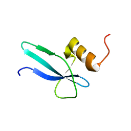 | | Solution structure of a ribosomal protein | | 分子名称: | Uncharacterized protein | | 著者 | Feng, Y. | | 登録日 | 2014-03-17 | | 公開日 | 2014-06-11 | | 最終更新日 | 2024-05-15 | | 実験手法 | SOLUTION NMR | | 主引用文献 | Structure determination of archaea-specific ribosomal protein L46a reveals a novel protein fold.
Biochem.Biophys.Res.Commun., 450, 2014
|
|
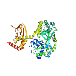
3HBN
 | | Crystal structure PseG-UDP complex from Campylobacter jejuni | | 分子名称: | CHLORIDE ION, GLYCEROL, UDP-sugar hydrolase, ... | | 著者 | Rangarajan, E.S, Proteau, A, Cygler, M, Matte, A, Sulea, T, Schoenhofen, I.C. | | 登録日 | 2009-05-04 | | 公開日 | 2009-05-26 | | 最終更新日 | 2023-11-22 | | 実験手法 | X-RAY DIFFRACTION (1.85 Å) | | 主引用文献 | Structural and functional analysis of Campylobacter jejuni PseG: a udp-sugar hydrolase from the pseudaminic acid biosynthetic pathway.
J.Biol.Chem., 284, 2009
|
|
3HBM
 | | Crystal Structure of PseG from Campylobacter jejuni | | 分子名称: | SULFATE ION, UDP-sugar hydrolase | | 著者 | Rangarajan, E.S, Proteau, A, Cygler, M, Matte, A, Sulea, T, Schoenhofen, I.C. | | 登録日 | 2009-05-04 | | 公開日 | 2009-05-26 | | 最終更新日 | 2021-10-13 | | 実験手法 | X-RAY DIFFRACTION (1.8 Å) | | 主引用文献 | Structural and functional analysis of Campylobacter jejuni PseG: a udp-sugar hydrolase from the pseudaminic acid biosynthetic pathway.
J.Biol.Chem., 284, 2009
|
|
7E6T
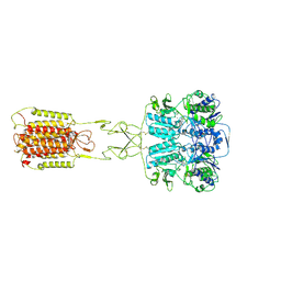 | | Structural insights into the activation of human calcium-sensing receptor | | 分子名称: | 2-acetamido-2-deoxy-beta-D-glucopyranose, CALCIUM ION, CYCLOMETHYLTRYPTOPHAN, ... | | 著者 | Geng, Y, Chen, X.C, Wang, L, Cui, Q.Q, Ding, Z.Y, Han, L, Kou, Y.J, Zhang, W.Q, Wang, H.N, Jia, X.M, Dai, M, Shi, Z.Z, Li, Y.Y, Li, X.Y. | | 登録日 | 2021-02-24 | | 公開日 | 2021-09-22 | | 実験手法 | ELECTRON MICROSCOPY (3 Å) | | 主引用文献 | Structural insights into the activation of human calcium-sensing receptor.
Elife, 10, 2021
|
|
7E6U
 | | the complex of inactive CaSR and NB2D11 | | 分子名称: | Extracellular calcium-sensing receptor, NB-2D11 | | 著者 | Geng, Y, Chen, X.C, Wang, L, Cui, Q.Q, Ding, Z.Y, Han, L, Kou, Y.J, Zhang, W.Q, Wang, H.N, Jia, X.M, Dai, M, Shi, Z.Z, Li, Y.Y, Li, X.Y. | | 登録日 | 2021-02-24 | | 公開日 | 2021-09-22 | | 実験手法 | ELECTRON MICROSCOPY (6 Å) | | 主引用文献 | Structural insights into the activation of human calcium-sensing receptor.
Elife, 10, 2021
|
|
7FAU
 | | Structure Determination of the NB1B11-RBD Complex | | 分子名称: | NB_1B11, Spike protein S1, ZINC ION | | 著者 | Shi, Z.Z, Li, X.X, Wang, L, Sun, Z.C, Zhang, H.W, Chen, X.C, Cui, Q.Q, Qiao, H.R, Lan, Z.Y, Zhang, X. | | 登録日 | 2021-07-07 | | 公開日 | 2022-06-01 | | 最終更新日 | 2023-11-29 | | 実験手法 | X-RAY DIFFRACTION (2.08 Å) | | 主引用文献 | Structural basis of nanobodies neutralizing SARS-CoV-2 variants.
Structure, 30, 2022
|
|
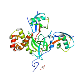
5ZMY
 | | Crystal structure of a cis-epoxysuccinate hydrolase producing D(-)-tartaric acids | | 分子名称: | Cis-epoxysuccinate hydrolase, D(-)-TARTARIC ACID, ZINC ION | | 著者 | Dong, S, Liu, X, Wang, X, Feng, Y. | | 登録日 | 2018-04-06 | | 公開日 | 2018-08-08 | | 最終更新日 | 2023-11-22 | | 実験手法 | X-RAY DIFFRACTION (1.87 Å) | | 主引用文献 | Structural insight into the catalytic mechanism of a cis-epoxysuccinate hydrolase producing enantiomerically pure d(-)-tartaric acid.
Chem. Commun. (Camb.), 54, 2018
|
|
5ZMU
 | | Crystal structure of a cis-epoxysuccinate hydrolase producing D(-)-tartaric acids | | 分子名称: | Cis-epoxysuccinate hydrolase, ZINC ION | | 著者 | Dong, S, Liu, X, Wang, X, Feng, Y. | | 登録日 | 2018-04-06 | | 公開日 | 2018-08-08 | | 最終更新日 | 2023-11-22 | | 実験手法 | X-RAY DIFFRACTION (1.5 Å) | | 主引用文献 | Structural insight into the catalytic mechanism of a cis-epoxysuccinate hydrolase producing enantiomerically pure d(-)-tartaric acid.
Chem. Commun. (Camb.), 54, 2018
|
|
3E7J
 | |


