5Z82
 
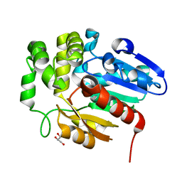 | |
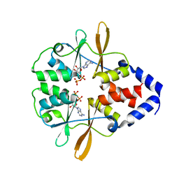
5Z89
 | | Structural basis for specific inhibition of highly sensitive ShHTL7 receptor | | 分子名称: | 2-(2-{2-[2-(2-{2-[2-(2-{2-[4-(1,1,3,3-TETRAMETHYL-BUTYL)-PHENOXY]-ETHOXY}-ETHOXY)-ETHOXY]-ETHOXY}-ETHOXY)-ETHOXY]-ETHOX Y}-ETHOXY)-ETHANOL, GLYCEROL, Hyposensitive to light 7, ... | | 著者 | Hameed, U.S, Arold, S.T. | | 登録日 | 2018-01-31 | | 公開日 | 2018-07-25 | | 最終更新日 | 2023-11-22 | | 実験手法 | X-RAY DIFFRACTION (1.42 Å) | | 主引用文献 | Structural basis for specific inhibition of the highly sensitive ShHTL7 receptor.
EMBO Rep., 19, 2018
|
|
5Z8P
 | |
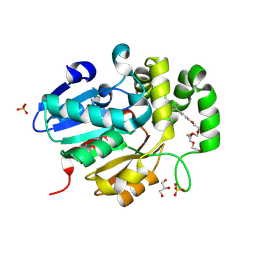
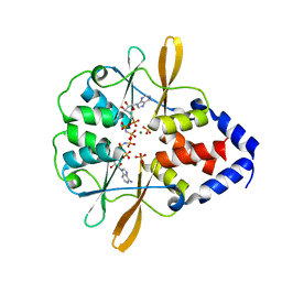
6SG4
 | | Structure of CDK2/cyclin A M246Q, S247EN | | 分子名称: | Cyclin-A2, Cyclin-dependent kinase 2 | | 著者 | Salamina, M, Basle, A, Massa, B, Noble, M.E.M, Endicott, J.A. | | 登録日 | 2019-08-02 | | 公開日 | 2021-01-27 | | 最終更新日 | 2024-01-24 | | 実験手法 | X-RAY DIFFRACTION (2.43 Å) | | 主引用文献 | Discriminative SKP2 Interactions with CDK-Cyclin Complexes Support a Cyclin A-Specific Role in p27KIP1 Degradation.
J.Mol.Biol., 433, 2021
|
|
3FWR
 | |
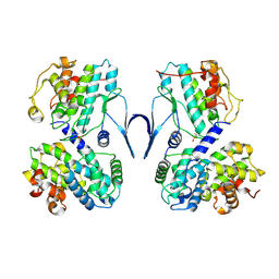
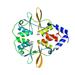
3FWS
 | | Crystal Structure of the CBS domains from the Bacillus subtilis CcpN repressor complexed with AppNp, phosphate and magnesium ions | | 分子名称: | MAGNESIUM ION, PHOSPHATE ION, PHOSPHOAMINOPHOSPHONIC ACID-ADENYLATE ESTER, ... | | 著者 | Chaix, D, Arold, S, Hoh, F, Declerck, N. | | 登録日 | 2009-01-19 | | 公開日 | 2010-01-26 | | 最終更新日 | 2023-11-01 | | 実験手法 | X-RAY DIFFRACTION (2.03 Å) | | 主引用文献 | Ligand recognition by the energy sensor domain of the CcpN repressor
To be Published
|
|
3FV6
 | |
3NXC
 | |
7W7Z
 | |


