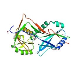
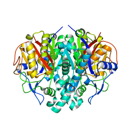
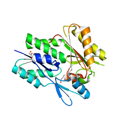
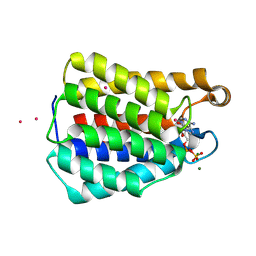
6DJ6
 
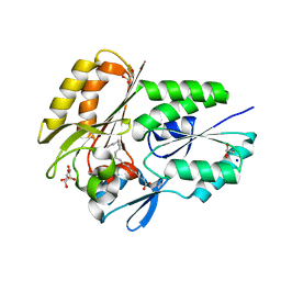 | | The X-ray crystal structure of the Streptococcus pneumoniae Fatty Acid Kinase (Fak) B2 protein loaded with cis-oleic acid to 1.9 Angstrom resolution | | 分子名称: | Fatty Acid Kinase (Fak) B2 protein (SPR1019), GLYCEROL, OLEIC ACID, ... | | 著者 | Cuypers, M.G, Subramanian, C, White, S.W, Rock, C.O. | | 登録日 | 2018-05-24 | | 公開日 | 2019-05-29 | | 最終更新日 | 2024-04-03 | | 実験手法 | X-RAY DIFFRACTION (1.9 Å) | | 主引用文献 | The X-ray crystal structure of the Streptococcus pneumoniae Fatty Acid Kinase (Fak) B2 protein (SPR1019) loaded with cis-oleic acid to 1.9 Angstrom resolution
To Be Published
|
|
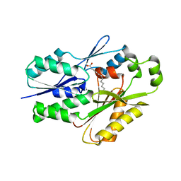
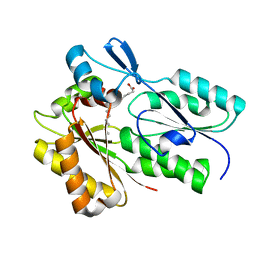
6CNG
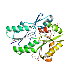 | | The X-ray crystal structure of the Streptococcus pneumoniae Fatty Acid Kinase (Fak) B3 protein loaded with linoleic acid to 1.47 Angstrom resolution | | 分子名称: | ACETATE ION, Fatty Acid Kinase (Fak) B3 protein, GLYCEROL, ... | | 著者 | Cuypers, M.G, Subramanian, C, White, S.W, Rock, C.O. | | 登録日 | 2018-03-08 | | 公開日 | 2019-07-03 | | 最終更新日 | 2023-10-04 | | 実験手法 | X-RAY DIFFRACTION (1.47 Å) | | 主引用文献 | The X-ray crystal structure of the Streptococcus pneumoniae Fatty Acid Kinase (Fak) B3 protein loaded with linoleic acid to 1.47 Angstrom resolution
To Be Published
|
|
3T7G
 | | Atg8 transfer from Atg7 to Atg3: a distinctive E1-E2 architecture and mechanism in the autophagy pathway | | 分子名称: | Autophagy-related protein 3, Ubiquitin-like modifier-activating enzyme ATG7 | | 著者 | Taherbhoy, A.M, Tait, S.W, Kaiser, S.E, Williams, A.H, Deng, A, Nourse, A, Hammel, M, Kurinov, I, Rock, C.O, Green, D.R, Schulman, B.A. | | 登録日 | 2011-07-30 | | 公開日 | 2011-11-23 | | 最終更新日 | 2023-09-13 | | 実験手法 | X-RAY DIFFRACTION (2.08 Å) | | 主引用文献 | Atg8 transfer from atg7 to atg3: a distinctive e1-e2 architecture and mechanism in the autophagy pathway.
Mol.Cell, 44, 2011
|
|
3T7H
 | | Atg8 transfer from Atg7 to Atg3: a distinctive E1-E2 architecture and mechanism in the autophagy pathway | | 分子名称: | Ubiquitin-like modifier-activating enzyme ATG7 | | 著者 | Taherbhoy, A.M, Tait, S.W, Kaiser, S.E, Williams, A.H, Deng, A, Nourse, A, Hammel, M, Kurinov, I, Rock, C.O, Green, D.R, Schulman, B.A. | | 登録日 | 2011-07-30 | | 公開日 | 2011-11-23 | | 最終更新日 | 2023-09-13 | | 実験手法 | X-RAY DIFFRACTION (1.6 Å) | | 主引用文献 | Atg8 transfer from atg7 to atg3: a distinctive e1-e2 architecture and mechanism in the autophagy pathway.
Mol.Cell, 44, 2011
|
|
3T7F
 | | Atg8 transfer from Atg7 to Atg3: a distinctive E1-E2 architecture and mechanism in the autophagy pathway | | 分子名称: | Ubiquitin-like modifier-activating enzyme ATG7 | | 著者 | Taherbhoy, A.M, Tait, S.W, Kaiser, S.E, Williams, A.H, Deng, A, Nourse, A, Hammel, M, Kurinov, I, Rock, C.O, Green, D.R, Schulman, B.A. | | 登録日 | 2011-07-30 | | 公開日 | 2011-11-23 | | 最終更新日 | 2024-11-27 | | 実験手法 | X-RAY DIFFRACTION (1.89 Å) | | 主引用文献 | Atg8 transfer from atg7 to atg3: a distinctive e1-e2 architecture and mechanism in the autophagy pathway.
Mol.Cell, 44, 2011
|
|
3T7E
 | | Atg8 transfer from Atg7 to Atg3: a distinctive E1-E2 architecture and mechanism in the autophagy pathway | | 分子名称: | Ubiquitin-like modifier-activating enzyme ATG7, ZINC ION | | 著者 | Taherbhoy, A.M, Tait, S.W, Kaiser, S.E, Williams, A.H, Deng, A, Nourse, A, Hammel, M, Kurinov, I, Rock, C.O, Green, D.R, Schulman, B.A. | | 登録日 | 2011-07-30 | | 公開日 | 2011-11-23 | | 最終更新日 | 2024-02-28 | | 実験手法 | X-RAY DIFFRACTION (2.25 Å) | | 主引用文献 | Atg8 transfer from atg7 to atg3: a distinctive e1-e2 architecture and mechanism in the autophagy pathway.
Mol.Cell, 44, 2011
|
|
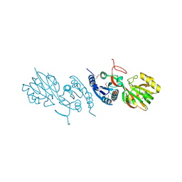
1M7E
 | | Crystal structure of the phosphotyrosine binding domain(PTB) of mouse Disabled 2(Dab2):implications for Reeling signaling | | 分子名称: | Disabled homolog 2, NGYENPTYK peptide | | 著者 | Yun, M, Keshvara, L, Park, C.-G, Zhang, Y.-M, Dickerson, J.B, Zheng, J, Rock, C.O, Curran, T, Park, H.-W. | | 登録日 | 2002-07-19 | | 公開日 | 2003-08-05 | | 最終更新日 | 2024-05-22 | | 実験手法 | X-RAY DIFFRACTION (2.45 Å) | | 主引用文献 | Crystal structures of the Dab homology domains of mouse disabled 1 and 2
J.Biol.Chem., 278, 2003
|
|
1OX0
 | |
1OQN
 | | Crystal structure of the phosphotyrosine binding domain (PTB) of mouse Disabled 1 (Dab1) | | 分子名称: | Alzheimer's disease amyloid A4 protein homolog, D-MYO-INOSITOL-1,4,5-TRIPHOSPHATE, Disabled homolog 1 | | 著者 | Yun, M, Keshvara, L, Park, C.-G, Zhang, Y.-M, Dickerson, J.B, Zheng, J, Rock, C.O, Curran, T, Park, H.-W. | | 登録日 | 2003-03-10 | | 公開日 | 2003-08-05 | | 最終更新日 | 2023-08-16 | | 実験手法 | X-RAY DIFFRACTION (2.3 Å) | | 主引用文献 | Crystal structures of the Dab homology domains of mouse disabled 1 and 2
J.Biol.Chem., 278, 2003
|
|
1OXH
 | |
1P3R
 | | Crystal structure of the phosphotyrosin binding domain(PTB) of mouse Disabled 1(Dab1) | | 分子名称: | Disabled homolog 2 | | 著者 | Yun, M, Keshvara, L, Park, C.G, Zhang, Y.M, Dickerson, J.B, Zheng, J, Rock, C.O, Curran, T, Park, H.W. | | 登録日 | 2003-04-18 | | 公開日 | 2003-08-05 | | 最終更新日 | 2023-08-16 | | 実験手法 | X-RAY DIFFRACTION (2.1 Å) | | 主引用文献 | Crystal structures of the Dab homology domains of mouse disabled 1 and 2.
J.Biol.Chem., 278, 2003
|
|
4Q9N
 | | Crystal structure of Chlamydia trachomatis enoyl-ACP reductase (FabI) in complex with NADH and AFN-1252 | | 分子名称: | 1,4-DIHYDRONICOTINAMIDE ADENINE DINUCLEOTIDE, Enoyl-[acyl-carrier-protein] reductase [NADH], N-methyl-N-[(3-methyl-1-benzofuran-2-yl)methyl]-3-(7-oxo-5,6,7,8-tetrahydro-1,8-naphthyridin-3-yl)propanamide | | 著者 | Yao, J, Abdelrahman, Y, Robertson, R.M, Cox, J.V, Belland, R.J, White, S.W, Rock, C.O. | | 登録日 | 2014-05-01 | | 公開日 | 2014-06-25 | | 最終更新日 | 2023-09-20 | | 実験手法 | X-RAY DIFFRACTION (1.795 Å) | | 主引用文献 | Type II Fatty Acid Synthesis Is Essential for the Replication of Chlamydia trachomatis.
J.Biol.Chem., 289, 2014
|
|
6MH9
 | | The crystal structure of the Staphylococcus aureus Fatty acid Kinase (Fak) B1 protein A121I mutant to 2.02 Angstrom resolution | | 分子名称: | Fatty Acid Kinase (Fak) B1 protein, PALMITIC ACID | | 著者 | Cuypers, M.G, Ericson, M, Subramanian, C, White, S.W, Rock, C.O. | | 登録日 | 2018-09-17 | | 公開日 | 2019-11-13 | | 最終更新日 | 2023-10-11 | | 実験手法 | X-RAY DIFFRACTION (2.02 Å) | | 主引用文献 | Identification of structural transitions in bacterial fatty acid binding proteins that permit ligand entry and exit at membranes.
J.Biol.Chem., 298, 2022
|
|
6NYU
 | | The X-ray crystal structure of Staphylococcus aureus Fatty Acid Kinase (Fak) B1 F263T mutant protein to 2.18 Angstrom resolution | | 分子名称: | DegV domain-containing protein, GLYCEROL, MYRISTIC ACID, ... | | 著者 | Cuypers, M.G, Gullett, J.M, Subramanian, C, White, S.W, Rock, C.O. | | 登録日 | 2019-02-12 | | 公開日 | 2020-02-12 | | 最終更新日 | 2023-10-11 | | 実験手法 | X-RAY DIFFRACTION (2.183 Å) | | 主引用文献 | The X-ray crystal structure of STAPHYLOCOCCUS AUREUS Fatty Acid Kinase (Fak) B1 protein F263T mutant to 2.18 Angstrom resolution
To Be Published
|
|
6NM1
 | | The crystal structure of the Staphylococcus aureus Fatty acid Kinase (Fak) B1 protein A158L mutant to 2.33 Angstrom resolution exhibits a conformation change compared to the wild type form | | 分子名称: | Fatty acid Kinase (Fak) B1 protein, MYRISTIC ACID | | 著者 | Cuypers, M.G, Gullett, J.M, Subramanian, C, Ericson, M, White, S.W, Rock, C.O. | | 登録日 | 2019-01-10 | | 公開日 | 2020-01-15 | | 最終更新日 | 2023-10-11 | | 実験手法 | X-RAY DIFFRACTION (2.33 Å) | | 主引用文献 | Identification of structural transitions in bacterial fatty acid binding proteins that permit ligand entry and exit at membranes.
J.Biol.Chem., 298, 2022
|
|
6NR1
 | | The X-ray crystal structure of Streptococcus pneumoniae Fatty Acid Kinase (Fak) B2 protein loaded with Vaccenic acid (C18:1 delta11) to 2.1 Angstrom resolution | | 分子名称: | Fatty Acid Kinase (Fak) B2 protein (SPR1019), GLYCEROL, VACCENIC ACID | | 著者 | Cuypers, M.G, Gullett, J.M, Subramanian, C, White, S.W, Rock, C.O. | | 登録日 | 2019-01-22 | | 公開日 | 2019-08-21 | | 最終更新日 | 2023-10-11 | | 実験手法 | X-RAY DIFFRACTION (2.1 Å) | | 主引用文献 | The X-ray crystal structure of Streptococcus pneumoniae Fatty Acid Kinase (Fak) B2 protein loaded with Vaccenic acid (C18:1 delta11) to 2.1 Angstrom resolution
To Be Published
|
|
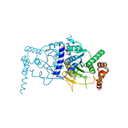
7RM7
 | | The X-ray crystal structure of the N-terminal domain of Staphylococcus aureus Fatty Acid Kinase A (FakA, residues 1-208) in complex with ADP to 1.025 Angstrom resolution | | 分子名称: | ADENOSINE-5'-DIPHOSPHATE, Fatty Acid Kinase A, GLYCEROL, ... | | 著者 | Cuypers, M.G, Subramanian, C, Rock, C.O, White, S.W. | | 登録日 | 2021-07-26 | | 公開日 | 2022-03-02 | | 最終更新日 | 2025-02-26 | | 実験手法 | X-RAY DIFFRACTION (1.025 Å) | | 主引用文献 | Domain architecture and catalysis of the Staphylococcus aureus fatty acid kinase.
J.Biol.Chem., 298, 2022
|
|
7RZK
 | | Co-soak crystal structure of the N-terminal domain of Staphylococcus aureus Fatty Acid Kinase A (FakA, residues 1-208) in complex with ADP to 1.9 Angstrom resolution - SAD data | | 分子名称: | ADENOSINE-5'-DIPHOSPHATE, COBALT (II) ION, Fatty Acid Kinase A, ... | | 著者 | Cuypers, M.G, Subramanian, C, Rock, C.O, White, S.W. | | 登録日 | 2021-08-27 | | 公開日 | 2022-03-02 | | 最終更新日 | 2025-02-26 | | 実験手法 | X-RAY DIFFRACTION (1.9 Å) | | 主引用文献 | Domain architecture and catalysis of the Staphylococcus aureus fatty acid kinase.
J.Biol.Chem., 298, 2022
|
|
6NOK
 | | The X-ray crystal structure of Streptococcus pneumoniae Fatty Acid Kinase (Fak) B1 protein loaded with myristic acid (C14:0) to 1.69 Angstrom resolution | | 分子名称: | Fatty Acid Kinase (Fak) B1 protein, MYRISTIC ACID, SODIUM ION | | 著者 | Cuypers, M.G, Gullett, J.M, Subramanian, C, White, S.W, Rock, C.O. | | 登録日 | 2019-01-16 | | 公開日 | 2019-08-21 | | 最終更新日 | 2023-10-11 | | 実験手法 | X-RAY DIFFRACTION (1.69 Å) | | 主引用文献 | The X-ray crystal structure of Streptococcus pneumoniae Fatty Acid Kinase (Fak) B1 protein loaded with myristic acid (C14:0) to 1.69 Angstrom resolution
To Be Published
|
|
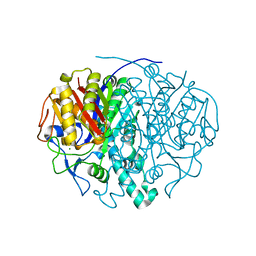
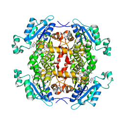
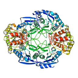
2P2D
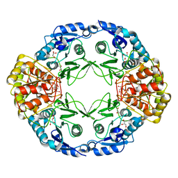 | | Crystal Structure and Allosteric Regulation of the Cytoplasmic Escherichia coli L-Asparaginase I | | 分子名称: | GLYCEROL, L-ASPARAGINASE I | | 著者 | Yun, M.-K, Nourse, A, White, S.W, Rock, C.O, Heath, R.J. | | 登録日 | 2007-03-07 | | 公開日 | 2007-05-15 | | 最終更新日 | 2023-08-30 | | 実験手法 | X-RAY DIFFRACTION (1.89 Å) | | 主引用文献 | Crystal Structure and Allosteric Regulation of the Cytoplasmic Escherichia colil-Asparaginase I
J.Mol.Biol., 369, 2007
|
|
2QVL
 | |
2P2N
 | | Crystal Structure and Allosteric Regulation of the Cytoplasmic Escherichia coli L-Asparaginase I | | 分子名称: | 1,2-ETHANEDIOL, ASPARAGINE, ASPARTIC ACID, ... | | 著者 | Yun, M.-K, Nourse, A, White, S.W, Rock, C.O, Heath, R.J. | | 登録日 | 2007-03-07 | | 公開日 | 2007-05-15 | | 最終更新日 | 2024-11-20 | | 実験手法 | X-RAY DIFFRACTION (1.9 Å) | | 主引用文献 | Crystal Structure and Allosteric Regulation of the Cytoplasmic Escherichia colil-Asparaginase I
J.Mol.Biol., 369, 2007
|
|
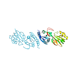
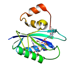
2QV7
 | | Crystal Structure of Diacylglycerol Kinase DgkB in complex with ADP and Mg | | 分子名称: | ADENOSINE-5'-DIPHOSPHATE, Diacylglycerol Kinase DgkB, MAGNESIUM ION | | 著者 | Miller, D.J, Jerga, A, Rock, C.O, White, S.W. | | 登録日 | 2007-08-07 | | 公開日 | 2008-06-17 | | 最終更新日 | 2024-11-20 | | 実験手法 | X-RAY DIFFRACTION (2.3 Å) | | 主引用文献 | Analysis of the Staphylococcus aureus DgkB Structure Reveals a Common Catalytic Mechanism for the Soluble Diacylglycerol Kinases.
Structure, 16, 2008
|
|
4M5J
 | | The Identification, Analysis and Structure-Based Development of Novel Inhibitors of 6-hydroxymethyl-7,8-dihydropterin pyrophosphokinase | | 分子名称: | 2-amino-4-hydroxy-6-hydroxymethyldihydropteridine pyrophosphokinase, 2-amino-8-{[2-(4-methoxyphenyl)-2-oxoethyl]sulfanyl}-1,9-dihydro-6H-purin-6-one, CHLORIDE ION, ... | | 著者 | Yun, M, Hoagland, D, Kumar, G, Waddell, B, Rock, C.O, Lee, R.E, White, S.W. | | 登録日 | 2013-08-08 | | 公開日 | 2014-04-02 | | 最終更新日 | 2023-09-20 | | 実験手法 | X-RAY DIFFRACTION (1.696 Å) | | 主引用文献 | The identification, analysis and structure-based development of novel inhibitors of 6-hydroxymethyl-7,8-dihydropterin pyrophosphokinase.
Bioorg.Med.Chem., 22, 2014
|
|
4M5K
 | | The Identification, Analysis and Structure-Based Development of Novel Inhibitors of 6-hydroxymethyl-7,8-dihydropterin pyrophosphokinase | | 分子名称: | 2-amino-4-hydroxy-6-hydroxymethyldihydropteridine pyrophosphokinase, 2-amino-8-{[2-(3-methylphenyl)-2-oxoethyl]sulfanyl}-1,9-dihydro-6H-purin-6-one, CALCIUM ION, ... | | 著者 | Yun, M, Hoagland, D, Kumar, G, Waddell, B, Rock, C.O, Lee, R.E, White, S.W. | | 登録日 | 2013-08-08 | | 公開日 | 2014-04-02 | | 最終更新日 | 2023-09-20 | | 実験手法 | X-RAY DIFFRACTION (1.296 Å) | | 主引用文献 | The identification, analysis and structure-based development of novel inhibitors of 6-hydroxymethyl-7,8-dihydropterin pyrophosphokinase.
Bioorg.Med.Chem., 22, 2014
|
|


