1VAI
 
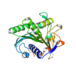
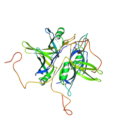
 | | Structure of e. coli cyclophilin B K163T mutant bound to n-acetyl-ala-ala-pro-ala-7-amino-4-methylcoumarin | | 分子名称: | (ACE)AAPA(MCM), cyclophilin B | | 著者 | Konno, M, Sano, Y, Okudaira, K, Kawaguchi, Y, Yamagishi-Ohmori, Y, Fushinobu, S, Matsuzawa, H. | | 登録日 | 2004-02-17 | | 公開日 | 2004-09-21 | | 最終更新日 | 2023-10-25 | | 実験手法 | X-RAY DIFFRACTION (1.8 Å) | | 主引用文献 | Escherichia coli cyclophilin B binds a highly distorted form of trans-prolyl peptide isomer
Eur.J.Biochem., 271, 2004
|
|
1J2A
 | | Structure of E. coli cyclophilin B K163T mutant | | 分子名称: | cyclophilin B | | 著者 | Konno, M, Sano, Y, Okudaira, K, Kawaguchi, Y, Yamagishi-Ohmori, Y, Fushinobu, S, Matsuzawa, H. | | 登録日 | 2002-12-26 | | 公開日 | 2004-02-10 | | 最終更新日 | 2023-10-25 | | 実験手法 | X-RAY DIFFRACTION (1.8 Å) | | 主引用文献 | Escherichia coli cyclophilin B binds a highly distorted form of trans-prolyl peptide isomer
Eur.J.Biochem., 271, 2004
|
|
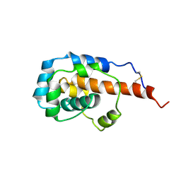
2RO0
 | | Solution structure of the knotted tudor domain of the yeast histone acetyltransferase, Esa1 | | 分子名称: | Histone acetyltransferase ESA1 | | 著者 | Shimojo, H, Sano, N, Moriwaki, Y, Okuda, M, Horikoshi, M, Nishimura, Y. | | 登録日 | 2008-03-01 | | 公開日 | 2008-04-29 | | 最終更新日 | 2024-05-29 | | 実験手法 | SOLUTION NMR | | 主引用文献 | Novel structural and functional mode of a knot essential for RNA binding activity of the Esa1 presumed chromodomain
J.Mol.Biol., 378, 2008
|
|
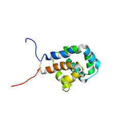
2RNZ
 | | Solution structure of the presumed chromodomain of the yeast histone acetyltransferase, Esa1 | | 分子名称: | Histone acetyltransferase ESA1 | | 著者 | Shimojo, H, Sano, N, Moriwaki, Y, Okuda, M, Horikoshi, M, Nishimura, Y. | | 登録日 | 2008-03-01 | | 公開日 | 2008-04-29 | | 最終更新日 | 2024-05-29 | | 実験手法 | SOLUTION NMR | | 主引用文献 | Novel structural and functional mode of a knot essential for RNA binding activity of the Esa1 presumed chromodomain
J.Mol.Biol., 378, 2008
|
|
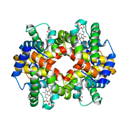
1LGY
 | | LIPASE II FROM RHIZOPUS NIVEUS | | 分子名称: | TRIACYLGLYCEROL LIPASE | | 著者 | Kohno, M, Funatsu, J, Mikami, B, Kugimiya, W, Matsuo, T, Morita, Y. | | 登録日 | 1996-05-23 | | 公開日 | 1996-12-23 | | 最終更新日 | 2024-06-05 | | 実験手法 | X-RAY DIFFRACTION (2.2 Å) | | 主引用文献 | The crystal structure of lipase II from Rhizopus niveus at 2.2 A resolution.
J.Biochem.(Tokyo), 120, 1996
|
|
3O0R
 | | Crystal structure of nitric oxide reductase from Pseudomonas aeruginosa in complex with antibody fragment | | 分子名称: | CALCIUM ION, FE (III) ION, HEME C, ... | | 著者 | Hino, T, Matsumoto, Y, Nagano, S, Sugimoto, H, Fukumori, Y, Murata, T, Iwata, S, Shiro, Y. | | 登録日 | 2010-07-20 | | 公開日 | 2010-12-29 | | 最終更新日 | 2013-10-16 | | 実験手法 | X-RAY DIFFRACTION (2.7 Å) | | 主引用文献 | Structural basis of biological N2O generation by bacterial nitric oxide reductase
Science, 330, 2010
|
|
6KJO
 | | The microtubule-binding domains of yeast cytoplasmic dynein in the low affinity state | | 分子名称: | Dynein heavy chain, cytoplasmic | | 著者 | Nishida, N, Komori, Y, Takarada, O, Watanabe, A, Tamura, S, Kubo, S, Shimada, I, Kikkawa, M. | | 登録日 | 2019-07-22 | | 公開日 | 2020-03-18 | | 最終更新日 | 2023-06-14 | | 実験手法 | SOLUTION NMR | | 主引用文献 | Structural basis for two-way communication between dynein and microtubules.
Nat Commun, 11, 2020
|
|
6KJN
 | | The microtubule-binding domains of yeast cytoplasmic dynein in the high affinity state | | 分子名称: | Dynein heavy chain, cytoplasmic | | 著者 | Nishida, N, Komori, Y, Takarada, O, Watanabe, A, Tamura, S, Kubo, S, Shimada, I, Kikkawa, M. | | 登録日 | 2019-07-22 | | 公開日 | 2020-03-18 | | 最終更新日 | 2023-06-14 | | 実験手法 | SOLUTION NMR | | 主引用文献 | Structural basis for two-way communication between dynein and microtubules.
Nat Commun, 11, 2020
|
|
6KYE
 | | The crystal structure of recombinant human adult hemoglobin | | 分子名称: | CARBON MONOXIDE, Hemoglobin subunit alpha, Hemoglobin subunit beta, ... | | 著者 | Kihira, K, Funaki, R, Okamoto, W, Endo, C, Morita, Y, Komatsu, T. | | 登録日 | 2019-09-18 | | 公開日 | 2020-01-01 | | 最終更新日 | 2023-11-22 | | 実験手法 | X-RAY DIFFRACTION (2.28 Å) | | 主引用文献 | Genetically engineered haemoglobin wrapped covalently with human serum albumins as an artificial O2carrier.
J Mater Chem B, 8, 2020
|
|
8JVG
 | | Crystal structure of dephospho-coenzyme A kinase | | 分子名称: | GTP-dependent dephospho-CoA kinase | | 著者 | Kita, A, Ishida, Y, Shimosaka, T, Michimori, Y, Makarova, K, Koonin, E, Atomi, H, Miki, K. | | 登録日 | 2023-06-28 | | 公開日 | 2024-06-19 | | 実験手法 | X-RAY DIFFRACTION (2.5 Å) | | 主引用文献 | Crystal structure of GTP-dependent dephospho-coenzyme A kinase from the hyperthermophilic archaeon, Thermococcus kodakarensis.
Proteins, 92, 2024
|
|
8JVC
 | | Crystal structure of dephospho-coenzyme A kinase | | 分子名称: | GTP-dependent dephospho-CoA kinase | | 著者 | Kita, A, Ishida, Y, Shimosaka, T, Michimori, Y, Makarova, K, Koonin, E, Atomi, H, Miki, K. | | 登録日 | 2023-06-28 | | 公開日 | 2024-06-19 | | 実験手法 | X-RAY DIFFRACTION (2.15 Å) | | 主引用文献 | Crystal structure of GTP-dependent dephospho-coenzyme A kinase from the hyperthermophilic archaeon, Thermococcus kodakarensis.
Proteins, 92, 2024
|
|
8JVF
 | | Crystal structure of dephospho-coenzyme A kinase | | 分子名称: | GTP-dependent dephospho-CoA kinase, GUANOSINE-5'-TRIPHOSPHATE, MAGNESIUM ION | | 著者 | Kita, A, Ishida, Y, Shimosaka, T, Michimori, Y, Makarova, K, Koonin, E, Atomi, H, Miki, K. | | 登録日 | 2023-06-28 | | 公開日 | 2024-06-19 | | 実験手法 | X-RAY DIFFRACTION (2.4 Å) | | 主引用文献 | Crystal structure of GTP-dependent dephospho-coenzyme A kinase from the hyperthermophilic archaeon, Thermococcus kodakarensis.
Proteins, 92, 2024
|
|
1EEX
 | | CRYSTAL STRUCTURE OF THE DIOL DEHYDRATASE-ADENINYLPENTYLCOBALAMIN COMPLEX FROM KLEBSIELLA OXYTOCA | | 分子名称: | CO-(ADENIN-9-YL-PENTYL)-COBALAMIN, POTASSIUM ION, PROPANEDIOL DEHYDRATASE, ... | | 著者 | Shibata, N, Masuda, J, Toraya, T, Morimoto, Y, Yasuoka, N. | | 登録日 | 2000-02-04 | | 公開日 | 2001-02-07 | | 最終更新日 | 2024-03-13 | | 実験手法 | X-RAY DIFFRACTION (1.7 Å) | | 主引用文献 | How a protein generates a catalytic radical from coenzyme B(12): X-ray structure of a diol-dehydratase-adeninylpentylcobalamin complex.
Structure Fold.Des., 8, 2000
|
|
1EGV
 | | CRYSTAL STRUCTURE OF THE DIOL DEHYDRATASE-ADENINYLPENTYLCOBALAMIN COMPLEX FROM KLEBSELLA OXYTOCA UNDER THE ILLUMINATED CONDITION. | | 分子名称: | CO-(ADENIN-9-YL-PENTYL)-COBALAMIN, POTASSIUM ION, PROPANEDIOL DEHYDRATASE, ... | | 著者 | Masuda, J, Shibata, N, Toraya, T, Morimoto, Y, Yasuoka, N. | | 登録日 | 2000-02-17 | | 公開日 | 2001-02-21 | | 最終更新日 | 2024-03-13 | | 実験手法 | X-RAY DIFFRACTION (1.75 Å) | | 主引用文献 | How a protein generates a catalytic radical from coenzyme B(12): X-ray structure of a diol-dehydratase-adeninylpentylcobalamin complex.
Structure Fold.Des., 8, 2000
|
|
1EGM
 | | CRYSTAL STRUCTURE OF DIOL DEHYDRATASE-CYANOCOBALAMIN COMPLEX AT 100K. | | 分子名称: | CYANOCOBALAMIN, POTASSIUM ION, PROPANEDIOL DEHYDRATASE, ... | | 著者 | Masuda, J, Shibata, N, Toraya, T, Morimoto, Y, Yasuoka, N. | | 登録日 | 2000-02-15 | | 公開日 | 2000-09-20 | | 最終更新日 | 2024-02-07 | | 実験手法 | X-RAY DIFFRACTION (1.85 Å) | | 主引用文献 | How a protein generates a catalytic radical from coenzyme B(12): X-ray structure of a diol-dehydratase-adeninylpentylcobalamin complex.
Structure Fold.Des., 8, 2000
|
|
2OWF
 | | Crystal structure of PH0725 from Pyrococcus horikoshii OT3 | | 分子名称: | S-ADENOSYL-L-HOMOCYSTEINE, diphthine synthase | | 著者 | Sugahara, M, Morikawa, Y, Matsuura, Y, Shimada, H, Kunishima, N, RIKEN Structural Genomics/Proteomics Initiative (RSGI) | | 登録日 | 2007-02-16 | | 公開日 | 2007-08-21 | | 最終更新日 | 2023-10-25 | | 実験手法 | X-RAY DIFFRACTION (2.2 Å) | | 主引用文献 | Crystal structure of PH0725 from Pyrococcus horikoshii OT3
To be Published
|
|
2P30
 | | Crystal structure of TTHB049 from Thermus thermophilus HB8 | | 分子名称: | Alpha-ribazole-5'-phosphate phosphatase | | 著者 | Sugahara, M, Taketa, M, Morikawa, Y, Matsuura, Y, Kunishima, N, RIKEN Structural Genomics/Proteomics Initiative (RSGI) | | 登録日 | 2007-03-08 | | 公開日 | 2007-09-11 | | 最終更新日 | 2023-10-25 | | 実験手法 | X-RAY DIFFRACTION (1.85 Å) | | 主引用文献 | Crystal structure of TTHB049 from Thermus thermophilus HB8
To be Published
|
|
2PCA
 | | Crystal structure of PH0725 from Pyrococcus horikoshii OT3 | | 分子名称: | Probable diphthine synthase, S-ADENOSYL-L-HOMOCYSTEINE, SODIUM ION | | 著者 | Sugahara, M, Taketa, M, Morikawa, Y, Matsuura, Y, Kunishima, N, RIKEN Structural Genomics/Proteomics Initiative (RSGI) | | 登録日 | 2007-03-29 | | 公開日 | 2007-10-02 | | 最終更新日 | 2023-10-25 | | 実験手法 | X-RAY DIFFRACTION (2 Å) | | 主引用文献 | Crystal structure of PH0725 from Pyrococcus horikoshii OT3
To be Published
|
|
2P6M
 | | Crystal structure of TTHB049 from Thermus thermophilus HB8 | | 分子名称: | Alpha-ribazole-5'-phosphate phosphatase, GLYCEROL, SODIUM ION | | 著者 | Sugahara, M, Matsuura, Y, Morikawa, Y, Shimada, H, Kunishima, N, RIKEN Structural Genomics/Proteomics Initiative (RSGI) | | 登録日 | 2007-03-19 | | 公開日 | 2007-09-25 | | 最終更新日 | 2023-10-25 | | 実験手法 | X-RAY DIFFRACTION (1.9 Å) | | 主引用文献 | Crystal structure of TTHB049 from Thermus thermophilus HB8
To be Published
|
|
2P2Y
 | | Crystal structure of TTHB049 from Thermus thermophilus HB8 | | 分子名称: | Alpha-ribazole-5'-phosphate phosphatase, CHLORIDE ION | | 著者 | Sugahara, M, Morikawa, Y, Taketa, M, Matsuura, Y, Kunishima, N, RIKEN Structural Genomics/Proteomics Initiative (RSGI) | | 登録日 | 2007-03-08 | | 公開日 | 2007-09-11 | | 最終更新日 | 2023-10-25 | | 実験手法 | X-RAY DIFFRACTION (1.95 Å) | | 主引用文献 | Crystal structure of TTHB049 from Thermus thermophilus HB8
To be Published
|
|
2P6D
 | | Crystal structure of PH0725 from Pyrococcus horikoshii OT3 | | 分子名称: | S-ADENOSYL-L-HOMOCYSTEINE, diphthine synthase | | 著者 | Yamamoto, H, Taketa, M, Morikawa, Y, Matsuura, Y, Kunishima, N, RIKEN Structural Genomics/Proteomics Initiative (RSGI) | | 登録日 | 2007-03-17 | | 公開日 | 2007-09-18 | | 最終更新日 | 2023-10-25 | | 実験手法 | X-RAY DIFFRACTION (2.4 Å) | | 主引用文献 | Crystal structure of PH0725 from Pyrococcus horikoshii OT3
To be Published
|
|
1QVC
 | | CRYSTAL STRUCTURE ANALYSIS OF SINGLE STRANDED DNA BINDING PROTEIN (SSB) FROM E.COLI | | 分子名称: | SINGLE STRANDED DNA BINDING PROTEIN MONOMER | | 著者 | Matsumoto, T, Morimoto, Y, Shibata, N, Shimamoto, N, Tsukihara, T, Yasuoka, N. | | 登録日 | 1999-07-07 | | 公開日 | 2000-06-05 | | 最終更新日 | 2024-02-14 | | 実験手法 | X-RAY DIFFRACTION (2.2 Å) | | 主引用文献 | Roles of functional loops and the C-terminal segment of a single-stranded DNA binding protein elucidated by X-Ray structure analysis.
J.Biochem.(Tokyo), 127, 2000
|
|
1IRU
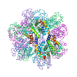
 | | Crystal Structure of the mammalian 20S proteasome at 2.75 A resolution | | 分子名称: | 20S proteasome, MAGNESIUM ION | | 著者 | Unno, M, Mizushima, T, Morimoto, Y, Tomisugi, Y, Tanaka, K, Yasuoka, N, Tsukihara, T. | | 登録日 | 2001-10-24 | | 公開日 | 2002-05-22 | | 最終更新日 | 2023-12-27 | | 実験手法 | X-RAY DIFFRACTION (2.75 Å) | | 主引用文献 | The structure of the mammalian 20S proteasome at 2.75 A resolution.
Structure, 10, 2002
|
|
2D2M
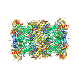
 | | Structure of an extracellular giant hemoglobin of the gutless beard worm Oligobrachia mashikoi | | 分子名称: | Giant hemoglobin, A1(b) globin chain, A2(a5) globin chain, ... | | 著者 | Numoto, N, Nakagawa, T, Kita, A, Sasayama, Y, Fukumori, Y, Miki, K. | | 登録日 | 2005-09-12 | | 公開日 | 2005-10-25 | | 最終更新日 | 2011-07-13 | | 実験手法 | X-RAY DIFFRACTION (2.85 Å) | | 主引用文献 | Structure of an extracellular giant hemoglobin of the gutless beard worm Oligobrachia mashikoi.
Proc.Natl.Acad.Sci.USA, 102, 2005
|
|
2D2N
 | | Structure of an extracellular giant hemoglobin of the gutless beard worm Oligobrachia mashikoi | | 分子名称: | Giant hemoglobin, A1(b) globin chain, A2(a5) globin chain, ... | | 著者 | Numoto, N, Nakagawa, T, Kita, A, Sasayama, Y, Fukumori, Y, Miki, K. | | 登録日 | 2005-09-12 | | 公開日 | 2005-10-25 | | 最終更新日 | 2011-07-13 | | 実験手法 | X-RAY DIFFRACTION (3.2 Å) | | 主引用文献 | Structure of an extracellular giant hemoglobin of the gutless beard worm Oligobrachia mashikoi.
Proc.Natl.Acad.Sci.USA, 102, 2005
|
|


