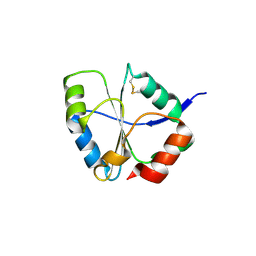
2ORY
 
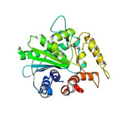 | |
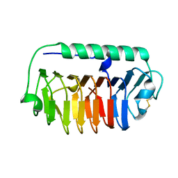
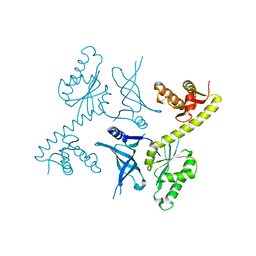
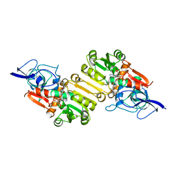
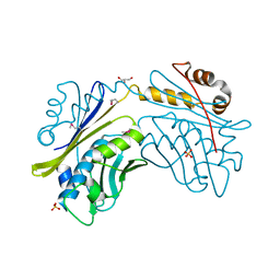
4RHE
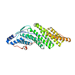
 | | Crystal structure of UbiX, an aromatic acid decarboxylase from the Colwellia psychrerythraea 34H | | 分子名称: | 3-octaprenyl-4-hydroxybenzoate carboxy-lyase, FLAVIN MONONUCLEOTIDE, SULFATE ION | | 著者 | Do, H, Kim, S.J, Lee, C.W, Kim, H.-W, Park, H.H, Kim, H.M, Park, H, Park, H.J, Lee, J.H. | | 登録日 | 2014-10-02 | | 公開日 | 2015-02-18 | | 最終更新日 | 2024-02-28 | | 実験手法 | X-RAY DIFFRACTION (2.003 Å) | | 主引用文献 | Crystal structure of UbiX, an aromatic acid decarboxylase from the psychrophilic bacterium Colwellia psychrerythraea that undergoes FMN-induced conformational changes.
Sci Rep, 5, 2015
|
|
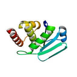
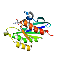
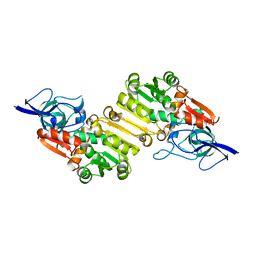
4RHF
 | | Crystal structure of UbiX mutant V47S from Colwellia psychrerythraea 34H | | 分子名称: | 3-octaprenyl-4-hydroxybenzoate carboxy-lyase, SULFATE ION | | 著者 | Do, H, Kim, S.J, Lee, C.W, Kim, H.-W, Park, H.H, Kim, H.M, Park, H, Park, H.J, Lee, J.H. | | 登録日 | 2014-10-02 | | 公開日 | 2015-02-18 | | 最終更新日 | 2024-02-28 | | 実験手法 | X-RAY DIFFRACTION (1.764 Å) | | 主引用文献 | Crystal structure of UbiX, an aromatic acid decarboxylase from the psychrophilic bacterium Colwellia psychrerythraea that undergoes FMN-induced conformational changes.
Sci Rep, 5, 2015
|
|
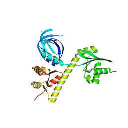
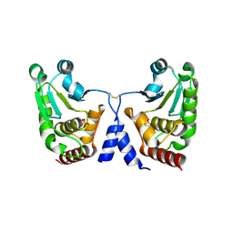
5CRV
 | | Crystal structure of the Bro domain of HD-PTP in a complex with the core region of STAM2 | | 分子名称: | GLYCEROL, Signal transducing adapter molecule 2, Tyrosine-protein phosphatase non-receptor type 23 | | 著者 | Lee, J, Ku, B, Kim, S.J. | | 登録日 | 2015-07-23 | | 公開日 | 2016-02-24 | | 最終更新日 | 2023-11-08 | | 実験手法 | X-RAY DIFFRACTION (2.001 Å) | | 主引用文献 | Structural Study of the HD-PTP Bro1 Domain in a Complex with the Core Region of STAM2, a Subunit of ESCRT-0
Plos One, 11, 2016
|
|
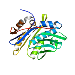
5CRU
 | | Crystal structure of the Bro domain of HD-PTP | | 分子名称: | Tyrosine-protein phosphatase non-receptor type 23 | | 著者 | Lee, J, Ku, B, Kim, S.J. | | 登録日 | 2015-07-23 | | 公開日 | 2016-02-24 | | 最終更新日 | 2023-11-08 | | 実験手法 | X-RAY DIFFRACTION (2.4 Å) | | 主引用文献 | Structural Study of the HD-PTP Bro1 Domain in a Complex with the Core Region of STAM2, a Subunit of ESCRT-0
Plos One, 11, 2016
|
|
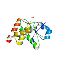
2NT2
 | | Crystal Structure of Slingshot phosphatase 2 | | 分子名称: | Protein phosphatase Slingshot homolog 2, SULFATE ION | | 著者 | Jung, S.K, Jeong, D.G, Yoon, T.S, Kim, J.H, Ryu, S.E, Kim, S.J. | | 登録日 | 2006-11-06 | | 公開日 | 2007-06-05 | | 最終更新日 | 2023-08-30 | | 実験手法 | X-RAY DIFFRACTION (2.1 Å) | | 主引用文献 | Crystal structure of human slingshot phosphatase 2.
Proteins, 68, 2007
|
|
4B04
 | | Crystal structure of the Catalytic Domain of Human DUSP26 (C152S) | | 分子名称: | DUAL SPECIFICITY PROTEIN PHOSPHATASE 26 | | 著者 | Won, E.-Y, Lee, D.Y, Park, S.G, Yokoyama, S, Kim, S.J, Chi, S.-W. | | 登録日 | 2012-06-28 | | 公開日 | 2013-05-29 | | 最終更新日 | 2023-12-20 | | 実験手法 | X-RAY DIFFRACTION (2.205 Å) | | 主引用文献 | High-Resolution Crystal Structure of the Catalytic Domain of Human Dual-Specificity Phosphatase 26
Acta Crystallogr.,Sect.D, 69, 2013
|
|
7CUY
 | | Crystal structure of Primo-1 | | 分子名称: | 4-(2-HYDROXYETHYL)-1-PIPERAZINE ETHANESULFONIC ACID, Low molecular weight phosphotyrosine protein phosphatase 1 | | 著者 | Lee, H.S, Kim, S.J, Ku, B. | | 登録日 | 2020-08-25 | | 公開日 | 2021-01-13 | | 最終更新日 | 2023-11-29 | | 実験手法 | X-RAY DIFFRACTION (2.081 Å) | | 主引用文献 | Structural and Biochemical Characterization of the Two Drosophila Low Molecular Weight-Protein Tyrosine Phosphatases DARP and Primo-1.
Mol.Cells, 43, 2020
|
|
1UC7
 | | Crystal structure of DsbDgamma | | 分子名称: | Thiol:disulfide interchange protein dsbD | | 著者 | Kim, J.H, Kim, S.J, Jeong, D.G, Son, J.H, Ryu, S.E. | | 登録日 | 2003-04-09 | | 公開日 | 2004-04-27 | | 最終更新日 | 2023-12-27 | | 実験手法 | X-RAY DIFFRACTION (1.9 Å) | | 主引用文献 | Crystal structure of DsbDgamma reveals the mechanism of redox potential shift and substrate specificity(1)
FEBS LETT., 543, 2003
|
|
6JM4
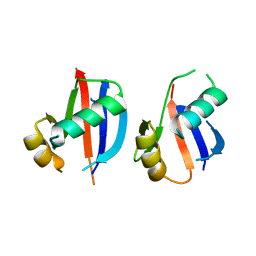 | |
4NUH
 | | Crystal structure of mLeIBP, a capping head region swapped mutant of ice-binding protein | | 分子名称: | DI(HYDROXYETHYL)ETHER, ice-binding protein | | 著者 | Do, H, Kim, S.J, Lee, S.G, Park, H, Kim, H.J, Lee, J.H. | | 登録日 | 2013-12-03 | | 公開日 | 2014-04-16 | | 最終更新日 | 2023-11-08 | | 実験手法 | X-RAY DIFFRACTION (1.34 Å) | | 主引用文献 | Structure-based characterization and antifreeze properties of a hyperactive ice-binding protein from the Antarctic bacterium Flavobacterium frigoris PS1
Acta Crystallogr.,Sect.D, 70, 2014
|
|
4NU3
 | | Crystal structure of mFfIBP, a capping head region swapped mutant of ice-binding protein | | 分子名称: | SODIUM ION, SULFATE ION, ice-binding protein | | 著者 | Do, H, Kim, S.J, Lee, S.G, Park, H, Kim, H.J, Lee, J.H. | | 登録日 | 2013-12-03 | | 公開日 | 2014-04-16 | | 最終更新日 | 2023-11-08 | | 実験手法 | X-RAY DIFFRACTION (1.399 Å) | | 主引用文献 | Structure-based characterization and antifreeze properties of a hyperactive ice-binding protein from the Antarctic bacterium Flavobacterium frigoris PS1
Acta Crystallogr.,Sect.D, 70, 2014
|
|
4NU2
 | | Crystal structure of an ice-binding protein (FfIBP) from the Antarctic bacterium, Flavobacterium frigoris PS1 | | 分子名称: | Antifreeze protein | | 著者 | Do, H, Kim, S.J, Lee, S.G, Park, H, Kim, H.J, Lee, J.H. | | 登録日 | 2013-12-03 | | 公開日 | 2014-04-16 | | 最終更新日 | 2023-11-08 | | 実験手法 | X-RAY DIFFRACTION (2.1 Å) | | 主引用文献 | Structure-based characterization and antifreeze properties of a hyperactive ice-binding protein from the Antarctic bacterium Flavobacterium frigoris PS1
Acta Crystallogr.,Sect.D, 70, 2014
|
|
3LJ8
 | | Crystal Structure of MKP-4 | | 分子名称: | Tyrosine-protein phosphatase | | 著者 | Jeong, D.G, Yoon, T.S, Jung, S.-K, Park, H.S, Ryu, S.E, Kim, S.J. | | 登録日 | 2010-01-26 | | 公開日 | 2010-12-29 | | 最終更新日 | 2023-11-01 | | 実験手法 | X-RAY DIFFRACTION (2.7 Å) | | 主引用文献 | Exploring binding sites other than the catalytic core in the crystal structure of the catalytic domain of MKP-4
Acta Crystallogr.,Sect.D, 67, 2011
|
|
3OBY
 | | Crystal structure of Archaeoglobus fulgidus Pelota reveals inter-domain structural plasticity | | 分子名称: | Protein pelota homolog | | 著者 | Lee, H.H, Jang, J.Y, Yoon, H.-J, Kim, S.J, Suh, S.W. | | 登録日 | 2010-08-09 | | 公開日 | 2010-09-01 | | 最終更新日 | 2024-03-20 | | 実験手法 | X-RAY DIFFRACTION (2.9 Å) | | 主引用文献 | Crystal structures of two archaeal Pelotas reveal inter-domain structural plasticity
Biochem.Biophys.Res.Commun., 399, 2010
|
|
3M7M
 | | Crystal structure of monomeric hsp33 | | 分子名称: | 33 kDa chaperonin | | 著者 | Chi, S.W, Jeong, D.G, Woo, J.R, Park, B.C, Ryu, S.E, Kim, S.J. | | 登録日 | 2010-03-16 | | 公開日 | 2011-01-26 | | 最終更新日 | 2023-11-01 | | 実験手法 | X-RAY DIFFRACTION (2.9 Å) | | 主引用文献 | Crystal structure of monomeric hsp33
To be Published
|
|
3OBW
 | | Crystal structure of two archaeal Pelotas reveal inter-domain structural plasticity | | 分子名称: | Protein pelota homolog | | 著者 | Lee, H.H, Jang, J.Y, Yoon, H.-J, Kim, S.J, Suh, S.W. | | 登録日 | 2010-08-09 | | 公開日 | 2010-09-01 | | 最終更新日 | 2024-03-20 | | 実験手法 | X-RAY DIFFRACTION (2.6 Å) | | 主引用文献 | Crystal structures of two archaeal Pelotas reveal inter-domain structural plasticity
Biochem.Biophys.Res.Commun., 399, 2010
|
|
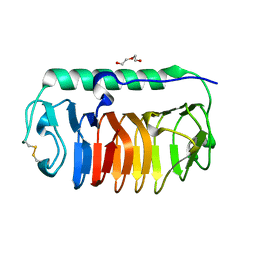
5XC3
 | |
2J3I
 | | Crystal structure of Arabidopsis thaliana Double Bond Reductase (AT5G16970)-Binary Complex | | 分子名称: | NADP NICOTINAMIDE-ADENINE-DINUCLEOTIDE PHOSPHATE, NADP-DEPENDENT OXIDOREDUCTASE P1 | | 著者 | Youn, B, Kim, S.J, Moinuddin, S.G, Lee, C, Bedgar, D.L, Harper, A.R, Davin, L.B, Lewis, N.G, Kang, C. | | 登録日 | 2006-08-21 | | 公開日 | 2006-10-05 | | 最終更新日 | 2011-07-13 | | 実験手法 | X-RAY DIFFRACTION (2.8 Å) | | 主引用文献 | Mechanistic and Structural Studies of Apoform, Binary, and Ternary Complexes of the Arabidopsis Alkenal Double Bond Reductase at5G16970.
J.Biol.Chem., 281, 2006
|
|
2J3K
 | | Crystal structure of Arabidopsis thaliana Double Bond Reductase (AT5G16970)-Ternary Complex II | | 分子名称: | (2E,4R)-4-HYDROXYNON-2-ENAL, NADP NICOTINAMIDE-ADENINE-DINUCLEOTIDE PHOSPHATE, NADPH-dependent oxidoreductase 2-alkenal reductase | | 著者 | Youn, B, Kim, S.J, Moinuddin, S.G, Lee, C, Bedgar, D.L, Harper, A.R, Davin, L.B, Lewis, N.G, Kang, C. | | 登録日 | 2006-08-22 | | 公開日 | 2006-10-05 | | 最終更新日 | 2024-05-08 | | 実験手法 | X-RAY DIFFRACTION (2.8 Å) | | 主引用文献 | Mechanistic and structural studies of apoform, binary, and ternary complexes of the Arabidopsis alkenal double bond reductase At5g16970.
J. Biol. Chem., 281, 2006
|
|
2J3J
 | | Crystal structure of Arabidopsis thaliana Double Bond Reductase (AT5G16970)-Ternary Complex I | | 分子名称: | 4'-HYDROXYCINNAMIC ACID, NADP NICOTINAMIDE-ADENINE-DINUCLEOTIDE PHOSPHATE, NADPH-dependent oxidoreductase 2-alkenal reductase | | 著者 | Youn, B, Kim, S.J, Moinuddin, S.G, Lee, C, Bedgar, D.L, Harper, A.R, Davin, L.B, Lewis, N.G, Kang, C. | | 登録日 | 2006-08-21 | | 公開日 | 2006-10-05 | | 最終更新日 | 2024-05-08 | | 実験手法 | X-RAY DIFFRACTION (2.8 Å) | | 主引用文献 | Mechanistic and structural studies of apoform, binary, and ternary complexes of the Arabidopsis alkenal double bond reductase At5g16970.
J. Biol. Chem., 281, 2006
|
|
2J3H
 | | Crystal structure of Arabidopsis thaliana Double Bond Reductase (AT5G16970)-Apo form | | 分子名称: | NADP-DEPENDENT OXIDOREDUCTASE P1 | | 著者 | Youn, B, Kim, S.J, Moinuddin, S.G, Lee, C, Bedgar, D.L, Harper, A.R, Davin, L.B, Lewis, N.G, Kang, C. | | 登録日 | 2006-08-21 | | 公開日 | 2006-10-05 | | 最終更新日 | 2024-05-08 | | 実験手法 | X-RAY DIFFRACTION (2.5 Å) | | 主引用文献 | Mechanistic and Structural Studies of Apoform, Binary, and Ternary Complexes of the Arabidopsis Alkenal Double Bond Reductase at5G16970.
J.Biol.Chem., 281, 2006
|
|
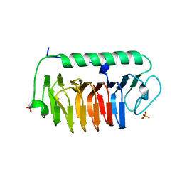
5GTJ
 | |
3S4E
 | | Crystal Structrue of a Novel Mitogen-activated Protein Kinase Phosphatase, SKRP1 | | 分子名称: | Dual specificity protein phosphatase 19, PHOSPHATE ION, SULFATE ION | | 著者 | Wei, C.H, Ryu, S.Y, Jeon, Y.H, Jeong, D.G, Kim, S.J, Ryu, S.E. | | 登録日 | 2011-05-19 | | 公開日 | 2012-04-04 | | 最終更新日 | 2023-11-01 | | 実験手法 | X-RAY DIFFRACTION (1.26 Å) | | 主引用文献 | Crystal structure of a novel mitogen-activated protein kinase phosphatase, SKRP1.
Proteins, 79, 2011
|
|
1I7F
 | | CRYSTAL STRUCTURE OF THE HSP33 DOMAIN WITH CONSTITUTIVE CHAPERONE ACTIVITY | | 分子名称: | GLYCEROL, HEAT SHOCK PROTEIN 33, SULFATE ION | | 著者 | Kim, S.-J, Jeong, D.-G, Chi, S.-W, Lee, J.-S, Ryu, S.-E. | | 登録日 | 2001-03-09 | | 公開日 | 2001-05-09 | | 最終更新日 | 2021-10-27 | | 実験手法 | X-RAY DIFFRACTION (2.7 Å) | | 主引用文献 | Crystal structure of proteolytic fragments of the redox-sensitive Hsp33 with constitutive chaperone activity
Nat.Struct.Biol., 8, 2001
|
|


