1SZZ
 
 | | Crystal structure of peptide deformylase from Leptospira Interrogans complexed with inhibitor actinonin | | 分子名称: | ACTINONIN, Peptide deformylase, ZINC ION | | 著者 | Zhou, Z, Song, X, Li, Y, Gong, W. | | 登録日 | 2004-04-06 | | 公開日 | 2005-08-16 | | 最終更新日 | 2023-10-25 | | 実験手法 | X-RAY DIFFRACTION (3.3 Å) | | 主引用文献 | Novel conformational states of peptide deformylase from pathogenic bacterium Leptospira interrogans: implications for population shift
J.Biol.Chem., 280, 2005
|
|
1T8P
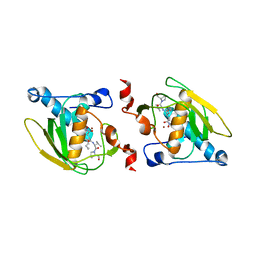
 | | Crystal structure of Human erythrocyte 2,3-bisphosphoglycerate mutase | | 分子名称: | Bisphosphoglycerate mutase | | 著者 | Wang, Y, Wei, Z, Bian, Q, Cheng, Z, Wan, M, Liu, L, Gong, W. | | 登録日 | 2004-05-13 | | 公開日 | 2004-08-10 | | 最終更新日 | 2023-10-25 | | 実験手法 | X-RAY DIFFRACTION (2.5 Å) | | 主引用文献 | Crystal structure of human bisphosphoglycerate mutase
J.Biol.Chem., 279, 2004
|
|
1CLK
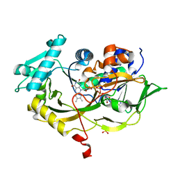
 | | CRYSTAL STRUCTURE OF STREPTOMYCES DIASTATICUS NO.7 STRAIN M1033 XYLOSE ISOMERASE AT 1.9 A RESOLUTION WITH PSEUDO-I222 SPACE GROUP | | 分子名称: | COBALT (II) ION, MAGNESIUM ION, XYLOSE ISOMERASE | | 著者 | Niu, L, Teng, M, Zhu, X, Gong, W. | | 登録日 | 1999-04-29 | | 公開日 | 2000-05-03 | | 最終更新日 | 2023-08-09 | | 実験手法 | X-RAY DIFFRACTION (1.9 Å) | | 主引用文献 | Structure of xylose isomerase from Streptomyces diastaticus no. 7 strain M1033 at 1.85 A resolution.
Acta Crystallogr.,Sect.D, 56, 2000
|
|
3JUI
 | | Crystal Structure of the C-terminal Domain of Human Translation Initiation Factor eIF2B epsilon Subunit | | 分子名称: | GLYCEROL, Translation initiation factor eIF-2B subunit epsilon | | 著者 | Wei, J, Xu, H, Zhang, C, Wang, M, Gao, F, Gong, W. | | 登録日 | 2009-09-15 | | 公開日 | 2009-10-06 | | 最終更新日 | 2021-11-10 | | 実験手法 | X-RAY DIFFRACTION (2 Å) | | 主引用文献 | Crystal Structure of the C-terminal Domain of Human Translation Initiation Factor eIF2B epsilon Subunit
To be published
|
|
4Q5W
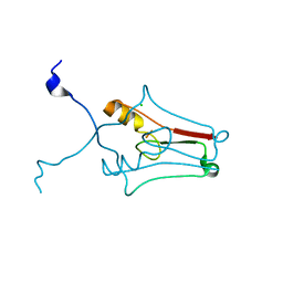
 | | Crystal structure of extended-Tudor 9 of Drosophila melanogaster | | 分子名称: | 4-(2-HYDROXYETHYL)-1-PIPERAZINE ETHANESULFONIC ACID, Maternal protein tudor | | 著者 | Ren, R, Liu, H, Wang, W, Wang, M, Yang, N, Dong, Y, Gong, W, Lehmann, R, Xu, R.M. | | 登録日 | 2014-04-17 | | 公開日 | 2014-05-21 | | 最終更新日 | 2024-03-20 | | 実験手法 | X-RAY DIFFRACTION (1.801 Å) | | 主引用文献 | Structure and domain organization of Drosophila Tudor
Cell Res., 24, 2014
|
|
4Q5Y
 | | Crystal structure of extended-Tudor 10-11 of Drosophila melanogaster | | 分子名称: | Maternal protein tudor | | 著者 | Liu, H, Ren, R, Wang, W, Wang, M, Yang, N, Dong, Y, Gong, W, Lehmann, R, Xu, R.M. | | 登録日 | 2014-04-18 | | 公開日 | 2014-05-21 | | 最終更新日 | 2023-11-08 | | 実験手法 | X-RAY DIFFRACTION (3 Å) | | 主引用文献 | Structure and domain organization of Drosophila Tudor
Cell Res., 24, 2014
|
|
4RSL
 | | Structure of fructosyl peptide oxidase from E. terrenum | | 分子名称: | FLAVIN-ADENINE DINUCLEOTIDE, Fructosyl peptide oxidase, PHOSPHATE ION | | 著者 | Gan, W, Gao, F, Xing, K, Jia, M, Liu, H, Gong, W. | | 登録日 | 2014-11-08 | | 公開日 | 2015-05-20 | | 最終更新日 | 2024-03-20 | | 実験手法 | X-RAY DIFFRACTION (1.9 Å) | | 主引用文献 | Structural basis of the substrate specificity of the FPOD/FAOD family revealed by fructosyl peptide oxidase from Eupenicillium terrenum
Acta Crystallogr.,Sect.F, 71, 2015
|
|
1BSJ
 | | COBALT DEFORMYLASE INHIBITOR COMPLEX FROM E.COLI | | 分子名称: | (S)-2-(PHOSPHONOXY)CAPROYL-L-LEUCYL-P-NITROANILIDE, COBALT (II) ION, PHOSPHATE ION, ... | | 著者 | Hao, B, Gong, W, Rajagopalan, P.T, Hu, Y, Pei, D, Chan, M.K. | | 登録日 | 1998-08-28 | | 公開日 | 2000-04-15 | | 最終更新日 | 2023-08-09 | | 実験手法 | X-RAY DIFFRACTION (3 Å) | | 主引用文献 | Structural basis for the design of antibiotics targeting peptide deformylase.
Biochemistry, 38, 1999
|
|
1BSK
 | | ZINC DEFORMYLASE INHIBITOR COMPLEX FROM E.COLI | | 分子名称: | (S)-2-(PHOSPHONOXY)CAPROYL-L-LEUCYL-P-NITROANILIDE, PHOSPHATE ION, PROTEIN (PEPTIDE DEFORMYLASE), ... | | 著者 | Hao, B, Gong, W, Rajagopalan, P.T, Hu, Y, Pei, D, Chan, M.K. | | 登録日 | 1998-08-28 | | 公開日 | 2000-04-15 | | 最終更新日 | 2023-08-09 | | 実験手法 | X-RAY DIFFRACTION (3 Å) | | 主引用文献 | Structural basis for the design of antibiotics targeting peptide deformylase.
Biochemistry, 38, 1999
|
|
2M9N
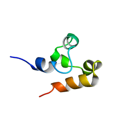 | | Solution Structure of (HhH)2 domain of human FAAP24 | | 分子名称: | Fanconi anemia-associated protein of 24 kDa | | 著者 | Wu, F, Han, X, Shi, C, Gong, W, Tian, C. | | 登録日 | 2013-06-18 | | 公開日 | 2013-09-18 | | 最終更新日 | 2024-05-15 | | 実験手法 | SOLUTION NMR | | 主引用文献 | Structure analysis of FAAP24 reveals single-stranded DNA-binding activity and domain functions in DNA damage response.
Cell Res., 23, 2013
|
|
2O9Q
 | | The crystal structure of Bovine Trypsin complexed with a small inhibition peptide ORB2K | | 分子名称: | CALCIUM ION, Cationic trypsin, ORB2K, ... | | 著者 | Li, J, Zhang, C, Xu, X, Wang, J, Gong, W, Lai, R. | | 登録日 | 2006-12-14 | | 公開日 | 2007-12-25 | | 最終更新日 | 2023-10-25 | | 実験手法 | X-RAY DIFFRACTION (1.7 Å) | | 主引用文献 | From protease inhibitor to antibiotics: single point mutation makes tremendous functional shift
To be Published
|
|
2PL5
 | | Crystal Structure of Homoserine O-acetyltransferase from Leptospira interrogans | | 分子名称: | GLYCEROL, Homoserine O-acetyltransferase | | 著者 | Liu, L, Wang, M, Wang, Y, Wei, Z, Xu, H, Gong, W. | | 登録日 | 2007-04-19 | | 公開日 | 2007-11-20 | | 最終更新日 | 2024-03-13 | | 実験手法 | X-RAY DIFFRACTION (2.2 Å) | | 主引用文献 | Crystal structure of homoserine O-acetyltransferase from Leptospira interrogans
Biochem.Biophys.Res.Commun., 363, 2007
|
|
2R13
 | | Crystal structure of human mitoNEET reveals a novel [2Fe-2S] cluster coordination | | 分子名称: | CHLORIDE ION, FE2/S2 (INORGANIC) CLUSTER, Zinc finger CDGSH domain-containing protein 1 | | 著者 | Hou, X, Liu, R, Ross, S, Smart, E.J, Zhu, H, Gong, W. | | 登録日 | 2007-08-22 | | 公開日 | 2007-09-11 | | 最終更新日 | 2024-03-13 | | 実験手法 | X-RAY DIFFRACTION (1.8 Å) | | 主引用文献 | Crystallographic studies of human MitoNEET
J.Biol.Chem., 282, 2007
|
|
2DHO
 | | Crystal structure of human IPP isomerase I in space group P212121 | | 分子名称: | CHLORIDE ION, Isopentenyl-diphosphate delta-isomerase 1, MANGANESE (II) ION, ... | | 著者 | Zhang, C, Wei, Z, Gong, W. | | 登録日 | 2006-03-24 | | 公開日 | 2007-06-05 | | 最終更新日 | 2021-11-10 | | 実験手法 | X-RAY DIFFRACTION (1.6 Å) | | 主引用文献 | Crystal structures of human IPP isomerase: new insights into the catalytic mechanism
J.Mol.Biol., 366, 2007
|
|
6D26
 | | Crystal structure of the prostaglandin D2 receptor CRTH2 with fevipiprant | | 分子名称: | OLEIC ACID, Prostaglandin D2 receptor 2, Endolysin chimera, ... | | 著者 | Wang, L, Yao, D, Deepak, K, Liu, H, Gong, W, Fan, H, Wei, Z, Zhang, C. | | 登録日 | 2018-04-13 | | 公開日 | 2018-10-03 | | 最終更新日 | 2023-10-04 | | 実験手法 | X-RAY DIFFRACTION (2.798 Å) | | 主引用文献 | Structures of the Human PGD2Receptor CRTH2 Reveal Novel Mechanisms for Ligand Recognition.
Mol. Cell, 72, 2018
|
|
2FQD
 | | Crystal Structures of E. coli Laccase CueO under different copper binding situations | | 分子名称: | Blue copper oxidase cueO, CITRIC ACID, COPPER (II) ION, ... | | 著者 | Li, X, Wei, Z, Zhang, M, Teng, M, Gong, W. | | 登録日 | 2006-01-18 | | 公開日 | 2007-01-30 | | 最終更新日 | 2024-03-13 | | 実験手法 | X-RAY DIFFRACTION (2.4 Å) | | 主引用文献 | Crystal structures of E. coli laccase CueO at different copper concentrations.
Biochem.Biophys.Res.Commun., 354, 2007
|
|
2FQE
 | | Crystal Structures of E. coli Laccase CueO under different copper binding situations | | 分子名称: | Blue copper oxidase cueO, CITRIC ACID, COPPER (II) ION, ... | | 著者 | Li, X, Wei, Z, Zhang, M, Teng, M, Gong, W. | | 登録日 | 2006-01-18 | | 公開日 | 2007-01-30 | | 最終更新日 | 2024-03-13 | | 実験手法 | X-RAY DIFFRACTION (1.92 Å) | | 主引用文献 | Crystal structures of E. coli laccase CueO at different copper concentrations.
Biochem.Biophys.Res.Commun., 354, 2007
|
|
2FQG
 | | Crystal Structures of E. coli Laccase CueO under different copper binding situations | | 分子名称: | Blue copper oxidase cueO, CITRIC ACID, COPPER (II) ION, ... | | 著者 | Li, X, Wei, Z, Zhang, M, Teng, M, Gong, W. | | 登録日 | 2006-01-18 | | 公開日 | 2007-01-30 | | 最終更新日 | 2024-03-13 | | 実験手法 | X-RAY DIFFRACTION (2.3 Å) | | 主引用文献 | Crystal structures of E. coli laccase CueO at different copper concentrations.
Biochem.Biophys.Res.Commun., 354, 2007
|
|
6D27
 | | Crystal structure of the prostaglandin D2 receptor CRTH2 with CAY10471 | | 分子名称: | 2-(N-MORPHOLINO)-ETHANESULFONIC ACID, DI(HYDROXYETHYL)ETHER, OLEIC ACID, ... | | 著者 | Wang, L, Yao, D, Deepak, K, Liu, H, Gong, W, Fan, H, Wei, Z, Zhang, C. | | 登録日 | 2018-04-13 | | 公開日 | 2018-10-03 | | 最終更新日 | 2023-10-04 | | 実験手法 | X-RAY DIFFRACTION (2.738 Å) | | 主引用文献 | Structures of the Human PGD2Receptor CRTH2 Reveal Novel Mechanisms for Ligand Recognition.
Mol. Cell, 72, 2018
|
|
2FQF
 | | Crystal Structures of E. coli Laccase CueO under different copper binding situations | | 分子名称: | Blue copper oxidase cueO, CITRIC ACID, COPPER (II) ION, ... | | 著者 | Li, X, Wei, Z, Zhang, M, Teng, M, Gong, W. | | 登録日 | 2006-01-18 | | 公開日 | 2007-01-30 | | 最終更新日 | 2024-03-13 | | 実験手法 | X-RAY DIFFRACTION (2 Å) | | 主引用文献 | Crystal structures of E. coli laccase CueO at different copper concentrations.
Biochem.Biophys.Res.Commun., 354, 2007
|
|
2FUL
 | | Crystal Structure of the C-terminal Domain of S. cerevisiae eIF5 | | 分子名称: | Eukaryotic translation initiation factor 5, SULFATE ION | | 著者 | Wei, Z, Xue, Y, Xu, H, Gong, W. | | 登録日 | 2006-01-27 | | 公開日 | 2006-05-23 | | 最終更新日 | 2024-03-13 | | 実験手法 | X-RAY DIFFRACTION (1.5 Å) | | 主引用文献 | Crystal Structure of the C-terminal Domain of S.cerevisiae eIF5
J.Mol.Biol., 359, 2006
|
|
2F90
 | | Crystal structure of bisphosphoglycerate mutase in complex with 3-phosphoglycerate and AlF4- | | 分子名称: | 3-PHOSPHOGLYCERIC ACID, Bisphosphoglycerate mutase, TETRAFLUOROALUMINATE ION | | 著者 | Wang, Y, Liu, L, Wei, Z, Gong, W. | | 登録日 | 2005-12-05 | | 公開日 | 2006-10-24 | | 最終更新日 | 2024-03-13 | | 実験手法 | X-RAY DIFFRACTION (2 Å) | | 主引用文献 | Seeing the process of histidine phosphorylation in human bisphosphoglycerate mutase
J.Biol.Chem., 281, 2006
|
|
2F0X
 | | Crystal structure and function of human thioesterase superfamily member 2(THEM2) | | 分子名称: | SULFATE ION, Thioesterase superfamily member 2 | | 著者 | Cheng, Z, Song, F, Shan, X, Wang, Y, Wei, Z, Gong, W. | | 登録日 | 2005-11-14 | | 公開日 | 2006-10-10 | | 最終更新日 | 2017-10-18 | | 実験手法 | X-RAY DIFFRACTION (2.3 Å) | | 主引用文献 | Crystal structure of human thioesterase superfamily member 2
Biochem.Biophys.Res.Commun., 349, 2006
|
|
2LCW
 | |
2H4Z
 | |


