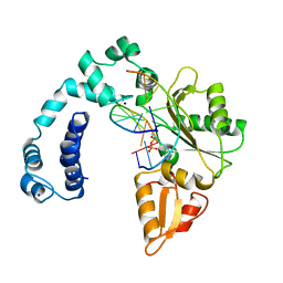
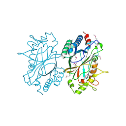
8ICM
 
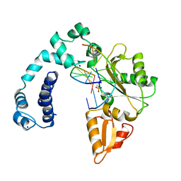 | | DNA POLYMERASE BETA (POL B) (E.C.2.7.7.7) COMPLEXED WITH SEVEN BASE PAIRS OF DNA; SOAKED IN THE PRESENCE OF DATP (1 MILLIMOLAR), MNCL2 (5 MILLIMOLAR), AND AMMONIUM SULFATE (75 MILLIMOLAR) | | 分子名称: | DNA (5'-D(*CP*AP*TP*TP*AP*GP*AP*A)-3'), DNA (5'-D(*TP*CP*TP*AP*AP*TP*G)-3'), PROTEIN (DNA POLYMERASE BETA (E.C.2.7.7.7)), ... | | 著者 | Pelletier, H, Sawaya, M.R. | | 登録日 | 1996-01-04 | | 公開日 | 1996-11-15 | | 最終更新日 | 2023-08-02 | | 実験手法 | X-RAY DIFFRACTION (2.9 Å) | | 主引用文献 | A structural basis for metal ion mutagenicity and nucleotide selectivity in human DNA polymerase beta.
Biochemistry, 35, 1996
|
|
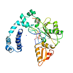
8ICR
 | |
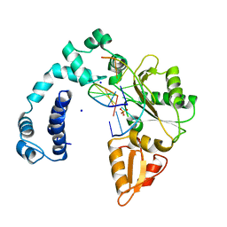
8ICJ
 | |
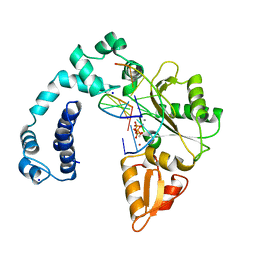
8ICS
 | |
8ICQ
 | |
8ICN
 | |
8ICP
 | |
6LEK
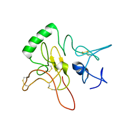 | | Tertiary structure of Barnacle cement protein MrCP20 | | 分子名称: | Cement protein-20k | | 著者 | Mohanram, H. | | 登録日 | 2019-11-25 | | 公開日 | 2020-01-15 | | 最終更新日 | 2024-10-16 | | 実験手法 | SOLUTION NMR | | 主引用文献 | Three-dimensional structure of Megabalanus rosa Cement Protein 20 revealed by multi-dimensional NMR and molecular dynamics simulations.
Philos.Trans.R.Soc.Lond.B Biol.Sci., 374, 2019
|
|
2NAF
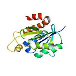 | | Solution structure of peptidyl-tRNA hydrolase from Mycobacterium smegmatis | | 分子名称: | Peptidyl-tRNA hydrolase | | 著者 | Yadav, R, Pathak, P, Fatma, F, Kabra, A, Pulavarti, S, Jain, A, Kumar, A, Shukla, V, Arora, A. | | 登録日 | 2015-12-23 | | 公開日 | 2017-01-11 | | 最終更新日 | 2024-05-15 | | 実験手法 | SOLUTION NMR | | 主引用文献 | Structural characterization of peptidyl-tRNA hydrolase from Mycobacterium smegmatis by NMR spectroscopy.
Biochim.Biophys.Acta, 1864, 2016
|
|
2GLZ
 | |
2QTP
 | |
2GVI
 | |
2ETS
 | |
2G36
 | |
2FG0
 | |
2EVR
 | |
9F00
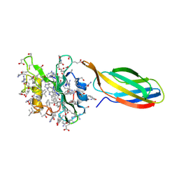 | | Complex between D-SH2 domain of ABL with monobody DAM27 | | 分子名称: | monobody DAM27, synthetic D-SH2 domain | | 著者 | Essen, L.-O, Hantschel, O, Schmidt, N, Korf, L. | | 登録日 | 2024-04-14 | | 公開日 | 2024-12-25 | | 最終更新日 | 2025-01-22 | | 実験手法 | X-RAY DIFFRACTION (2.91 Å) | | 主引用文献 | Development of mirror-image monobodies targeting the oncogenic BCR::ABL1 kinase.
Nat Commun, 15, 2024
|
|
9F01
 | | Complex between D-SH2 domain of ABL with monobody 'DAM21 | | 分子名称: | CHLORIDE ION, nanobody DAM21.3, synthetic D-SH2 domain NS1-10 | | 著者 | Essen, L.-O, Hantschel, O, Schmidt, N, Korf, L. | | 登録日 | 2024-04-14 | | 公開日 | 2024-12-25 | | 最終更新日 | 2025-01-22 | | 実験手法 | X-RAY DIFFRACTION (2.73 Å) | | 主引用文献 | Development of mirror-image monobodies targeting the oncogenic BCR::ABL1 kinase.
Nat Commun, 15, 2024
|
|
2GHR
 | |
2HBW
 | |
2GVK
 | |
2H1T
 | |
2HAG
 | |
2HUJ
 | |
2OOC
 | |


