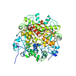
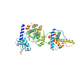
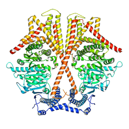
6IXJ
 
 | | The crystal structure of sulfoacetaldehyde reductase from Klebsiella oxytoca | | 分子名称: | 2-hydroxyethylsulfonic acid, NADPH DIHYDRO-NICOTINAMIDE-ADENINE-DINUCLEOTIDE PHOSPHATE, Sulfoacetaldehyde reductase | | 著者 | Zhou, Y, Xu, T, Lin, L, Zhang, Y, Yuchi, Z. | | 登録日 | 2018-12-10 | | 公開日 | 2019-02-13 | | 最終更新日 | 2023-11-22 | | 実験手法 | X-RAY DIFFRACTION (2.8 Å) | | 主引用文献 | Biochemical and structural investigation of sulfoacetaldehyde reductase fromKlebsiella oxytoca.
Biochem. J., 476, 2019
|
|
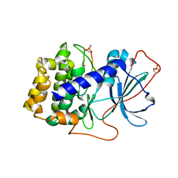
6J6P
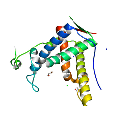
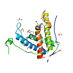 | | Crystal structure of diamondback moth ryanodine receptor phosphorylation domain(2836-3050) mutant S2946D | | 分子名称: | 4-(2-HYDROXYETHYL)-1-PIPERAZINE ETHANESULFONIC ACID, CHLORIDE ION, GLYCEROL, ... | | 著者 | Xu, T, Lin, L, Yuchi, Z. | | 登録日 | 2019-01-15 | | 公開日 | 2019-09-04 | | 最終更新日 | 2023-11-22 | | 実験手法 | X-RAY DIFFRACTION (1.53 Å) | | 主引用文献 | Crystal structure of diamondback moth ryanodine receptor Repeat34 domain reveals insect-specific phosphorylation sites.
Bmc Biol., 17, 2019
|
|
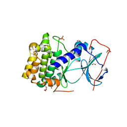
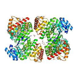
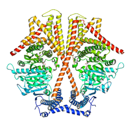
6JKP
 | | Crystal structure of sulfoacetaldehyde reductase from Bifidobacterium kashiwanohense in complex with NAD+ | | 分子名称: | Methanol dehydrogenase, NICOTINAMIDE-ADENINE-DINUCLEOTIDE, ZINC ION | | 著者 | Zhou, Y, Xu, T, Lin, L, Zhang, Y, Yuchi, Z. | | 登録日 | 2019-03-01 | | 公開日 | 2019-06-12 | | 最終更新日 | 2023-11-22 | | 実験手法 | X-RAY DIFFRACTION (3.008 Å) | | 主引用文献 | Identification and characterization of a new sulfoacetaldehyde reductase from the human gut bacteriumBifidobacterium kashiwanohense.
Biosci.Rep., 39, 2019
|
|
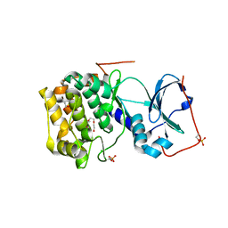
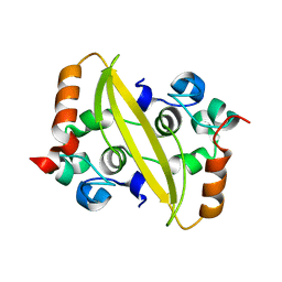
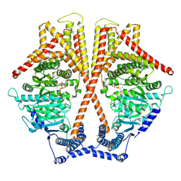
6JKO
 | | Crystal structure of sulfoacetaldehyde reductase from Bifidobacterium kashiwanohense | | 分子名称: | Methanol dehydrogenase, ZINC ION | | 著者 | Zhou, Y, Xu, T, Lin, L, Zhang, Y, Yuchi, Z. | | 登録日 | 2019-03-01 | | 公開日 | 2019-06-12 | | 最終更新日 | 2023-11-22 | | 実験手法 | X-RAY DIFFRACTION (1.9 Å) | | 主引用文献 | Identification and characterization of a new sulfoacetaldehyde reductase from the human gut bacteriumBifidobacterium kashiwanohense.
Biosci.Rep., 39, 2019
|
|
7E12
 | | Crystal structure of PKAc-A11E complex | | 分子名称: | MAGNESIUM ION, PHOSPHOAMINOPHOSPHONIC ACID-ADENYLATE ESTER, THR-ARG-SER-GLU-ILE-ARG-ARG-ALA-SER-THR-ILE-GLU, ... | | 著者 | Qin, J, Lin, L, Yuchi, Z. | | 登録日 | 2021-01-28 | | 公開日 | 2022-04-27 | | 最終更新日 | 2023-11-29 | | 実験手法 | X-RAY DIFFRACTION (2.796 Å) | | 主引用文献 | Structures of PKA-phospholamban complexes reveal a mechanism of familial dilated cardiomyopathy.
Elife, 11, 2022
|
|
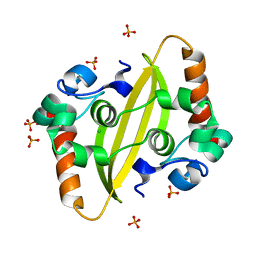
6JIX
 | | The cyrstal structure of taurine:2-oxoglutarate aminotransferase from Bifidobacterium kashiwanohense, in complex with PLP and glutamate | | 分子名称: | GLUTAMIC ACID, PYRIDOXAL-5'-PHOSPHATE, taurine:2-oxoglutarate aminotransferase | | 著者 | Li, M, Lin, L, Zhang, Y, Yuchi, Z. | | 登録日 | 2019-02-23 | | 公開日 | 2020-01-01 | | 最終更新日 | 2023-11-29 | | 実験手法 | X-RAY DIFFRACTION (2.647 Å) | | 主引用文献 | Biochemical and structural investigation of taurine:2-oxoglutarate aminotransferase fromBifidobacterium kashiwanohense.
Biochem.J., 476, 2019
|
|
6KIM
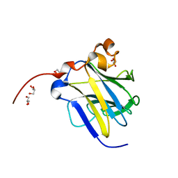 | |
6J6O
 | | Crystal structure of diamondback moth ryanodine receptor phosphorylation domain(2836-3050) | | 分子名称: | CHLORIDE ION, GLYCEROL, Ryanodine receptor, ... | | 著者 | Xu, T, Lin, L, Yuchi, Z. | | 登録日 | 2019-01-15 | | 公開日 | 2019-09-04 | | 最終更新日 | 2023-11-22 | | 実験手法 | X-RAY DIFFRACTION (1.848 Å) | | 主引用文献 | Crystal structure of diamondback moth ryanodine receptor Repeat34 domain reveals insect-specific phosphorylation sites.
Bmc Biol., 17, 2019
|
|
7VUA
 | | Anaerobic hydroxyproline degradation involving C-N cleavage by a glycyl radical enzyme | | 分子名称: | (4S)-4-hydroxy-D-proline, HplG | | 著者 | Duan, Y, Lu, Q, Yuchi, Z, Zhang, Y. | | 登録日 | 2021-11-01 | | 公開日 | 2022-06-01 | | 最終更新日 | 2024-04-03 | | 実験手法 | X-RAY DIFFRACTION (2.695 Å) | | 主引用文献 | Anaerobic Hydroxyproline Degradation Involving C-N Cleavage by a Glycyl Radical Enzyme.
J.Am.Chem.Soc., 144, 2022
|
|
7E7L
 | | The crystal structure of arylacetate decarboxylase from Olsenella scatoligenes. | | 分子名称: | 4-HYDROXYPHENYLACETATE, Hydroxyphenylacetic acid decarboxylase | | 著者 | Lu, Q, Duan, Y, Zhang, Y, Yuchi, Z. | | 登録日 | 2021-02-26 | | 公開日 | 2021-07-28 | | 最終更新日 | 2023-11-29 | | 実験手法 | X-RAY DIFFRACTION (3.53 Å) | | 主引用文献 | The Glycyl Radical Enzyme Arylacetate Decarboxylase from Olsenella scatoligenes
Acs Catalysis, 11, 2021
|
|
7EEV
 | | Structure of UTP cyclohydrolase | | 分子名称: | DEOXYURIDINE-5'-TRIPHOSPHATE, GTP cyclohydrolase II, ZINC ION | | 著者 | Zhang, H, Zhang, Y, Yuchi, Z. | | 登録日 | 2021-03-19 | | 公開日 | 2021-07-28 | | 最終更新日 | 2022-09-21 | | 実験手法 | X-RAY DIFFRACTION (2.15 Å) | | 主引用文献 | Structural and biochemical investigation of UTP cyclohydrolase
Acs Catalysis, 2021
|
|
7EJ3
 | | UTP cyclohydrolase | | 分子名称: | DIPHOSPHATE, GLYCINE, GTP cyclohydrolase II, ... | | 著者 | Zhang, H, Zhang, Y, Yuchi, Z. | | 登録日 | 2021-04-01 | | 公開日 | 2022-09-21 | | 最終更新日 | 2024-05-29 | | 実験手法 | X-RAY DIFFRACTION (1.6 Å) | | 主引用文献 | Structural and biochemical investigation of UTP cyclohydrolase
Acs Catalysis, 2021
|
|
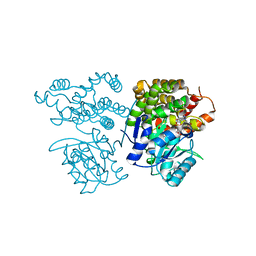
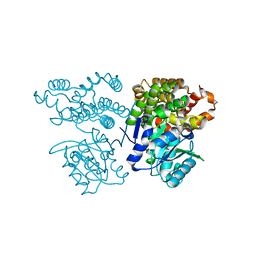
7CF9
 | | Structure of RyR1 (Ca2+/CHL) | | 分子名称: | 5-bromanyl-N-[4-chloranyl-2-methyl-6-(methylcarbamoyl)phenyl]-2-(3-chloranylpyridin-2-yl)pyrazole-3-carboxamide, CALCIUM ION, Peptidyl-prolyl cis-trans isomerase FKBP1B, ... | | 著者 | Ma, R, Haji-Ghassemi, O, Ma, D, Lin, L, Samurkas, A, Van Petegem, F, Yuchi, Z. | | 登録日 | 2020-06-24 | | 公開日 | 2020-09-02 | | 最終更新日 | 2024-03-27 | | 実験手法 | ELECTRON MICROSCOPY (4.7 Å) | | 主引用文献 | Structural basis for diamide modulation of ryanodine receptor.
Nat.Chem.Biol., 16, 2020
|
|
6MM5
 | |
6MM7
 | |
6MM6
 | |
6MM8
 | |
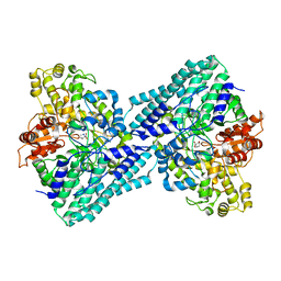
5ZXL
 | | Structure of GldA from E.coli | | 分子名称: | CHLORIDE ION, GLYCEROL, Glycerol dehydrogenase, ... | | 著者 | Zhang, J, Lin, L. | | 登録日 | 2018-05-21 | | 公開日 | 2019-03-20 | | 最終更新日 | 2023-11-22 | | 実験手法 | X-RAY DIFFRACTION (2.794 Å) | | 主引用文献 | Structure of glycerol dehydrogenase (GldA) from Escherichia coli.
Acta Crystallogr F Struct Biol Commun, 75, 2019
|
|
7F2B
 | |
7F2E
 | |
7WJM
 | |
7WJO
 | | CryoEM structure of chitin synthase 1 from Phytophthora sojae complexed with nikkomycin Z | | 分子名称: | (2S)-{[(2S,3S,4S)-2-amino-4-hydroxy-4-(5-hydroxypyridin-2-yl)-3-methylbutanoyl]amino}[(2R,3S,4R,5R)-5-(2,4-dioxo-3,4-dihydropyrimidin-1(2H)-yl)-3,4-dihydroxyoxolan-2-yl]acetic acid (non-preferred name), Chitin synthase | | 著者 | Chen, W, Cao, P, Gong, Y, Yang, Q. | | 登録日 | 2022-01-07 | | 公開日 | 2022-09-28 | | 最終更新日 | 2024-06-26 | | 実験手法 | ELECTRON MICROSCOPY (3.2 Å) | | 主引用文献 | Structural basis for directional chitin biosynthesis.
Nature, 610, 2022
|
|
7WJN
 | | CryoEM structure of chitin synthase 1 mutant E495A from Phytophthora sojae complexed with UDP-GlcNAc | | 分子名称: | Chitin synthase, MANGANESE (II) ION, URIDINE-DIPHOSPHATE-N-ACETYLGLUCOSAMINE | | 著者 | Chen, W, Cao, P, Gong, Y, Yang, Q. | | 登録日 | 2022-01-07 | | 公開日 | 2022-09-28 | | 最終更新日 | 2024-06-26 | | 実験手法 | ELECTRON MICROSCOPY (3.3 Å) | | 主引用文献 | Structural basis for directional chitin biosynthesis.
Nature, 610, 2022
|
|
7X05
 | | CryoEM structure of chitin synthase 1 from Phytophthora sojae complexed with the nascent chitooligosaccharide | | 分子名称: | 2-acetamido-2-deoxy-beta-D-glucopyranose-(1-4)-2-acetamido-2-deoxy-beta-D-glucopyranose-(1-4)-2-acetamido-2-deoxy-beta-D-glucopyranose, Chitin synthase, MANGANESE (II) ION, ... | | 著者 | Chen, W, Cao, P, Gong, Y, Yang, Q. | | 登録日 | 2022-02-21 | | 公開日 | 2022-09-28 | | 最終更新日 | 2024-06-26 | | 実験手法 | ELECTRON MICROSCOPY (3.9 Å) | | 主引用文献 | Structural basis for directional chitin biosynthesis.
Nature, 610, 2022
|
|
7X06
 | | CryoEM structure of chitin synthase 1 from Phytophthora sojae complexed with UDP | | 分子名称: | Chitin synthase, MAGNESIUM ION, URIDINE-5'-DIPHOSPHATE | | 著者 | Chen, W, Cao, P, Gong, Y, Yang, Q. | | 登録日 | 2022-02-21 | | 公開日 | 2022-09-28 | | 最終更新日 | 2024-06-26 | | 実験手法 | ELECTRON MICROSCOPY (3.1 Å) | | 主引用文献 | Structural basis for directional chitin biosynthesis.
Nature, 610, 2022
|
|


