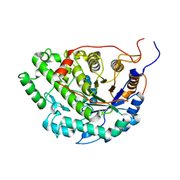
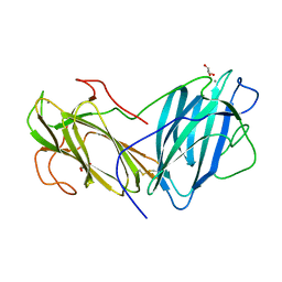
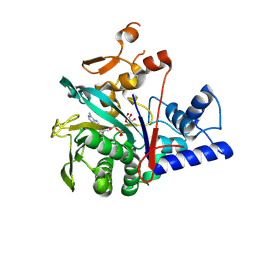
1P4M
 
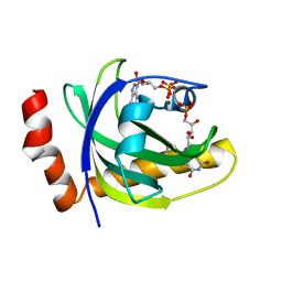 | | CRYSTAL STRUCTURE OF RIBOFLAVIN KINASE | | 分子名称: | ADENOSINE-5'-DIPHOSPHATE, FLAVIN MONONUCLEOTIDE, MAGNESIUM ION, ... | | 著者 | Karthikeyan, S, Zhou, Q, Mseeh, F, Grishin, N.V, Osterman, A.L, Zhang, H. | | 登録日 | 2003-04-23 | | 公開日 | 2003-05-13 | | 最終更新日 | 2023-08-16 | | 実験手法 | X-RAY DIFFRACTION (1.8 Å) | | 主引用文献 | Crystal Structure of Human Riboflavin Kinase Reveals a Beta Barrel Fold and a Novel Active Site Arch
Structure, 11, 2003
|
|
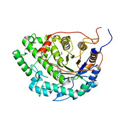
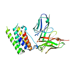
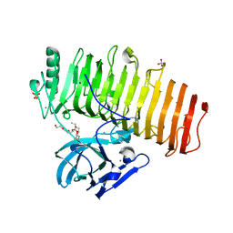
1R2Q
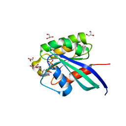 | | Crystal Structure of Human Rab5a GTPase Domain at 1.05 A resolution | | 分子名称: | GLYCEROL, MAGNESIUM ION, PHOSPHOAMINOPHOSPHONIC ACID-GUANYLATE ESTER, ... | | 著者 | Terzyan, S, Zhu, G, Li, G, Zhang, X.C. | | 登録日 | 2003-09-29 | | 公開日 | 2003-12-23 | | 最終更新日 | 2023-08-23 | | 実験手法 | X-RAY DIFFRACTION (1.05 Å) | | 主引用文献 | Refinement of the structure of human Rab5a GTPase domain at 1.05 A resolution.
Acta Crystallogr.,Sect.D, 60, 2004
|
|
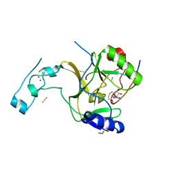
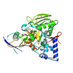
1RQX
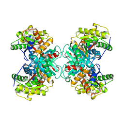 | | Crystal structure of ACC Deaminase complexed with Inhibitor | | 分子名称: | 1-AMINOCYCLOPROPYLPHOSPHONATE, 1-aminocyclopropane-1-carboxylate deaminase, PYRIDOXAL-5'-PHOSPHATE | | 著者 | Karthikeyan, S, Zhao, Z, Kao, C.L, Zhou, Q, Tao, Z, Zhang, H, Liu, H.W. | | 登録日 | 2003-12-07 | | 公開日 | 2004-08-17 | | 最終更新日 | 2023-08-23 | | 実験手法 | X-RAY DIFFRACTION (2.5 Å) | | 主引用文献 | Structural analysis of 1-aminocyclopropane-1-carboxylate deaminase: observation of an aminyl intermediate and identification of Tyr 294 as the active-site nucleophile.
Angew.Chem.Int.Ed.Engl., 43, 2004
|
|
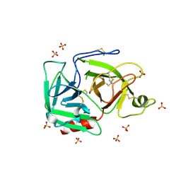
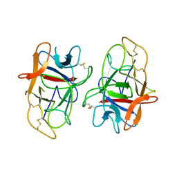
2RJ2
 | | Crystal Structure of the Sugar Recognizing SCF Ubiquitin Ligase at 1.7 Resolution | | 分子名称: | CHLORIDE ION, F-box only protein 2, NICKEL (II) ION | | 著者 | Vaijayanthimala, S, Velmurugan, D, Mizushima, T, Yamane, T, Yoshida, Y, Tanaka, K. | | 登録日 | 2007-10-14 | | 公開日 | 2008-10-14 | | 最終更新日 | 2023-11-08 | | 実験手法 | X-RAY DIFFRACTION (1.7 Å) | | 主引用文献 | Crystal Structure of the Sugar Recognizing SCF Ubiquitin Ligase at 1.7 Resolution
To be Published
|
|
1RJX
 | | Human PLASMINOGEN CATALYTIC DOMAIN, K698M MUTANT | | 分子名称: | Plasminogen, SULFATE ION | | 著者 | Terzyan, S, Wakeham, N, Zhai, P, Rodgers, K, Zhang, X.C. | | 登録日 | 2003-11-20 | | 公開日 | 2003-12-02 | | 最終更新日 | 2023-08-23 | | 実験手法 | X-RAY DIFFRACTION (2.3 Å) | | 主引用文献 | Characterization of Lys-698 to met substitution in human plasminogen catalytic domain
Proteins, 56, 2004
|
|
5WQU
 | | Crystal structure of Sweet Potato Beta-Amylase complexed with Maltotetraose | | 分子名称: | Beta-amylase, alpha-D-glucopyranose-(1-4)-alpha-D-glucopyranose-(1-4)-alpha-D-glucopyranose-(1-4)-alpha-D-glucopyranose | | 著者 | Vajravijayan, S, Sergei, P, Nandhagopal, N, Gunasekaran, K. | | 登録日 | 2016-11-28 | | 公開日 | 2017-12-06 | | 最終更新日 | 2023-11-08 | | 実験手法 | X-RAY DIFFRACTION (2.49 Å) | | 主引用文献 | Structural insights on starch hydrolysis by plant beta-amylase and its evolutionary relationship with bacterial enzymes
Int. J. Biol. Macromol., 113, 2018
|
|
5WQS
 | | Crystal structure of Apo Beta-Amylase from Sweet potato | | 分子名称: | Beta-amylase, ISOPROPYL ALCOHOL | | 著者 | Vajravijayan, S, Sergei, P, Nandhagopal, N, Gunasekaran, K. | | 登録日 | 2016-11-28 | | 公開日 | 2017-12-06 | | 最終更新日 | 2023-11-08 | | 実験手法 | X-RAY DIFFRACTION (1.9 Å) | | 主引用文献 | Structural insights on starch hydrolysis by plant beta-amylase and its evolutionary relationship with bacterial enzymes
Int. J. Biol. Macromol., 113, 2018
|
|
5JJY
 | | Crystal structure of SETD2 bound to histone H3.3 K36M peptide | | 分子名称: | Histone H3.3, Histone-lysine N-methyltransferase SETD2, S-ADENOSYL-L-HOMOCYSTEINE, ... | | 著者 | Yang, S, Zheng, X, Li, H. | | 登録日 | 2016-04-25 | | 公開日 | 2016-11-02 | | 最終更新日 | 2024-04-03 | | 実験手法 | X-RAY DIFFRACTION (2.053 Å) | | 主引用文献 | Molecular basis for oncohistone H3 recognition by SETD2 methyltransferase
Genes Dev., 30, 2016
|
|
5YCZ
 | | Crystal structure of Alocasin, protease inhibitor from Giant Taro (Arum macrorrhizon) | | 分子名称: | Trypsin/chymotrypsin inhibitor | | 著者 | Vajravijayan, S, Pletnev, S, Nandhagopal, N, Gunasekaran, K. | | 登録日 | 2017-09-08 | | 公開日 | 2018-06-13 | | 最終更新日 | 2018-11-28 | | 実験手法 | X-RAY DIFFRACTION (2.502 Å) | | 主引用文献 | Crystal structure of a novel Kunitz type inhibitor, alocasin with anti-Aedes aegypti activity targeting midgut proteases.
Pest Manag. Sci., 74, 2018
|
|
2JLN
 | | Structure of Mhp1, a nucleobase-cation-symport-1 family transporter | | 分子名称: | MERCURY (II) ION, MHP1, SODIUM ION | | 著者 | Weyand, S, Shimamura, T, Yajima, S, Suzuki, S, Mirza, O, Krusong, K, Carpenter, E.P, Rutherford, N.G, Hadden, J.M, O'Reilly, J, Ma, P, Saidijam, M, Patching, S.G, Hope, R.J, Norbertczak, H.T, Roach, P.C.J, Iwata, S, Henderson, P.J.F, Cameron, A.D. | | 登録日 | 2008-09-11 | | 公開日 | 2008-10-28 | | 最終更新日 | 2024-05-08 | | 実験手法 | X-RAY DIFFRACTION (2.85 Å) | | 主引用文献 | Structure and Molecular Mechanism of a Nucleobase-Cation-Symport-1 Family Transporter.
Science, 322, 2008
|
|
6LIK
 | | Crystal Structure of Lectin from Pleurotus ostreatus in complex with Galactose | | 分子名称: | 2-acetamido-2-deoxy-beta-D-glucopyranose-(1-4)-2-acetamido-2-deoxy-beta-D-glucopyranose, CALCIUM ION, GLYCEROL, ... | | 著者 | Vajravijayan, S, Pletnev, S, Luo, Z, Iqbal, S, Nandhagopal, N, Gunasekaran, K. | | 登録日 | 2019-12-11 | | 公開日 | 2020-08-05 | | 最終更新日 | 2023-11-22 | | 実験手法 | X-RAY DIFFRACTION (2.4 Å) | | 主引用文献 | Crystallographic and calorimetric analysis on Pleurotus ostreatus lectin and its sugar complexes - promiscuous binding driven by geometry.
Int.J.Biol.Macromol., 152, 2020
|
|
1BIY
 | | STRUCTURE OF DIFERRIC BUFFALO LACTOFERRIN | | 分子名称: | CARBONATE ION, FE (III) ION, LACTOFERRIN | | 著者 | Karthikeyan, S, Yadav, S, Singh, T.P. | | 登録日 | 1998-06-21 | | 公開日 | 1999-01-13 | | 最終更新日 | 2024-04-03 | | 実験手法 | X-RAY DIFFRACTION (3.37 Å) | | 主引用文献 | Structure of buffalo lactoferrin at 3.3 A resolution at 277 K.
Acta Crystallogr.,Sect.D, 56, 2000
|
|
1CE2
 | | STRUCTURE OF DIFERRIC BUFFALO LACTOFERRIN AT 2.5A RESOLUTION | | 分子名称: | CARBONATE ION, FE (III) ION, PROTEIN (LACTOFERRIN) | | 著者 | Karthikeyan, S, Paramasivam, M, Yadav, S, Srinivasan, A, Singh, T.P. | | 登録日 | 1999-03-13 | | 公開日 | 1999-03-19 | | 最終更新日 | 2023-08-09 | | 実験手法 | X-RAY DIFFRACTION (2.5 Å) | | 主引用文献 | Structure of buffalo lactoferrin at 2.5 A resolution using crystals grown at 303 K shows different orientations of the N and C lobes.
Acta Crystallogr.,Sect.D, 55, 1999
|
|
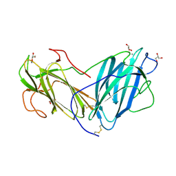
3UX9
 | | Structural insights into a human anti-IFN antibody exerting therapeutic potential for systemic lupus erythematosus | | 分子名称: | Interferon alpha-1/13, ScFv antibody | | 著者 | Ouyang, S, Zhao, L.X, Liang, W, Shaw, N, Liu, Z.-J, Liang, M.-F. | | 登録日 | 2011-12-04 | | 公開日 | 2012-02-29 | | 実験手法 | X-RAY DIFFRACTION (2.8 Å) | | 主引用文献 | Structural insights into a human anti-IFN antibody exerting therapeutic potential for systemic lupus erythematosus
J.Mol.Med., 2012
|
|
2KAP
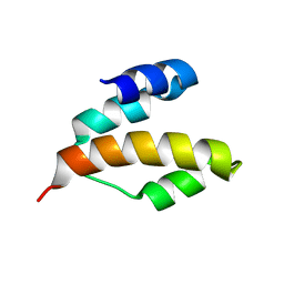 | | Solution structure of DLC1-SAM | | 分子名称: | Rho GTPase-activating protein 7 | | 著者 | Yang, S, Yang, D. | | 登録日 | 2008-11-12 | | 公開日 | 2009-10-20 | | 最終更新日 | 2024-05-29 | | 実験手法 | SOLUTION NMR | | 主引用文献 | Characterization of DLC1-SAM equilibrium unfolding at the amino acid residue level
Biochemistry, 48, 2009
|
|
8IPJ
 | | Crystal structure of the Legionella effector protein MavL with ADPR-Ub | | 分子名称: | MavL, Ubiquitin, [(2R,3S,4R,5R)-5-(6-AMINOPURIN-9-YL)-3,4-DIHYDROXY-OXOLAN-2-YL]METHYL [HYDROXY-[[(2R,3S,4R,5S)-3,4,5-TRIHYDROXYOXOLAN-2-YL]METHOXY]PHOSPHORYL] HYDROGEN PHOSPHATE | | 著者 | Ouyang, S, Guan, H. | | 登録日 | 2023-03-14 | | 公開日 | 2024-03-20 | | 実験手法 | X-RAY DIFFRACTION (2.003 Å) | | 主引用文献 | Crystal structure of the Legionella effector protein MavL with ADPR-Ub
To Be Published
|
|
8IPW
 | | The sturecture of Legionella effector protein MavL with ADPR | | 分子名称: | MavL, [(2R,3S,4R,5R)-5-(6-AMINOPURIN-9-YL)-3,4-DIHYDROXY-OXOLAN-2-YL]METHYL [HYDROXY-[[(2R,3S,4R,5S)-3,4,5-TRIHYDROXYOXOLAN-2-YL]METHOXY]PHOSPHORYL] HYDROGEN PHOSPHATE | | 著者 | Ouyang, S, Hongxin, G. | | 登録日 | 2023-03-15 | | 公開日 | 2024-03-20 | | 実験手法 | X-RAY DIFFRACTION (2.35 Å) | | 主引用文献 | The sturecture of Legionella effector protein MavL with ADPR
To Be Published
|
|
5ZRU
 | | Crystal structure of Agl-KA catalytic domain | | 分子名称: | 3,6,9,12,15,18,21,24-OCTAOXAHEXACOSAN-1-OL, Alpha-1,3-glucanase, CALCIUM ION, ... | | 著者 | Yano, S, Makabe, K. | | 登録日 | 2018-04-25 | | 公開日 | 2019-10-23 | | 最終更新日 | 2024-03-27 | | 実験手法 | X-RAY DIFFRACTION (1.833 Å) | | 主引用文献 | Crystal structure of the catalytic unit of GH 87-type alpha-1,3-glucanase Agl-KA from Bacillus circulans.
Sci Rep, 9, 2019
|
|
6KBQ
 | | Crystal Structure of Lectin from Pleurotus ostreatus in complex with Glycerol | | 分子名称: | 2-acetamido-2-deoxy-beta-D-glucopyranose-(1-4)-2-acetamido-2-deoxy-beta-D-glucopyranose, CALCIUM ION, GLYCEROL, ... | | 著者 | Vajravijayan, S, Pletnev, S, Luo, Z, Gunasekaran, K, Nandhagopal, N. | | 登録日 | 2019-06-26 | | 公開日 | 2020-08-05 | | 最終更新日 | 2023-11-22 | | 実験手法 | X-RAY DIFFRACTION (2.299 Å) | | 主引用文献 | Crystallographic and calorimetric analysis on Pleurotus ostreatus lectin and its sugar complexes - promiscuous binding driven by geometry.
Int.J.Biol.Macromol., 152, 2020
|
|
6KBJ
 | | Structure of Lectin from Pleurotus ostreatus in complex with malonate | | 分子名称: | 2-acetamido-2-deoxy-beta-D-glucopyranose-(1-4)-2-acetamido-2-deoxy-beta-D-glucopyranose, CALCIUM ION, Lectin, ... | | 著者 | Vajravijayan, S, Pletnev, S, Luo, Z, Gunasekaran, K, Nandhagopal, N. | | 登録日 | 2019-06-25 | | 公開日 | 2020-08-12 | | 最終更新日 | 2023-11-22 | | 実験手法 | X-RAY DIFFRACTION (2.2 Å) | | 主引用文献 | Crystallographic and calorimetric analysis on Pleurotus ostreatus lectin and its sugar complexes - promiscuous binding driven by geometry.
Int.J.Biol.Macromol., 152, 2020
|
|
6LI7
 | | Crystal Structure of Lectin from Pleurotus ostreatus in complex with GalNAc | | 分子名称: | 2-acetamido-2-deoxy-beta-D-galactopyranose, 2-acetamido-2-deoxy-beta-D-glucopyranose-(1-4)-2-acetamido-2-deoxy-beta-D-glucopyranose, CALCIUM ION, ... | | 著者 | Vajravijayan, S, Pletnev, S, Luo, Z, Ankur, T, Gunasekaran, K, Nandhagopal, N. | | 登録日 | 2019-12-10 | | 公開日 | 2020-08-05 | | 最終更新日 | 2023-11-22 | | 実験手法 | X-RAY DIFFRACTION (2.098 Å) | | 主引用文献 | Crystallographic and calorimetric analysis on Pleurotus ostreatus lectin and its sugar complexes - promiscuous binding driven by geometry.
Int.J.Biol.Macromol., 152, 2020
|
|
5XTY
 | |
2KET
 | |
7WQY
 | |
4EF4
 | | Crystal structure of STING CTD complex with c-di-GMP | | 分子名称: | 9,9'-[(2R,3R,3aS,5S,7aR,9R,10R,10aS,12S,14aR)-3,5,10,12-tetrahydroxy-5,12-dioxidooctahydro-2H,7H-difuro[3,2-d:3',2'-j][1,3,7,9,2,8]tetraoxadiphosphacyclododecine-2,9-diyl]bis(2-amino-1,9-dihydro-6H-purin-6-one), CALCIUM ION, Transmembrane protein 173 | | 著者 | Ouyang, S, Ru, H, Shaw, N, Jiang, Y, Niu, F, Zhu, Y, Qiu, W, Li, Y, Liu, Z.-J. | | 登録日 | 2012-03-29 | | 公開日 | 2012-05-16 | | 最終更新日 | 2024-03-20 | | 実験手法 | X-RAY DIFFRACTION (2.147 Å) | | 主引用文献 | Structural analysis of the STING adaptor protein reveals a hydrophobic dimer interface and mode of cyclic di-GMP binding
Immunity, 36, 2012
|
|


