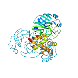
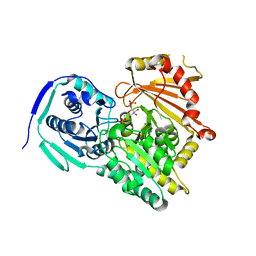
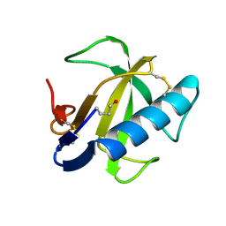
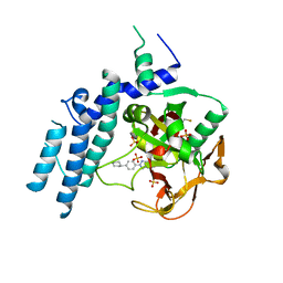
6DNQ
 
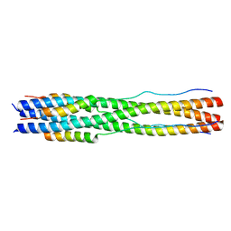
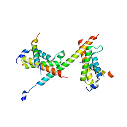 | | HBZ77 in complex with KIX and c-Myb | | 分子名称: | 1,2-ETHANEDIOL, BZIP factor, CREB-binding protein, ... | | 著者 | Yang, K, Wright, P.E, Stanfield, R.L. | | 登録日 | 2018-06-07 | | 公開日 | 2018-09-19 | | 最終更新日 | 2023-10-11 | | 実験手法 | X-RAY DIFFRACTION (2.35 Å) | | 主引用文献 | Structural basis for cooperative regulation of KIX-mediated transcription pathways by the HTLV-1 HBZ activation domain.
Proc. Natl. Acad. Sci. U.S.A., 115, 2018
|
|
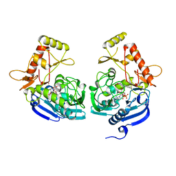
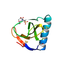
3BF3
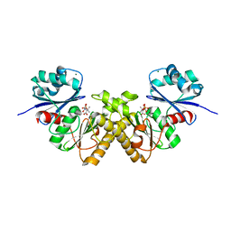 | | Type III pantothenate kinase from Thermotoga maritima complexed with product phosphopantothenate | | 分子名称: | MAGNESIUM ION, N-[(2R)-2-hydroxy-3,3-dimethyl-4-(phosphonooxy)butanoyl]-beta-alanine, Type III pantothenate kinase | | 著者 | Yang, K, Huerta, C, Strauss, E, Zhang, H. | | 登録日 | 2007-11-20 | | 公開日 | 2008-06-24 | | 最終更新日 | 2024-02-21 | | 実験手法 | X-RAY DIFFRACTION (1.63 Å) | | 主引用文献 | Structural basis for substrate binding and the catalytic mechanism of type III pantothenate kinase.
Biochemistry, 47, 2008
|
|
3BEX
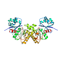 | | Type III pantothenate kinase from Thermotoga maritima complexed with pantothenate | | 分子名称: | PANTOTHENOIC ACID, PHOSPHATE ION, Type III pantothenate kinase | | 著者 | Yang, K, Huerta, C, Strauss, E, Zhang, H. | | 登録日 | 2007-11-20 | | 公開日 | 2008-06-24 | | 最終更新日 | 2023-08-30 | | 実験手法 | X-RAY DIFFRACTION (1.51 Å) | | 主引用文献 | Structural basis for substrate binding and the catalytic mechanism of type III pantothenate kinase.
Biochemistry, 47, 2008
|
|
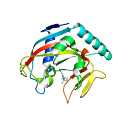
3E66
 | | Crystal structure of the beta-finger domain of yeast Prp8 | | 分子名称: | PRP8 | | 著者 | Yang, K, Zhang, L, Xu, T, Heroux, A, Zhao, R. | | 登録日 | 2008-08-14 | | 公開日 | 2008-10-14 | | 最終更新日 | 2024-02-21 | | 実験手法 | X-RAY DIFFRACTION (2.05 Å) | | 主引用文献 | Crystal structure of the beta-finger domain of Prp8 reveals analogy to ribosomal proteins.
Proc.Natl.Acad.Sci.Usa, 105, 2008
|
|
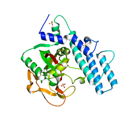
3BF1
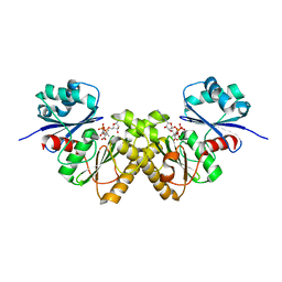 | | Type III pantothenate kinase from Thermotoga maritima complexed with pantothenate and ADP | | 分子名称: | ADENOSINE-5'-DIPHOSPHATE, PANTOTHENOIC ACID, Type III pantothenate kinase | | 著者 | Yang, K, Huerta, C, Strauss, E, Zhang, H. | | 登録日 | 2007-11-20 | | 公開日 | 2008-06-24 | | 最終更新日 | 2024-02-21 | | 実験手法 | X-RAY DIFFRACTION (2.3 Å) | | 主引用文献 | Structural basis for substrate binding and the catalytic mechanism of type III pantothenate kinase.
Biochemistry, 47, 2008
|
|
7JP0
 | | Crystal structure of Mpro with inhibitor r1 | | 分子名称: | 3C-like proteinase, DIMETHYL SULFOXIDE, N-[(benzyloxy)carbonyl]-L-valyl-N-{(2R)-1-hydroxy-3-[(3S)-2-oxopyrrolidin-3-yl]propan-2-yl}-L-leucinamide | | 著者 | Yang, K, Liu, W. | | 登録日 | 2020-08-07 | | 公開日 | 2021-10-13 | | 最終更新日 | 2023-11-15 | | 実験手法 | X-RAY DIFFRACTION (1.65 Å) | | 主引用文献 | Crystal structure of Mpro with inhibitor r1
To Be Published
|
|
7TIK
 | |
1P8X
 | |
6KNC
 | | PolD-PCNA-DNA (form B) | | 分子名称: | DNA polymerase D DP2 (DNA polymerase II large) subunit, DNA polymerase II small subunit, DNA polymerase sliding clamp 1, ... | | 著者 | Mayanagi, K, Oki, K, Miyazaki, N, Ishino, S, Yamagami, T, Iwasaki, K, Kohda, D, Morikawa, K, Shirai, T, Ishino, Y. | | 登録日 | 2019-08-05 | | 公開日 | 2020-08-05 | | 最終更新日 | 2024-03-27 | | 実験手法 | ELECTRON MICROSCOPY (9.3 Å) | | 主引用文献 | Two conformations of DNA polymerase D-PCNA-DNA, an archaeal replisome complex, revealed by cryo-electron microscopy.
Bmc Biol., 18, 2020
|
|
6KNB
 | | PolD-PCNA-DNA (form A) | | 分子名称: | DNA polymerase D DP2 (DNA polymerase II large) subunit, DNA polymerase II small subunit, DNA polymerase sliding clamp 1, ... | | 著者 | Mayanagi, K, Oki, K, Miyazaki, N, Ishino, S, Yamagami, T, Iwasaki, K, Kohda, D, Morikawa, K, Shirai, T, Ishino, Y. | | 登録日 | 2019-08-05 | | 公開日 | 2020-08-05 | | 最終更新日 | 2021-02-17 | | 実験手法 | ELECTRON MICROSCOPY (6.9 Å) | | 主引用文献 | Two conformations of DNA polymerase D-PCNA-DNA, an archaeal replisome complex, revealed by cryo-electron microscopy.
Bmc Biol., 18, 2020
|
|
8FA2
 | |
8FA1
 | |
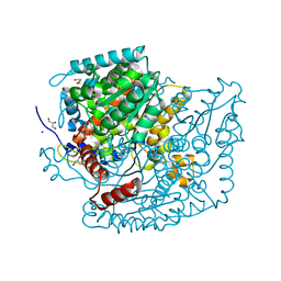
2GTD
 | | Crystal Structure of a Type III Pantothenate Kinase: Insight into the Catalysis of an Essential Coenzyme A Biosynthetic Enzyme Universally Distributed in Bacteria | | 分子名称: | Type III Pantothenate Kinase | | 著者 | Yang, K, Eyobo, Y, Brand, A.L, Martynowski, D, Tomchick, D. | | 登録日 | 2006-04-27 | | 公開日 | 2006-08-01 | | 最終更新日 | 2024-02-14 | | 実験手法 | X-RAY DIFFRACTION (2 Å) | | 主引用文献 | Crystal Structure of a Type III Pantothenate Kinase: Insight into the Mechanism of an Essential Coenzyme A Biosynthetic Enzyme Universally Distributed in Bacteria.
J.Bacteriol., 188, 2006
|
|
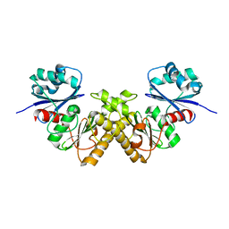
5HQP
 | | Crystal structure of the ERp44-peroxiredoxin 4 complex | | 分子名称: | Endoplasmic reticulum resident protein 44, Peroxiredoxin-4 | | 著者 | Yang, K, Li, D.F, Wang, X, Wang, C.C. | | 登録日 | 2016-01-22 | | 公開日 | 2016-10-12 | | 最終更新日 | 2023-11-08 | | 実験手法 | X-RAY DIFFRACTION (2.6 Å) | | 主引用文献 | Crystal Structure of the ERp44-Peroxiredoxin 4 Complex Reveals the Molecular Mechanisms of Thiol-Mediated Protein Retention.
Structure, 24, 2016
|
|
2A2Y
 | | NMR Structue of Sso10b2 from Sulfolobus solfataricus | | 分子名称: | DNA/RNA-binding protein Alba 2 | | 著者 | Biyani, K, Kahsai, M.A, Clark, A.T, Armstrong, T.L, Edmondson, S.P, Shriver, J.W. | | 登録日 | 2005-06-23 | | 公開日 | 2005-11-08 | | 最終更新日 | 2024-05-22 | | 実験手法 | SOLUTION NMR | | 主引用文献 | Solution structure, stability, and nucleic Acid binding of the hyperthermophile protein sso10b2.
Biochemistry, 44, 2005
|
|
7P5O
 | |
7PPR
 | |
7U34
 | | The structure of phosphoglucose isomerase from Aspergillus fumigatus | | 分子名称: | CHLORIDE ION, CITRATE ANION, GLYCEROL, ... | | 著者 | Yan, K, Kowalski, B, Fang, W, van Aalten, D. | | 登録日 | 2022-02-25 | | 公開日 | 2022-08-17 | | 最終更新日 | 2023-10-18 | | 実験手法 | X-RAY DIFFRACTION (1.56 Å) | | 主引用文献 | Phosphoglucose Isomerase Is Important for Aspergillus fumigatus Cell Wall Biogenesis.
Mbio, 13, 2022
|
|
5JZX
 | | Crystal Structure of UDP-N-acetylenolpyruvoylglucosamine reductase (MurB) from Mycobacterium tuberculosis | | 分子名称: | FLAVIN-ADENINE DINUCLEOTIDE, POTASSIUM ION, UDP-N-acetylenolpyruvoylglucosamine reductase | | 著者 | Dharavath, S, Eniyan, K, Bajpai, U, Gourinath, S. | | 登録日 | 2016-05-17 | | 公開日 | 2017-05-10 | | 最終更新日 | 2023-11-08 | | 実験手法 | X-RAY DIFFRACTION (2.2 Å) | | 主引用文献 | Crystal structure of UDP-N-acetylglucosamine-enolpyruvate reductase (MurB) from Mycobacterium tuberculosis
Biochim. Biophys. Acta, 1866, 2017
|
|
1FUS
 | |
1FUT
 | | CRYSTAL STRUCTURES OF RIBONUCLEASE F1 OF FUSARIUM MONILIFORME IN ITS FREE FORM AND IN COMPLEX WITH 2'GMP | | 分子名称: | GUANOSINE-2'-MONOPHOSPHATE, RIBONUCLEASE F1 | | 著者 | Katayanagi, K, Vassylyev, D.G, Ishikawa, K, Morikawa, K. | | 登録日 | 1993-01-18 | | 公開日 | 1993-10-31 | | 最終更新日 | 2019-12-25 | | 実験手法 | X-RAY DIFFRACTION (2 Å) | | 主引用文献 | Crystal structures of ribonuclease F1 of Fusarium moniliforme in its free form and in complex with 2'GMP.
J.Mol.Biol., 230, 1993
|
|
7KK6
 | |
7KKQ
 | |
7KK2
 | |
7KK5
 | |


