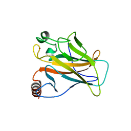
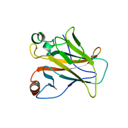
2CAY
 
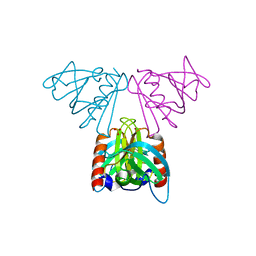 | | Vps36 N-terminal PH domain | | 分子名称: | SULFATE ION, VACUOLAR PROTEIN SORTING PROTEIN 36 | | 著者 | Teo, H, Williams, R.L, Perisic, O, Gill, D.J. | | 登録日 | 2005-12-23 | | 公開日 | 2006-04-07 | | 最終更新日 | 2024-05-08 | | 実験手法 | X-RAY DIFFRACTION (1.9 Å) | | 主引用文献 | Escrt-I Core and Escrt-II Glue Domain Structures Reveal Role for Glue in Linking to Escrt-I and Membranes.
Cell(Cambridge,Mass.), 125, 2006
|
|
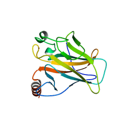
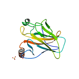
1W2F
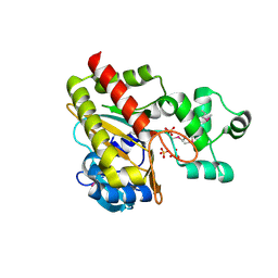 | | Human Inositol (1,4,5)-trisphosphate 3-kinase substituted with selenomethionine | | 分子名称: | INOSITOL-TRISPHOSPHATE 3-KINASE A, SULFATE ION | | 著者 | Gonzalez, B, Schell, M.J, Irvine, R.F, Williams, R.L. | | 登録日 | 2004-07-01 | | 公開日 | 2004-09-09 | | 最終更新日 | 2011-07-13 | | 実験手法 | X-RAY DIFFRACTION (1.8 Å) | | 主引用文献 | Structure of a Human Inositol 1,4,5-Trisphosphate 3-Kinase; Substrate Binding Reveals Why It is not a Phosphoinositide 3-Kinase
Mol.Cell, 15, 2004
|
|
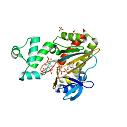
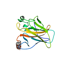
1W2D
 | | Human Inositol (1,4,5)-trisphosphate 3-kinase complexed with Mn2+/ADP/Ins(1,3,4,5)P4 | | 分子名称: | ADENOSINE-5'-DIPHOSPHATE, INOSITOL-(1,3,4,5)-TETRAKISPHOSPHATE, INOSITOL-TRISPHOSPHATE 3-KINASE A, ... | | 著者 | Gonzalez, B, Schell, M.J, Irvine, R.F, Williams, R.L. | | 登録日 | 2004-07-01 | | 公開日 | 2004-09-09 | | 最終更新日 | 2024-05-08 | | 実験手法 | X-RAY DIFFRACTION (1.94 Å) | | 主引用文献 | Structure of a Human Inositol 1,4,5-Trisphosphate 3-Kinase; Substrate Binding Reveals Why It is not a Phosphoinositide 3-Kinase
Mol.Cell, 15, 2004
|
|
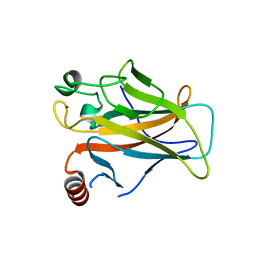
1W2C
 | | Human Inositol (1,4,5) trisphosphate 3-kinase complexed with Mn2+/AMPPNP/Ins(1,4,5)P3 | | 分子名称: | D-MYO-INOSITOL-1,4,5-TRIPHOSPHATE, INOSITOL-TRISPHOSPHATE 3-KINASE A, MANGANESE (II) ION, ... | | 著者 | Gonzalez, B, Schell, M.J, Irvine, R.F, Williams, R.L. | | 登録日 | 2004-07-01 | | 公開日 | 2004-09-09 | | 最終更新日 | 2024-05-08 | | 実験手法 | X-RAY DIFFRACTION (1.95 Å) | | 主引用文献 | Structure of a Human Inositol 1,4,5-Trisphosphate 3-Kinase; Substrate Binding Reveals Why It is not a Phosphoinositide 3-Kinase
Mol.Cell, 15, 2004
|
|
3O21
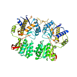 | | High resolution structure of GluA3 N-terminal domain (NTD) | | 分子名称: | 2-acetamido-2-deoxy-beta-D-glucopyranose, Glutamate receptor 3, PHOSPHATE ION | | 著者 | Rossmann, M, Sukumaran, M, Penn, A.C, Veprintsev, D.B, Babu, M.M, Jensen, M.H, Greger, I.H. | | 登録日 | 2010-07-22 | | 公開日 | 2011-03-09 | | 最終更新日 | 2020-07-29 | | 実験手法 | X-RAY DIFFRACTION (2.201 Å) | | 主引用文献 | Dynamics and allosteric potential of the AMPA receptor N-terminal domain
Embo J., 30, 2011
|
|
4AQP
 | | The structure of the AXH domain of ataxin-1. | | 分子名称: | ATAXIN-1, DI(HYDROXYETHYL)ETHER, SODIUM ION | | 著者 | Rees, M, Chen, Y.W, de Chiara, C, Pastore, A. | | 登録日 | 2012-04-19 | | 公開日 | 2013-03-27 | | 最終更新日 | 2023-12-20 | | 実験手法 | X-RAY DIFFRACTION (2.452 Å) | | 主引用文献 | Self-Assembly and Conformational Heterogeneity of the Axh Domain of Ataxin-1: An Unusual Example of a Chameleon Fold
Biophys.J., 104, 2013
|
|
4APT
 | | The structure of the AXH domain of ataxin-1. | | 分子名称: | ATAXIN-1, SODIUM ION | | 著者 | Rees, M, Chen, Y.W, de Chiara, C, Pastore, A. | | 登録日 | 2012-04-05 | | 公開日 | 2013-03-27 | | 最終更新日 | 2023-12-20 | | 実験手法 | X-RAY DIFFRACTION (2.5 Å) | | 主引用文献 | Self-Assembly and Conformational Heterogeneity of the Axh Domain of Ataxin-1: An Unusual Example of a Chameleon Fold
Biophys.J., 104, 2013
|
|
2J1X
 | |
2J1W
 | |
2J1Z
 | |
2J21
 | |
2J1Y
 | |
2J20
 | |


