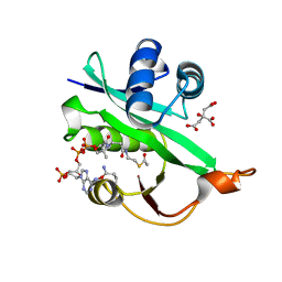
4V7G
 
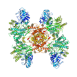
 | | Crystal Structure of Lumazine Synthase from Bacillus Anthracis | | 分子名称: | 6,7-dimethyl-8-ribityllumazine synthase, PHOSPHATE ION | | 著者 | Morgunova, E, Illarionov, B, Saller, S, Popov, A, Sambaiah, T, Bacher, A, Cushman, M, Fischer, M, Ladenstein, R. | | 登録日 | 2009-09-16 | | 公開日 | 2014-07-09 | | 最終更新日 | 2023-09-20 | | 実験手法 | X-RAY DIFFRACTION (3.5 Å) | | 主引用文献 | Structural study and thermodynamic characterization of inhibitor binding to lumazine synthase from Bacillus anthracis.
Acta Crystallogr.,Sect.D, 66, 2010
|
|
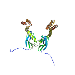
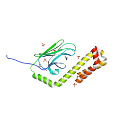
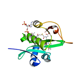
1SSO
 | | SOLUTION STRUCTURE AND DNA-BINDING PROPERTIES OF A THERMOSTABLE PROTEIN FROM THE ARCHAEON SULFOLOBUS SOLFATARICUS | | 分子名称: | SSO7D | | 著者 | Baumann, H, Knapp, S, Lundback, T, Ladenstein, R, Hard, T. | | 登録日 | 1995-03-31 | | 公開日 | 1995-05-08 | | 最終更新日 | 2024-05-22 | | 実験手法 | SOLUTION NMR | | 主引用文献 | Solution structure and DNA-binding properties of a thermostable protein from the archaeon Sulfolobus solfataricus.
Nat.Struct.Biol., 1, 1994
|
|
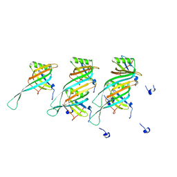
2VL6
 | | STRUCTURAL ANALYSIS OF THE SULFOLOBUS SOLFATARICUS MCM PROTEIN N- TERMINAL DOMAIN | | 分子名称: | MINICHROMOSOME MAINTENANCE PROTEIN MCM, ZINC ION | | 著者 | Liu, W, Pucci, B, Rossi, M, Pisani, F.M, Ladenstein, R. | | 登録日 | 2008-01-08 | | 公開日 | 2008-04-29 | | 最終更新日 | 2023-12-13 | | 実験手法 | X-RAY DIFFRACTION (2.8 Å) | | 主引用文献 | Structural Analysis of the Sulfolobus Solfataricus Mcm Protein N-Terminal Domain.
Nucleic Acids Res., 36, 2008
|
|
3EE6
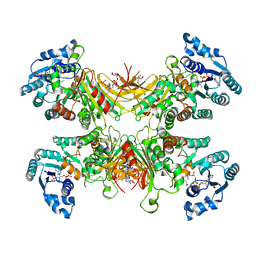
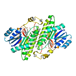
 | | Crystal Structure Analysis of Tripeptidyl peptidase -I | | 分子名称: | 2-acetamido-2-deoxy-beta-D-glucopyranose, CALCIUM ION, CHLORIDE ION, ... | | 著者 | Pal, A, Kraetzner, R, Grapp, M, Gruene, T, Schreiber, K, Granborg, M, Urlaub, H, Asif, A.R, Becker, S, Gartner, J, Sheldrick, G.M, Steinfeld, R. | | 登録日 | 2008-09-04 | | 公開日 | 2008-11-25 | | 最終更新日 | 2020-07-29 | | 実験手法 | X-RAY DIFFRACTION (2.35 Å) | | 主引用文献 | Structure of tripeptidyl-peptidase I provides insight into the molecular basis of late infantile neuronal ceroid lipofuscinosis
J.Biol.Chem., 284, 2009
|
|
6POF
 | |
6PPL
 | |
5JT2
 | | BRAFV600E Kinase Domain In Complex with Chemically Linked Vemurafenib Inhibitor VEM-BISAMIDE | | 分子名称: | 2,2'-oxybis(N-{[4-(3-{2,6-difluoro-3-[(propane-1-sulfonyl)amino]benzoyl}-1H-pyrrolo[2,3-b]pyridin-5-yl)phenyl]methyl}acetamide), BENZAMIDINE, Serine/threonine-protein kinase B-raf | | 著者 | Grasso, M.J, Marmorstein, R. | | 登録日 | 2016-05-09 | | 公開日 | 2016-09-14 | | 最終更新日 | 2023-10-18 | | 実験手法 | X-RAY DIFFRACTION (2.702 Å) | | 主引用文献 | Chemically Linked Vemurafenib Inhibitors Promote an Inactive BRAF(V600E) Conformation.
Acs Chem.Biol., 11, 2016
|
|
2IOO
 | | Crystal structure of the mouse p53 core domain | | 分子名称: | Cellular tumor antigen p53, ZINC ION | | 著者 | Ho, W.C, Luo, C, Zhao, K, Chai, X, Fitzgerald, M.X, Marmorstein, R. | | 登録日 | 2006-10-10 | | 公開日 | 2006-12-05 | | 最終更新日 | 2024-02-21 | | 実験手法 | X-RAY DIFFRACTION (2.02 Å) | | 主引用文献 | High-resolution structure of the p53 core domain: implications for binding small-molecule stabilizing compounds.
Acta Crystallogr.,Sect.D, 62, 2006
|
|
7STX
 | |
7SNI
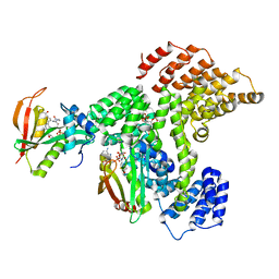
 | | Structure of G6PD-D200N tetramer bound to NADP+ and G6P | | 分子名称: | 6-O-phosphono-beta-D-glucopyranose, Glucose-6-phosphate 1-dehydrogenase, NADP NICOTINAMIDE-ADENINE-DINUCLEOTIDE PHOSPHATE | | 著者 | Wei, X, Marmorstein, R. | | 登録日 | 2021-10-28 | | 公開日 | 2022-07-13 | | 最終更新日 | 2024-06-05 | | 実験手法 | ELECTRON MICROSCOPY (2.5 Å) | | 主引用文献 | Allosteric role of a structural NADP + molecule in glucose-6-phosphate dehydrogenase activity.
Proc.Natl.Acad.Sci.USA, 119, 2022
|
|
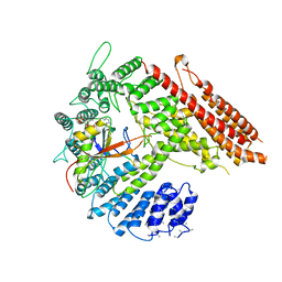
7SNG
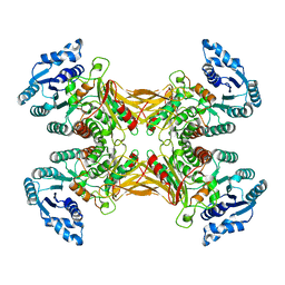 | | structure of G6PD-WT tetramer | | 分子名称: | Glucose-6-phosphate 1-dehydrogenase | | 著者 | Wei, X, Marmorstein, R. | | 登録日 | 2021-10-28 | | 公開日 | 2022-07-13 | | 最終更新日 | 2024-06-05 | | 実験手法 | ELECTRON MICROSCOPY (2.8 Å) | | 主引用文献 | Allosteric role of a structural NADP + molecule in glucose-6-phosphate dehydrogenase activity.
Proc.Natl.Acad.Sci.USA, 119, 2022
|
|
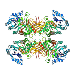
7SNH
 | | Structure of G6PD-D200N tetramer bound to NADP+ | | 分子名称: | Glucose-6-phosphate 1-dehydrogenase, NADP NICOTINAMIDE-ADENINE-DINUCLEOTIDE PHOSPHATE | | 著者 | Wei, X, Marmorstein, R. | | 登録日 | 2021-10-28 | | 公開日 | 2022-07-13 | | 最終更新日 | 2024-06-05 | | 実験手法 | ELECTRON MICROSCOPY (2.2 Å) | | 主引用文献 | Allosteric role of a structural NADP + molecule in glucose-6-phosphate dehydrogenase activity.
Proc.Natl.Acad.Sci.USA, 119, 2022
|
|
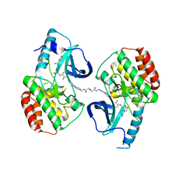
7SNF
 | | Structure of G6PD-WT dimer | | 分子名称: | Glucose-6-phosphate 1-dehydrogenase | | 著者 | Wei, X, Marmorstein, R. | | 登録日 | 2021-10-28 | | 公開日 | 2022-07-13 | | 最終更新日 | 2024-06-05 | | 実験手法 | ELECTRON MICROSCOPY (3.5 Å) | | 主引用文献 | Allosteric role of a structural NADP + molecule in glucose-6-phosphate dehydrogenase activity.
Proc.Natl.Acad.Sci.USA, 119, 2022
|
|
4R5J
 | | Crystal structure of the DnaK C-terminus (Dnak-SBD-A) | | 分子名称: | CALCIUM ION, Chaperone protein DnaK, PHOSPHATE ION | | 著者 | Leu, J.I, Zhang, P, Murphy, M.E, Marmorstein, R, George, D.L. | | 登録日 | 2014-08-21 | | 公開日 | 2014-09-10 | | 最終更新日 | 2024-02-28 | | 実験手法 | X-RAY DIFFRACTION (2.361 Å) | | 主引用文献 | Structural Basis for the Inhibition of HSP70 and DnaK Chaperones by Small-Molecule Targeting of a C-Terminal Allosteric Pocket.
Acs Chem.Biol., 9, 2014
|
|
4R5L
 | | Crystal structure of the DnaK C-terminus (Dnak-SBD-C) | | 分子名称: | CALCIUM ION, Chaperone protein DnaK, PHOSPHATE ION, ... | | 著者 | Leu, J.I, Zhang, P, Murphy, M.E, Marmorstein, R, George, D.L. | | 登録日 | 2014-08-21 | | 公開日 | 2014-09-10 | | 最終更新日 | 2024-02-28 | | 実験手法 | X-RAY DIFFRACTION (2.9701 Å) | | 主引用文献 | Structural Basis for the Inhibition of HSP70 and DnaK Chaperones by Small-Molecule Targeting of a C-Terminal Allosteric Pocket.
Acs Chem.Biol., 9, 2014
|
|
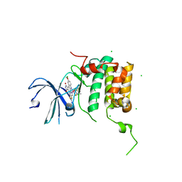
4R5G
 | | Crystal structure of the DnaK C-terminus with the inhibitor PET-16 | | 分子名称: | Chaperone protein DnaK, triphenyl(phenylethynyl)phosphonium | | 著者 | Leu, J.I, Zhang, P, Murphy, M.E, Marmorstein, R, George, D.L. | | 登録日 | 2014-08-21 | | 公開日 | 2014-09-10 | | 最終更新日 | 2023-09-20 | | 実験手法 | X-RAY DIFFRACTION (3.4501 Å) | | 主引用文献 | Structural Basis for the Inhibition of HSP70 and DnaK Chaperones by Small-Molecule Targeting of a C-Terminal Allosteric Pocket.
Acs Chem.Biol., 9, 2014
|
|
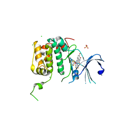
4R5I
 | | Crystal structure of the DnaK C-terminus with the substrate peptide NRLLLTG | | 分子名称: | Chaperone protein DnaK, HSP70/DnaK Substrate Peptide: NRLLLTG, PHOSPHATE ION, ... | | 著者 | Leu, J.I, Zhang, P, Murphy, M.E, Marmorstein, R, George, D.L. | | 登録日 | 2014-08-21 | | 公開日 | 2014-09-10 | | 最終更新日 | 2024-02-28 | | 実験手法 | X-RAY DIFFRACTION (1.9702 Å) | | 主引用文献 | Structural Basis for the Inhibition of HSP70 and DnaK Chaperones by Small-Molecule Targeting of a C-Terminal Allosteric Pocket.
Acs Chem.Biol., 9, 2014
|
|
7LJ9
 | |
4RLP
 | | Human p70s6k1 with ruthenium-based inhibitor FL772 | | 分子名称: | CHLORIDE ION, [(amino-kappaN)methanethiolato](3-fluoro-9-methoxypyrido[2,3-a]pyrrolo[3,4-c]carbazole-5,7(6H,12H)-dionato-kappa~2~N,N')(N-methyl-1,4,7-trithiecan-9-amine-kappa~3~S~1~,S~4~,S~7~)ruthenium, p70S6K1 | | 著者 | Domsic, J.F, Barber-Rotenberg, J, Salami, J, Qin, J, Marmorstein, R. | | 登録日 | 2014-10-17 | | 公開日 | 2015-01-21 | | 最終更新日 | 2023-09-20 | | 実験手法 | X-RAY DIFFRACTION (2.79 Å) | | 主引用文献 | Development of Organometallic S6K1 Inhibitors.
J.Med.Chem., 58, 2015
|
|
4RLO
 | | Human p70s6k1 with ruthenium-based inhibitor EM5 | | 分子名称: | CHLORIDE ION, DIMETHYL SULFOXIDE, GLYCEROL, ... | | 著者 | Domsic, J.F, Barber-Rotenberg, J, Salami, J, Qin, J, Marmorstein, R. | | 登録日 | 2014-10-17 | | 公開日 | 2015-01-21 | | 最終更新日 | 2023-09-20 | | 実験手法 | X-RAY DIFFRACTION (2.527 Å) | | 主引用文献 | Development of Organometallic S6K1 Inhibitors.
J.Med.Chem., 58, 2015
|
|
5WJD
 | | Crystal structure of Naa80 bound to acetyl-CoA | | 分子名称: | ACETYL COENZYME *A, CG8481, isoform B, ... | | 著者 | Goris, M, Magin, R.S, Marmorstein, R, Arnesen, T. | | 登録日 | 2017-07-21 | | 公開日 | 2018-03-28 | | 最終更新日 | 2023-10-04 | | 実験手法 | X-RAY DIFFRACTION (2.001 Å) | | 主引用文献 | Structural determinants and cellular environment define processed actin as the sole substrate of the N-terminal acetyltransferase NAA80.
Proc. Natl. Acad. Sci. U.S.A., 115, 2018
|
|
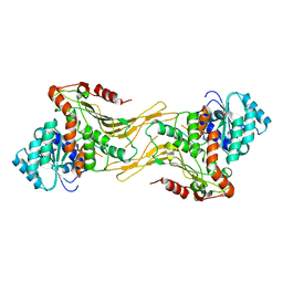
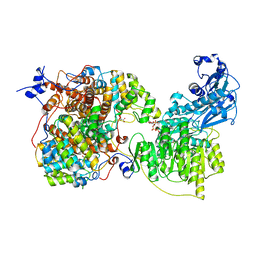
1MG5
 | | Crystal structure of Drosophila melanogaster alcohol dehydrogenase complexed with NADH and acetate at 1.6 A | | 分子名称: | 1,4-DIHYDRONICOTINAMIDE ADENINE DINUCLEOTIDE, ACETATE ION, alcohol dehydrogenase | | 著者 | Benach, J, Atrian, S, Gonzalez-Duarte, R, Ladenstein, R. | | 登録日 | 2002-08-14 | | 公開日 | 2003-10-14 | | 最終更新日 | 2024-02-14 | | 実験手法 | X-RAY DIFFRACTION (1.63 Å) | | 主引用文献 | Drosophila alcohol dehydrogenase: acetate-enzyme interactions and novel insights into the effects of electrostatics on catalysis
J.Mol.Biol., 345, 2005
|
|
5WJE
 | | Crystal structure of Naa80 bound to a bisubstrate analogue | | 分子名称: | Actin N-terminus peptide, CARBOXYMETHYL COENZYME *A, CG8481, ... | | 著者 | Goris, M, Magin, R.S, Marmorstein, R, Arnesen, T. | | 登録日 | 2017-07-21 | | 公開日 | 2018-03-28 | | 最終更新日 | 2023-10-04 | | 実験手法 | X-RAY DIFFRACTION (1.765 Å) | | 主引用文献 | Structural determinants and cellular environment define processed actin as the sole substrate of the N-terminal acetyltransferase NAA80.
Proc. Natl. Acad. Sci. U.S.A., 115, 2018
|
|
8QHS
 | | Cryo-EM structure of the monocin tail-tube, MttP. | | 分子名称: | Antigen A | | 著者 | Nadejda, S, Lichtenstein, R, Schlussel, S, Azulay, G, Borovok, I, Holdengraber, V, Elad, N, Wolf, S.G, Zalk, R, Zarivach, R, Frank, G.A, Herskovits, A.A. | | 登録日 | 2023-09-10 | | 公開日 | 2024-06-19 | | 実験手法 | ELECTRON MICROSCOPY (2.3 Å) | | 主引用文献 | Tailocin cell factories
To Be Published
|
|
6POE
 | |


