Loading
PDBj
メニューPDBj@FacebookPDBj@TwitterPDBj@YouTubewwPDB FoundationwwPDB
RCSB PDBPDBeBMRBAdv. SearchSearch help
Search by PDB author
4DI4
DownloadVisualize
BU of 4di4 by Molmil
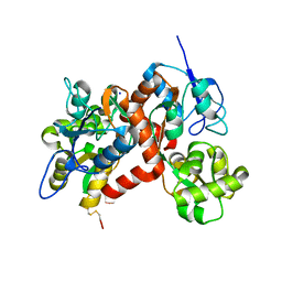
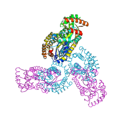
Crystal structure of a 3:1 complex of Treponema pallidum TatP(T) (Tp0957) bound to TatT (Tp0956)
分子名称: TETRAETHYLENE GLYCOL, TRIETHYLENE GLYCOL, TatP(T) (Tp0957), ...
著者Brautigam, C.A, Deka, R.K, Norgard, M.V.
登録日2012-01-30
公開日2012-05-23
最終更新日2023-09-13
実験手法X-RAY DIFFRACTION (2.714 Å)
主引用文献Structural and Thermodynamic Characterization of the Interaction between Two Periplasmic Treponema pallidum Lipoproteins that are Components of a TPR-Protein-Associated TRAP Transporter (TPAT).
J.Mol.Biol., 420, 2012
3H6G
DownloadVisualize
BU of 3h6g by Molmil
Crystal structure of the GluR6 amino terminal domain dimer assembly
分子名称: 2-acetamido-2-deoxy-beta-D-glucopyranose, CALCIUM ION, Glutamate receptor, ...
著者Kumar, J, Mayer, M.L.
登録日2009-04-23
公開日2009-05-26
最終更新日2023-09-06
実験手法X-RAY DIFFRACTION (2.697 Å)
主引用文献The N-terminal domain of GluR6-subtype glutamate receptor ion channels.
Nat.Struct.Mol.Biol., 16, 2009
3G3I
DownloadVisualize
BU of 3g3i by Molmil
Crystal structure of the GluR6 ligand binding domain dimer I442H K494E I749L Q753K mutant with glutamate and NaCl at 1.37 Angstrom resolution
分子名称: CHLORIDE ION, GLUTAMIC ACID, Glutamate receptor, ...
著者Chaudhry, C, Mayer, M.L.
登録日2009-02-02
公開日2009-06-02
最終更新日2021-10-20
実験手法X-RAY DIFFRACTION (1.371 Å)
主引用文献Stability of ligand-binding domain dimer assembly controls kainate receptor desensitization.
Embo J., 28, 2009
3G3J
DownloadVisualize
BU of 3g3j by Molmil
Crystal structure of the GluR6 ligand binding domain dimer I442H K494E K665R I749L Q753K mutant with glutamate and NaCl at 1.32 Angstrom resolution
分子名称: CHLORIDE ION, GLUTAMIC ACID, Glutamate receptor, ...
著者Chaudhry, C, Mayer, M.L.
登録日2009-02-02
公開日2009-06-02
最終更新日2021-10-20
実験手法X-RAY DIFFRACTION (1.321 Å)
主引用文献Stability of ligand-binding domain dimer assembly controls kainate receptor desensitization.
Embo J., 28, 2009
3G3G
DownloadVisualize
BU of 3g3g by Molmil
Crystal structure of the GluR6 ligand binding domain dimer K665R mutant with glutamate and NaCl at 1.3 Angstrom resolution
分子名称: CHLORIDE ION, GLUTAMIC ACID, Glutamate receptor, ...
著者Chaudhry, C, Mayer, M.L.
登録日2009-02-02
公開日2009-06-02
最終更新日2021-10-20
実験手法X-RAY DIFFRACTION (1.303 Å)
主引用文献Stability of ligand-binding domain dimer assembly controls kainate receptor desensitization.
Embo J., 28, 2009
3H6H
DownloadVisualize
BU of 3h6h by Molmil
Crystal structure of the GluR6 amino terminal domain dimer assembly MPD form
分子名称: 2-acetamido-2-deoxy-beta-D-glucopyranose, CALCIUM ION, Glutamate receptor, ...
著者Kumar, J, Mayer, M.L.
登録日2009-04-23
公開日2009-05-26
最終更新日2023-09-06
実験手法X-RAY DIFFRACTION (2.901 Å)
主引用文献The N-terminal domain of GluR6-subtype glutamate receptor ion channels.
Nat.Struct.Mol.Biol., 16, 2009
3QLV
DownloadVisualize
BU of 3qlv by Molmil
Crystal structure of the GluK2/GluK5 (GluR6/KA2) ATD tetramer assembly
分子名称: Glutamate receptor, ionotropic kainate 2, ionotropic kainate 5
著者Kumar, J, Mayer, M.L.
登録日2011-02-03
公開日2011-08-03
最終更新日2023-09-13
実験手法X-RAY DIFFRACTION (3.94 Å)
主引用文献Structure and assembly mechanism for heteromeric kainate receptors.
Neuron, 71, 2011
3QLU
DownloadVisualize
BU of 3qlu by Molmil
Crystal structure of the GluK2/GluK5 (GluR6/KA2) ATD dimer assembly
分子名称: 2-acetamido-2-deoxy-beta-D-glucopyranose, CHLORIDE ION, GLYCEROL, ...
著者Kumar, J, Mayer, M.L.
登録日2011-02-03
公開日2011-08-03
最終更新日2023-09-13
実験手法X-RAY DIFFRACTION (2.906 Å)
主引用文献Structure and assembly mechanism for heteromeric kainate receptors.
Neuron, 71, 2011
3QLT
DownloadVisualize
BU of 3qlt by Molmil
Crystal structure of a GluK2 (GluR6) glycan wedge homodimer assembly
分子名称: 2-acetamido-2-deoxy-beta-D-glucopyranose, Glutamate receptor, ionotropic kainate 2
著者Kumar, J, Mayer, M.L.
登録日2011-02-03
公開日2011-08-03
最終更新日2023-09-13
実験手法X-RAY DIFFRACTION (2.988 Å)
主引用文献Structure and assembly mechanism for heteromeric kainate receptors.
Neuron, 71, 2011
1QO3
DownloadVisualize
BU of 1qo3 by Molmil
Complex between NK cell receptor Ly49A and its MHC class I ligand H-2Dd
分子名称: 1,2-ETHANEDIOL, BETA-2-MICROGLOBULIN, HIV ENVELOPE GLYCOPROTEIN 120 PEPTIDE, ...
著者Tormo, J, Mariuzza, R.A.
登録日1999-11-01
公開日2000-01-02
最終更新日2023-12-13
実験手法X-RAY DIFFRACTION (2.3 Å)
主引用文献Crystal Structure of a Lectin-Like Natural Killer Cell Receptor Bound to its Mhc Class I Ligand
Nature, 402, 1999
<12


223166

件を2024-07-31に公開中

PDB statisticsPDBj update infoContact PDBjnumon