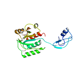
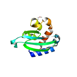
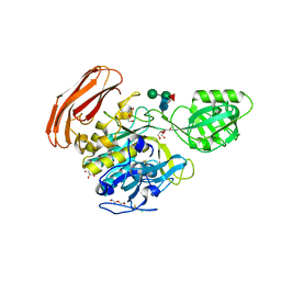
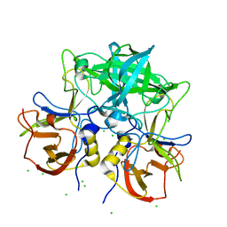
1IRQ
 
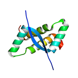 | | Crystal structure of omega transcriptional repressor at 1.5A resolution | | 分子名称: | omega transcriptional repressor | | 著者 | Murayama, K, Orth, P, De La Hoz, A.B, Alonso, J.C, Saenger, W. | | 登録日 | 2001-10-11 | | 公開日 | 2001-12-12 | | 最終更新日 | 2023-12-27 | | 実験手法 | X-RAY DIFFRACTION (1.5 Å) | | 主引用文献 | Crystal structure of omega transcriptional repressor encoded by Streptococcus pyogenes plasmid pSM19035 at 1.5 A resolution.
J.Mol.Biol., 314, 2001
|
|
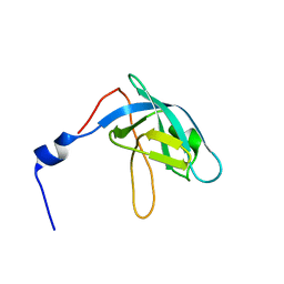
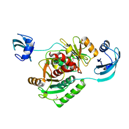
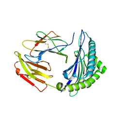
2DX1
 | | Crystal structure of RhoGEF protein Asef | | 分子名称: | Rho guanine nucleotide exchange factor 4 | | 著者 | Murayama, K, Kato-Murayama, M, Terada, T, Shirouzu, M, Yokoyama, S, RIKEN Structural Genomics/Proteomics Initiative (RSGI) | | 登録日 | 2006-08-22 | | 公開日 | 2007-01-02 | | 最終更新日 | 2011-07-13 | | 実験手法 | X-RAY DIFFRACTION (2.36 Å) | | 主引用文献 | Crystal structure of the rac activator, Asef, reveals its autoinhibitory mechanism
J.Biol.Chem., 282, 2007
|
|
2CWP
 | |
2Z0T
 | | Crystal structure of hypothetical protein PH0355 | | 分子名称: | Putative uncharacterized protein PH0355 | | 著者 | Murayama, K, Takemoto, C, Kato-Murayama, M, Kawazoe, M, Terada, T, Shirouzu, M, Yokoyama, S, RIKEN Structural Genomics/Proteomics Initiative (RSGI) | | 登録日 | 2007-05-07 | | 公開日 | 2007-11-13 | | 最終更新日 | 2024-03-13 | | 実験手法 | X-RAY DIFFRACTION (1.8 Å) | | 主引用文献 | Crystal structure of hypothetical protein PH0355
To be Published
|
|
8YK5
 | |
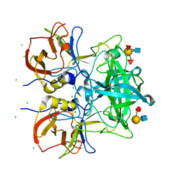
2Z0Y
 | | Crystal structure of TTHA0657-SAM complex | | 分子名称: | Putative uncharacterized protein TTHA0657, S-ADENOSYLMETHIONINE | | 著者 | Murayama, K, Terada, T, Kuramitsu, S, Shirouzu, M, Yokoyama, S, RIKEN Structural Genomics/Proteomics Initiative (RSGI) | | 登録日 | 2007-05-07 | | 公開日 | 2007-11-13 | | 最終更新日 | 2023-11-15 | | 実験手法 | X-RAY DIFFRACTION (2.8 Å) | | 主引用文献 | Crystal structure of TTHA0657-SAM complex
To be Published
|
|
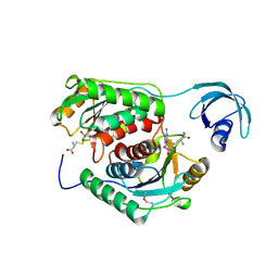
1UFO
 | | Crystal Structure of TT1662 from Thermus thermophilus | | 分子名称: | hypothetical protein TT1662 | | 著者 | Murayama, K, Shirouzu, M, Terada, T, Kuramitsu, S, Yokoyama, S, RIKEN Structural Genomics/Proteomics Initiative (RSGI) | | 登録日 | 2003-06-03 | | 公開日 | 2003-12-03 | | 最終更新日 | 2023-12-27 | | 実験手法 | X-RAY DIFFRACTION (1.6 Å) | | 主引用文献 | Crystal structure of TT1662 from Thermus thermophilus HB8: a member of the alpha/beta hydrolase fold enzymes.
Proteins, 58, 2005
|
|
1V6Z
 | |
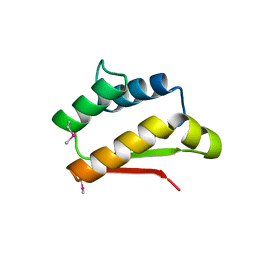
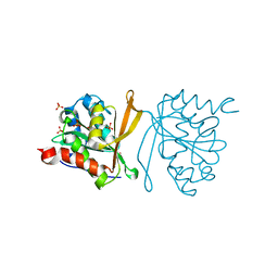
1WDZ
 | | Crystal structure of RCB domain of IRSp53 | | 分子名称: | insulin receptor substrate p53 | | 著者 | Murayama, K, Suetsugu, S, Seto, A, Shirouzu, M, Takenawa, T, Yokoyama, S, RIKEN Structural Genomics/Proteomics Initiative (RSGI) | | 登録日 | 2004-05-21 | | 公開日 | 2005-06-07 | | 最終更新日 | 2024-03-13 | | 実験手法 | X-RAY DIFFRACTION (2.63 Å) | | 主引用文献 | Crystal structure of RCB domain of IRSp53
TO BE PUBLISHED
|
|
2CX5
 | | Crystal structure of a putative trans-editing enzyme for prolyl tRNA synthetase | | 分子名称: | A PUTATIVE TRANS-EDITING ENZYME | | 著者 | Murayama, K, Nakagawa, N, Ebihara, A, Kuramitsu, S, Shirouzu, M, Yokoyama, S, RIKEN Structural Genomics/Proteomics Initiative (RSGI) | | 登録日 | 2005-06-28 | | 公開日 | 2005-12-28 | | 最終更新日 | 2024-03-13 | | 実験手法 | X-RAY DIFFRACTION (1.9 Å) | | 主引用文献 | Crystal structure of a putative trans-editing enzyme for prolyl tRNA synthetase
To be Published
|
|
2CX7
 | |
2CX6
 | |
2CX8
 | |
2CX9
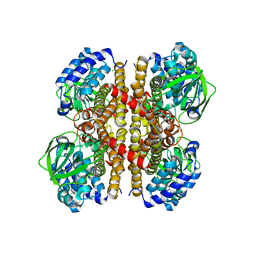 | |
4YN3
 | | Crystal structure of Cucumisin complex with pro-peptide | | 分子名称: | CHLORIDE ION, Cucumisin, DI(HYDROXYETHYL)ETHER, ... | | 著者 | Murayama, K, Kato-Murayama, M, Yokoyama, S, Arima, K, Shirouzu, M. | | 登録日 | 2015-03-09 | | 公開日 | 2016-03-09 | | 最終更新日 | 2023-11-08 | | 実験手法 | X-RAY DIFFRACTION (1.95 Å) | | 主引用文献 | Structural basis of cucumisin protease activity regulation by its propeptide
J. Biochem., 161, 2017
|
|
7EJL
 | |
7EJN
 | |
7EJM
 | |
7VS9
 | | Crystal structure of P domain from norovirus GI.9 capsid protein in complex with Lewis x antigen. | | 分子名称: | CHLORIDE ION, MAGNESIUM ION, VP1, ... | | 著者 | Murayama, K, Kato-Murayama, M, Shirouzu, M. | | 登録日 | 2021-10-26 | | 公開日 | 2022-08-31 | | 最終更新日 | 2023-11-29 | | 実験手法 | X-RAY DIFFRACTION (2.26 Å) | | 主引用文献 | Lewis fucose is a key moiety for the recognition of histo-blood group antigens by GI.9 norovirus, as revealed by structural analysis.
Febs Open Bio, 12, 2022
|
|
7VS8
 | | Crystal structure of P domain from norovirus GI.9 capsid protein in complex with Lewis b antigen. | | 分子名称: | CHLORIDE ION, MAGNESIUM ION, VP1, ... | | 著者 | Murayama, K, Kato-Murayama, M, Shirouzu, M. | | 登録日 | 2021-10-26 | | 公開日 | 2022-08-31 | | 最終更新日 | 2023-11-29 | | 実験手法 | X-RAY DIFFRACTION (2.4 Å) | | 主引用文献 | Lewis fucose is a key moiety for the recognition of histo-blood group antigens by GI.9 norovirus, as revealed by structural analysis.
Febs Open Bio, 12, 2022
|
|
2Z0Z
 | | Crystal structure of putative acetyltransferase | | 分子名称: | Putative uncharacterized protein TTHA1799, SULFATE ION | | 著者 | Murayama, K, Kato-Murayama, M, Terada, T, Kuramitsu, S, Shirouzu, M, Yokoyama, S, RIKEN Structural Genomics/Proteomics Initiative (RSGI) | | 登録日 | 2007-05-07 | | 公開日 | 2007-11-13 | | 最終更新日 | 2023-11-01 | | 実験手法 | X-RAY DIFFRACTION (2 Å) | | 主引用文献 | Genetic Encoding of 3-Iodo-l-Tyrosine in Escherichia coli for Single-Wavelength Anomalous Dispersion Phasing in Protein Crystallography
Structure, 17, 2009
|
|
2ZXV
 | | Crystal structure of putative acetyltransferase from T. thermophilus HB8 | | 分子名称: | Putative uncharacterized protein TTHA1799 | | 著者 | Murayama, K, Kato-Murayama, M, Terada, T, Kuramitsu, S, Shirouzu, M, Yokoyama, S, RIKEN Structural Genomics/Proteomics Initiative (RSGI) | | 登録日 | 2009-01-08 | | 公開日 | 2009-02-17 | | 最終更新日 | 2023-11-01 | | 実験手法 | X-RAY DIFFRACTION (2.3 Å) | | 主引用文献 | Genetic Encoding of 3-Iodo-l-Tyrosine in Escherichia coli for Single-Wavelength Anomalous Dispersion Phasing in Protein Crystallography
Structure, 17, 2009
|
|
2Z10
 | | Crystal structure of putative acetyltransferase | | 分子名称: | Ribosomal-protein-alanine acetyltransferase | | 著者 | Murayama, K, Kato-Murayama, M, Terada, T, Kuramitsu, S, Shirouzu, M, Yokoyama, S, RIKEN Structural Genomics/Proteomics Initiative (RSGI) | | 登録日 | 2007-05-07 | | 公開日 | 2007-11-13 | | 最終更新日 | 2011-07-13 | | 実験手法 | X-RAY DIFFRACTION (1.77 Å) | | 主引用文献 | Genetic Encoding of 3-Iodo-l-Tyrosine in Escherichia coli for Single-Wavelength Anomalous Dispersion Phasing in Protein Crystallography
Structure, 17, 2009
|
|
7YMQ
 | | Crystal structure of lysoplasmalogen specific phopholipase D, F211L mutant | | 分子名称: | Lysoplasmalogenase | | 著者 | Murayama, K, Kato-Murayama, M, Sugimori, D, Shirouzu, M, Hamana, H. | | 登録日 | 2022-07-29 | | 公開日 | 2023-02-08 | | 最終更新日 | 2023-11-29 | | 実験手法 | X-RAY DIFFRACTION (2.29 Å) | | 主引用文献 | Structural basis for the substrate specificity switching of lysoplasmalogen-specific phospholipase D from Thermocrispum sp. RD004668.
Biosci.Biotechnol.Biochem., 87, 2022
|
|
7YMR
 | | Complex structure of lysoplasmalogen specific phopholipase D, F211L mutant with LPC | | 分子名称: | Lysoplasmalogenase, [(2~{R})-2-oxidanyl-3-[oxidanyl-[2-(trimethyl-$l^{5}-azanyl)ethoxy]phosphoryl]oxy-propyl] hexadecanoate | | 著者 | Murayama, K, Kato-Murayama, M, Sugimori, D, Shirouzu, M, Hamana, H. | | 登録日 | 2022-07-29 | | 公開日 | 2023-02-08 | | 最終更新日 | 2023-11-29 | | 実験手法 | X-RAY DIFFRACTION (2.69 Å) | | 主引用文献 | Structural basis for the substrate specificity switching of lysoplasmalogen-specific phospholipase D from Thermocrispum sp. RD004668.
Biosci.Biotechnol.Biochem., 87, 2022
|
|


