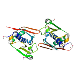
8IC1
 
 | | endo-alpha-D-arabinanase EndoMA1 D51N mutant from Microbacterium arabinogalactanolyticum in complex with arabinooligosaccharides | | 分子名称: | (3~{a}~{S},5~{R},6~{R},6~{a}~{S})-5-(hydroxymethyl)-2,2-dimethyl-3~{a},5,6,6~{a}-tetrahydrofuro[2,3-d][1,3]dioxol-6-ol, 2-(N-MORPHOLINO)-ETHANESULFONIC ACID, CALCIUM ION, ... | | 著者 | Li, J, Nakashima, C, Ishiwata, A, Fujita, K, Fushinobu, S. | | 登録日 | 2023-02-10 | | 公開日 | 2023-08-16 | | 最終更新日 | 2023-09-27 | | 実験手法 | X-RAY DIFFRACTION (1.8 Å) | | 主引用文献 | Identification and characterization of endo-alpha-, exo-alpha-, and exo-beta-D-arabinofuranosidases degrading lipoarabinomannan and arabinogalactan of mycobacteria.
Nat Commun, 14, 2023
|
|
3GP7
 | |
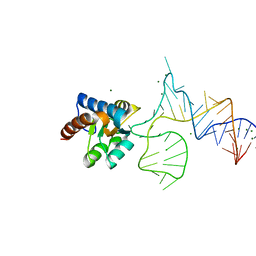
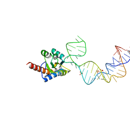
5DCV
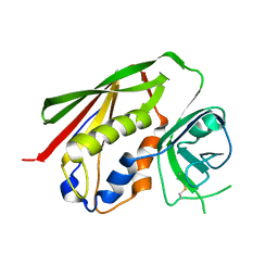
 | | Crystal structure of PhoRpp38-SL12M complex | | 分子名称: | 50S ribosomal protein L7Ae, RNA (47-MER) | | 著者 | Oshima, K, Tanaka, Y, Yao, M. | | 登録日 | 2015-08-24 | | 公開日 | 2016-07-06 | | 最終更新日 | 2023-11-08 | | 実験手法 | X-RAY DIFFRACTION (3.401 Å) | | 主引用文献 | Structural basis for recognition of a kink-turn motif by an archaeal homologue of human RNase P protein Rpp38
Biochem.Biophys.Res.Commun., 474, 2016
|
|
8WSM
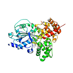 | | NLRP3 NACHT domain in complex with compound 32 | | 分子名称: | 2-[[2-methyl-5-(trifluoromethyl)phenyl]amino]-~{N}-(1,4-oxazepan-4-ylsulfonyl)-1,3-oxazole-4-carboxamide, ADENOSINE-5'-DIPHOSPHATE, MAGNESIUM ION, ... | | 著者 | Akai, S, Orita, T, Adachi, T. | | 登録日 | 2023-10-17 | | 公開日 | 2023-11-22 | | 最終更新日 | 2024-01-03 | | 実験手法 | X-RAY DIFFRACTION (2.7 Å) | | 主引用文献 | Discovery of Novel NLRP3 Inflammasome Inhibitors Composed of an Oxazole Scaffold Bearing an Acylsulfamide.
Acs Med.Chem.Lett., 14, 2023
|
|
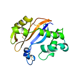
1J1F
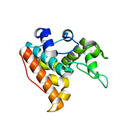
 | | Crystal structure of the RNase MC1 mutant N71T in complex with 5'-GMP | | 分子名称: | GUANOSINE-5'-MONOPHOSPHATE, RIBONUCLEASE MC1 | | 著者 | Numata, T, Suzuki, A, Kakuta, Y, Kimura, K, Yao, M, Tanaka, I, Yoshida, Y, Ueda, T, Kimura, M. | | 登録日 | 2002-12-03 | | 公開日 | 2003-05-20 | | 最終更新日 | 2023-10-25 | | 実験手法 | X-RAY DIFFRACTION (1.6 Å) | | 主引用文献 | Crystal Structures of the Ribonuclease MC1 Mutants N71T and N71S in Complex with 5'-GMP: Structural Basis for Alterations in Substrate Specificity
Biochemistry, 42, 2003
|
|
1J1G
 | | Crystal structure of the RNase MC1 mutant N71S in complex with 5'-GMP | | 分子名称: | GUANOSINE-5'-MONOPHOSPHATE, Ribonuclease MC1 | | 著者 | Numata, T, Suzuki, A, Kakuta, Y, Kimura, K, Yao, M, Tanaka, I, Yoshida, Y, Ueda, T, Kimura, M. | | 登録日 | 2002-12-04 | | 公開日 | 2003-05-20 | | 最終更新日 | 2023-10-25 | | 実験手法 | X-RAY DIFFRACTION (1.6 Å) | | 主引用文献 | Crystal Structures of the Ribonuclease MC1 Mutants N71T and N71S in Complex with 5'-GMP: Structural Basis for Alterations in Substrate Specificity
Biochemistry, 42, 2003
|
|
7F88
 | |
7XMH
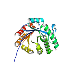 | | Crystal structure of a rice class IIIb chitinase, Oschib2 | | 分子名称: | 1,2-ETHANEDIOL, ACETATE ION, Putative class III chitinase | | 著者 | Jun, T, Tomoya, T, Tomoyuki, N, Takayuki, O. | | 登録日 | 2022-04-25 | | 公開日 | 2023-05-03 | | 最終更新日 | 2023-11-29 | | 実験手法 | X-RAY DIFFRACTION (1.18 Å) | | 主引用文献 | Characterization of two rice GH18 chitinases belonging to family 8 of plant pathogenesis-related proteins.
Plant Sci., 326, 2023
|
|
1BK7
 | |
5XTM
 | |
5Y7M
 | |
3AML
 | | Structure of the Starch Branching Enzyme I (BEI) from Oryza sativa L | | 分子名称: | 4-(2-HYDROXYETHYL)-1-PIPERAZINE ETHANESULFONIC ACID, ACETATE ION, BETA-MERCAPTOETHANOL, ... | | 著者 | Kakuta, Y, Chaen, K, Noguchi, J, Vu, N, Kimura, M. | | 登録日 | 2010-08-20 | | 公開日 | 2011-09-28 | | 最終更新日 | 2023-11-01 | | 実験手法 | X-RAY DIFFRACTION (1.7 Å) | | 主引用文献 | Crystal structure of the branching enzyme I (BEI) from Oryza sativa L with implications for catalysis and substrate binding.
Glycobiology, 21, 2011
|
|
3AMK
 | | Structure of the Starch Branching Enzyme I (BEI) from Oryza sativa L | | 分子名称: | GLYCEROL, Os06g0726400 protein, PHOSPHATE ION | | 著者 | Kakuta, Y, Chaen, K, Noguchi, J, Vu, N, Kimura, M. | | 登録日 | 2010-08-20 | | 公開日 | 2011-09-28 | | 最終更新日 | 2024-03-13 | | 実験手法 | X-RAY DIFFRACTION (1.9 Å) | | 主引用文献 | Crystal structure of the branching enzyme I (BEI) from Oryza sativa L with implications for catalysis and substrate binding.
Glycobiology, 21, 2011
|
|
1V7O
 | |
487D
 | |


