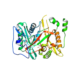
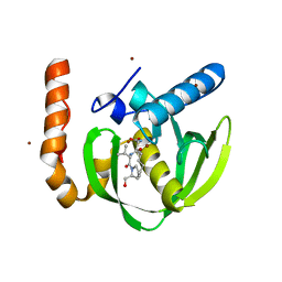
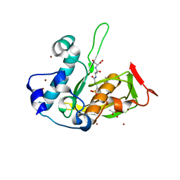
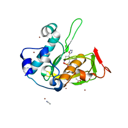
3M6Q
 
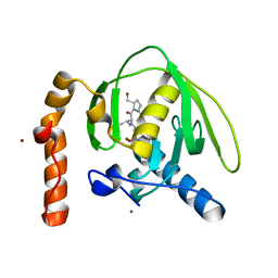 | |
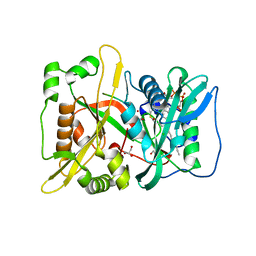
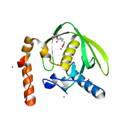
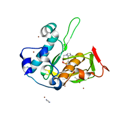
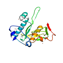
3M6P
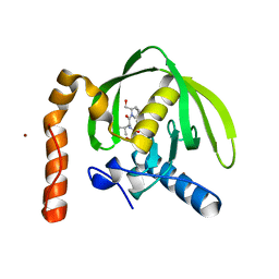 | |
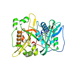
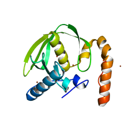
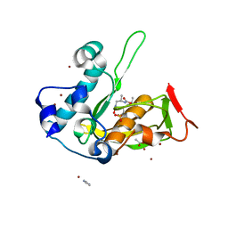
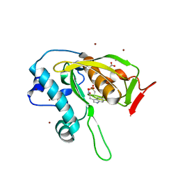
3M6O
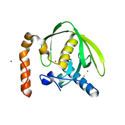 | |
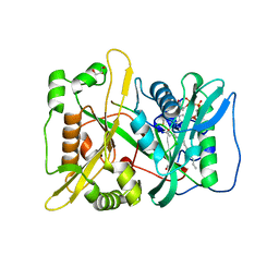
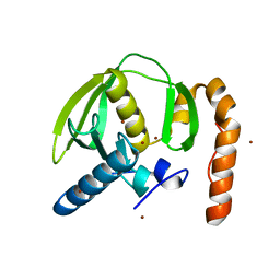
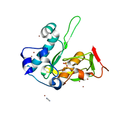
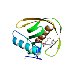
3M6R
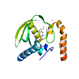 | |
5O9S
 | | HsNMT1 in complex with CoA and Myristoylated-GKSNSKLK octapeptide | | 分子名称: | CHLORIDE ION, GLYCEROL, Glycylpeptide N-tetradecanoyltransferase 1, ... | | 著者 | Dian, C, Meinnel, T, Giglione, C. | | 登録日 | 2017-06-20 | | 公開日 | 2018-06-27 | | 最終更新日 | 2024-01-17 | | 実験手法 | X-RAY DIFFRACTION (2.7 Å) | | 主引用文献 | Structural and genomic decoding of human and plant myristoylomes reveals a definitive recognition pattern.
Nat. Chem. Biol., 14, 2018
|
|
5O9T
 | | HsNMT1 in complex with CoA and acetylated-NCFSKPK peptide | | 分子名称: | 1IP-CYS-PHE-SER-LYS-PRO-ARG, CHLORIDE ION, GLYCEROL, ... | | 著者 | Dian, C, Meinnel, T, Giglione, C. | | 登録日 | 2017-06-20 | | 公開日 | 2018-06-27 | | 最終更新日 | 2024-01-31 | | 実験手法 | X-RAY DIFFRACTION (2.15 Å) | | 主引用文献 | Structural and genomic decoding of human and plant myristoylomes reveals a definitive recognition pattern.
Nat. Chem. Biol., 14, 2018
|
|
5O9U
 | | HsNMT1 in complex with CoA and Myristoylated-GCSVSKKK octapeptide | | 分子名称: | COENZYME A, Calcineurin B-like protein 4, GLYCEROL, ... | | 著者 | Dian, C, Meinnel, T, Giglione, C. | | 登録日 | 2017-06-20 | | 公開日 | 2018-06-27 | | 最終更新日 | 2024-01-17 | | 実験手法 | X-RAY DIFFRACTION (1.85 Å) | | 主引用文献 | Structural and genomic decoding of human and plant myristoylomes reveals a definitive recognition pattern.
Nat. Chem. Biol., 14, 2018
|
|
5O9V
 | | HsNMT1 in complex with CoA and Myristoylated-GGCFSKPK octapeptide | | 分子名称: | Apoptosis-inducing factor 3, CHLORIDE ION, COENZYME A, ... | | 著者 | Dian, C, Meinnel, T, Giglione, C. | | 登録日 | 2017-06-20 | | 公開日 | 2018-06-27 | | 最終更新日 | 2024-01-17 | | 実験手法 | X-RAY DIFFRACTION (2.201 Å) | | 主引用文献 | Structural and genomic decoding of human and plant myristoylomes reveals a definitive recognition pattern.
Nat. Chem. Biol., 14, 2018
|
|
5MTD
 | | Crystal structure of PDF from the Vibrio parahaemolyticus bacteriophage VP16T - crystal form II | | 分子名称: | NICKEL (II) ION, Putative uncharacterized protein orf60T, TRIETHYLENE GLYCOL, ... | | 著者 | Fieulaine, S, Grzela, R, Giglione, C, Meinnel, T. | | 登録日 | 2017-01-09 | | 公開日 | 2017-09-20 | | 最終更新日 | 2024-01-17 | | 実験手法 | X-RAY DIFFRACTION (1.5 Å) | | 主引用文献 | The C-terminal residue of phage Vp16 PDF, the smallest peptide deformylase, acts as an offset element locking the active conformation.
Sci Rep, 7, 2017
|
|
5MTC
 | | Crystal structure of PDF from the Vibrio parahaemolyticus bacteriophage VP16T - crystal form I | | 分子名称: | NICKEL (II) ION, Putative uncharacterized protein orf60T, ZINC ION | | 著者 | Fieulaine, S, Grzela, R, Giglione, C, Meinnel, T. | | 登録日 | 2017-01-09 | | 公開日 | 2017-09-20 | | 最終更新日 | 2024-01-17 | | 実験手法 | X-RAY DIFFRACTION (1.7 Å) | | 主引用文献 | The C-terminal residue of phage Vp16 PDF, the smallest peptide deformylase, acts as an offset element locking the active conformation.
Sci Rep, 7, 2017
|
|
3O3J
 | |
3PN2
 | |
3PN4
 | | Crystal structure of Arabidopsis thaliana petide deformylase 1B (AtPDF1B) in complex with actinonin (crystallized in PEG-550-MME) | | 分子名称: | ACTINONIN, Peptide deformylase 1B, chloroplastic, ... | | 著者 | Fieulaine, S, Meinnel, T, Giglione, C. | | 登録日 | 2010-11-18 | | 公開日 | 2011-06-08 | | 最終更新日 | 2023-09-06 | | 実験手法 | X-RAY DIFFRACTION (1.9 Å) | | 主引用文献 | Trapping conformational States along ligand-binding dynamics of Peptide deformylase: the impact of induced fit on enzyme catalysis.
Plos Biol., 9, 2011
|
|
3PN3
 | | Crystal structure of Arabidopsis thaliana petide deformylase 1B (AtPDF1B) in complex with inhibitor 21 | | 分子名称: | Peptide deformylase 1B, chloroplastic, ZINC ION, ... | | 著者 | Fieulaine, S, Meinnel, T, Giglione, C. | | 登録日 | 2010-11-18 | | 公開日 | 2011-06-08 | | 最終更新日 | 2023-09-06 | | 実験手法 | X-RAY DIFFRACTION (1.3 Å) | | 主引用文献 | Trapping conformational States along ligand-binding dynamics of Peptide deformylase: the impact of induced fit on enzyme catalysis.
Plos Biol., 9, 2011
|
|
3PN6
 | |
3PN5
 | |
5JEZ
 | | Crystal structure of type 2 PDF from Streptococcus agalactiae in complex with tripeptide Met-Ala-Ser | | 分子名称: | ACETATE ION, Met-Ala-Ser, Peptide deformylase, ... | | 著者 | Fieulaine, S, Giglione, C, Meinnel, T. | | 登録日 | 2016-04-19 | | 公開日 | 2016-11-30 | | 最終更新日 | 2024-01-10 | | 実験手法 | X-RAY DIFFRACTION (1.7 Å) | | 主引用文献 | A unique peptide deformylase platform to rationally design and challenge novel active compounds.
Sci Rep, 6, 2016
|
|
5JF7
 | | Crystal structure of type 2 PDF from Streptococcus agalactiae in complex with inhibitor SMP289 | | 分子名称: | 2-(3-benzyl-5-bromo-1H-indol-1-yl)-N-hydroxyacetamide, ACETATE ION, IMIDAZOLE, ... | | 著者 | Fieulaine, S, Giglione, C, Meinnel, T, Hamiche, K. | | 登録日 | 2016-04-19 | | 公開日 | 2016-11-30 | | 最終更新日 | 2024-01-10 | | 実験手法 | X-RAY DIFFRACTION (2.1 Å) | | 主引用文献 | A unique peptide deformylase platform to rationally design and challenge novel active compounds.
Sci Rep, 6, 2016
|
|
5JF8
 | | Crystal structure of type 2 PDF from Streptococcus agalactiae in complex with inhibitor RAS358 (21) | | 分子名称: | ACETATE ION, IMIDAZOLE, Peptide deformylase, ... | | 著者 | Fieulaine, S, Giglione, C, Meinnel, T. | | 登録日 | 2016-04-19 | | 公開日 | 2016-11-30 | | 最終更新日 | 2024-01-10 | | 実験手法 | X-RAY DIFFRACTION (1.8 Å) | | 主引用文献 | A unique peptide deformylase platform to rationally design and challenge novel active compounds.
Sci Rep, 6, 2016
|
|
5JF1
 | | Crystal structure of type 2 PDF from Streptococcus agalactiae in complex with actinonin | | 分子名称: | ACETATE ION, ACTINONIN, Peptide deformylase, ... | | 著者 | Fieulaine, S, Giglione, C, Meinnel, T. | | 登録日 | 2016-04-19 | | 公開日 | 2016-11-30 | | 最終更新日 | 2024-01-10 | | 実験手法 | X-RAY DIFFRACTION (2 Å) | | 主引用文献 | A unique peptide deformylase platform to rationally design and challenge novel active compounds.
Sci Rep, 6, 2016
|
|
5JEX
 | | Crystal structure of type 2 PDF from Streptococcus agalactiae, crystallized in imidazole buffer | | 分子名称: | IMIDAZOLE, Peptide deformylase, ZINC ION | | 著者 | Fieulaine, S, Giglione, C, Meinnel, T. | | 登録日 | 2016-04-19 | | 公開日 | 2016-11-30 | | 最終更新日 | 2024-01-10 | | 実験手法 | X-RAY DIFFRACTION (2 Å) | | 主引用文献 | A unique peptide deformylase platform to rationally design and challenge novel active compounds.
Sci Rep, 6, 2016
|
|
5JF3
 | | Crystal structure of type 2 PDF from Streptococcus agalactiae in complex with inhibitor AT018 | | 分子名称: | ACETATE ION, IMIDAZOLE, Peptide deformylase, ... | | 著者 | Fieulaine, S, Giglione, C, Meinnel, T. | | 登録日 | 2016-04-19 | | 公開日 | 2016-11-30 | | 最終更新日 | 2024-01-10 | | 実験手法 | X-RAY DIFFRACTION (1.6 Å) | | 主引用文献 | A unique peptide deformylase platform to rationally design and challenge novel active compounds.
Sci Rep, 6, 2016
|
|
5JEY
 | |
5JF6
 | | Crystal structure of type 2 PDF from Streptococcus agalactiae in complex with inhibitor 6b (AB47) | | 分子名称: | 2-(5-bromo-1H-indol-3-yl)-N-hydroxyacetamide, ACETATE ION, Peptide deformylase, ... | | 著者 | Fieulaine, S, Giglione, C, Meinnel, T, Hamiche, K. | | 登録日 | 2016-04-19 | | 公開日 | 2016-11-30 | | 最終更新日 | 2024-01-10 | | 実験手法 | X-RAY DIFFRACTION (1.7 Å) | | 主引用文献 | A unique peptide deformylase platform to rationally design and challenge novel active compounds.
Sci Rep, 6, 2016
|
|
5MTE
 | | Crystal structure of PDF from the Vibrio parahaemolyticus bacteriophage VP16T in complex with actinonin - crystal form II | | 分子名称: | ACTINONIN, NICKEL (II) ION, Putative uncharacterized protein orf60T, ... | | 著者 | Fieulaine, S, Grzela, R, Giglione, C, Meinnel, T. | | 登録日 | 2017-01-09 | | 公開日 | 2017-11-29 | | 最終更新日 | 2024-01-17 | | 実験手法 | X-RAY DIFFRACTION (1.4 Å) | | 主引用文献 | Peptide deformylases from Vibrio parahaemolyticus phage and bacteria display similar deformylase activity and inhibitor binding clefts.
Biochim. Biophys. Acta, 1866, 2018
|
|


