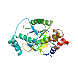
4H6X
 
 | |
4I3V
 | |
4I3U
 | |
4I3W
 | |
4I3T
 | |
4I3X
 | |
4H6V
 | |
3R95
 | |
3R9F
 | |
3R9E
 | |
3R96
 | |
3R9G
 | |
4R9F
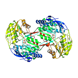
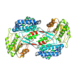
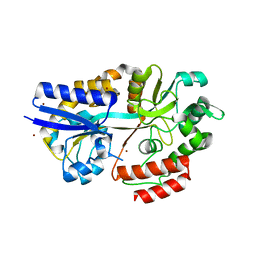
 | | CpMnBP1 with Mannobiose Bound | | 分子名称: | MBP1, beta-D-mannopyranose-(1-4)-beta-D-mannopyranose | | 著者 | Chekan, J.R, Agarwal, V, Nair, S.K. | | 登録日 | 2014-09-04 | | 公開日 | 2014-10-29 | | 最終更新日 | 2020-07-29 | | 実験手法 | X-RAY DIFFRACTION (1.4 Å) | | 主引用文献 | Structural and Biochemical Basis for Mannan Utilization by Caldanaerobius polysaccharolyticus Strain ATCC BAA-17.
J.Biol.Chem., 289, 2014
|
|
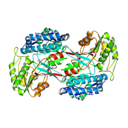
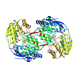
4R9G
 | | CpMnBP1 with Mannotriose Bound | | 分子名称: | MBP1, ZINC ION, beta-D-mannopyranose-(1-4)-beta-D-mannopyranose-(1-4)-alpha-D-mannopyranose | | 著者 | Chekan, J.R, Agarwal, V, Nair, S.K. | | 登録日 | 2014-09-04 | | 公開日 | 2014-10-29 | | 最終更新日 | 2024-02-28 | | 実験手法 | X-RAY DIFFRACTION (2.2 Å) | | 主引用文献 | Structural and Biochemical Basis for Mannan Utilization by Caldanaerobius polysaccharolyticus Strain ATCC BAA-17.
J.Biol.Chem., 289, 2014
|
|
3UQ5
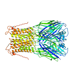 | | X-ray structure of a pentameric ligand gated ion channel from Erwinia chrysanthemi (ELIC) mutant L240A F247L (L9A F16L) in the presence of 10 mM cysteamine | | 分子名称: | Gamma-aminobutyric-acid receptor subunit beta-1, SODIUM ION | | 著者 | Gonzalez-Gutierrez, G, Lukk, T, Agarwal, V, Papke, D, Nair, S.K, Grosman, C. | | 登録日 | 2011-11-19 | | 公開日 | 2012-04-04 | | 最終更新日 | 2023-09-13 | | 実験手法 | X-RAY DIFFRACTION (4.2 Å) | | 主引用文献 | Mutations that stabilize the open state of the Erwinia chrisanthemi ligand-gated ion channel fail to change the conformation of the pore domain in crystals.
Proc.Natl.Acad.Sci.USA, 109, 2012
|
|
3UQ4
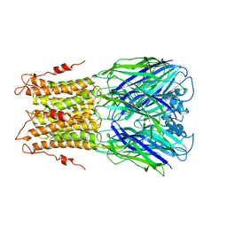 | | X-ray structure of a pentameric ligand gated ion channel from Erwinia chrysanthemi (ELIC) mutant F247L (F16L) | | 分子名称: | Gamma-aminobutyric-acid receptor subunit beta-1, SODIUM ION | | 著者 | Gonzalez-Gutierrez, G, Lukk, T, Agarwal, V, Papke, D, Nair, S.K, Grosman, C. | | 登録日 | 2011-11-19 | | 公開日 | 2012-04-04 | | 最終更新日 | 2023-09-13 | | 実験手法 | X-RAY DIFFRACTION (3.5 Å) | | 主引用文献 | Mutations that stabilize the open state of the Erwinia chrisanthemi ligand-gated ion channel fail to change the conformation of the pore domain in crystals.
Proc.Natl.Acad.Sci.USA, 109, 2012
|
|
3UQ7
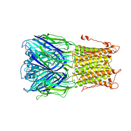 | | X-ray structure of a pentameric ligand gated ion channel from Erwinia chrysanthemi (ELIC) mutant L240S F247L (L9S F16L) in presence of 10 mM cysteamine | | 分子名称: | Gamma-aminobutyric-acid receptor subunit beta-1 | | 著者 | Gonzalez-Gutierrez, G, Lukk, T, Agarwal, V, Papke, D, Nair, S.K, Grosman, C. | | 登録日 | 2011-11-19 | | 公開日 | 2012-04-04 | | 最終更新日 | 2023-09-13 | | 実験手法 | X-RAY DIFFRACTION (3.8 Å) | | 主引用文献 | Mutations that stabilize the open state of the Erwinia chrisanthemi ligand-gated ion channel fail to change the conformation of the pore domain in crystals.
Proc.Natl.Acad.Sci.USA, 109, 2012
|
|
5TV8
 | | A. aeolicus BioW with AMP-CPP and pimelate | | 分子名称: | 6-carboxyhexanoate--CoA ligase, DIPHOSPHOMETHYLPHOSPHONIC ACID ADENOSYL ESTER, MAGNESIUM ION, ... | | 著者 | Estrada, P, Manandhar, M, Dong, S.-H, Deveryshetty, J, Agarwal, V, Cronan, J.E, Nair, S.K. | | 登録日 | 2016-11-08 | | 公開日 | 2016-12-07 | | 最終更新日 | 2017-05-31 | | 実験手法 | X-RAY DIFFRACTION (2.55 Å) | | 主引用文献 | The pimeloyl-CoA synthetase BioW defines a new fold for adenylate-forming enzymes.
Nat. Chem. Biol., 13, 2017
|
|
5TVA
 | | A. aeolicus BioW with AMP and CoA | | 分子名称: | 6-carboxyhexanoate--CoA ligase, ADENOSINE MONOPHOSPHATE, COENZYME A | | 著者 | Estrada, P, Manandhar, M, Dong, S.-H, Deveryshetty, J, Agarwal, V, Cronan, J.E, Nair, S.K. | | 登録日 | 2016-11-08 | | 公開日 | 2016-12-07 | | 最終更新日 | 2017-05-31 | | 実験手法 | X-RAY DIFFRACTION (2.25 Å) | | 主引用文献 | The pimeloyl-CoA synthetase BioW defines a new fold for adenylate-forming enzymes.
Nat. Chem. Biol., 13, 2017
|
|
5TV5
 | | BioW from Aquifex aeoulicus | | 分子名称: | 6-carboxyhexanoate--CoA ligase | | 著者 | Estrada, P, Manandhar, M, Dong, S.-H, Deveryshetty, J, Agarwal, V, Cronan, J.E, Nair, S.K. | | 登録日 | 2016-11-08 | | 公開日 | 2016-12-07 | | 最終更新日 | 2017-05-31 | | 実験手法 | X-RAY DIFFRACTION (2.5 Å) | | 主引用文献 | The pimeloyl-CoA synthetase BioW defines a new fold for adenylate-forming enzymes.
Nat. Chem. Biol., 13, 2017
|
|
5TV6
 | | A. aeolicus BioW with pimelate | | 分子名称: | 6-carboxyhexanoate--CoA ligase, PIMELIC ACID | | 著者 | Estrada, P, Manandhar, M, Dong, S.-H, Deveryshetty, J, Agarwal, V, Cronan, J.E, Nair, S.K. | | 登録日 | 2016-11-08 | | 公開日 | 2016-12-07 | | 最終更新日 | 2017-05-31 | | 実験手法 | X-RAY DIFFRACTION (2.456 Å) | | 主引用文献 | The pimeloyl-CoA synthetase BioW defines a new fold for adenylate-forming enzymes.
Nat. Chem. Biol., 13, 2017
|
|
8TB1
 | |
6POO
 | |
4KVZ
 | |
4KWC
 | |


