8Q4S
 
 | |
8QOB
 | |
2ON8
 | |
2ONQ
 | |
4MML
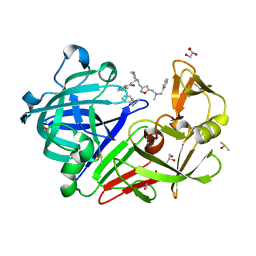
 | | D40A Hfq from Pseudomonas aeruginosa | | 分子名称: | MAGNESIUM ION, Protein hfq, SULFATE ION, ... | | 著者 | Murina, V.N, Filimonov, V.V, Melnik, B.S, Uhlein, M, Mueller, U, Weiss, M, Nikulin, A.D. | | 登録日 | 2013-09-09 | | 公開日 | 2014-07-09 | | 最終更新日 | 2024-10-16 | | 実験手法 | X-RAY DIFFRACTION (1.801 Å) | | 主引用文献 | Effect of conserved intersubunit amino Acid substitutions on hfq protein structure and stability.
Biochemistry Mosc., 79, 2014
|
|
4MMK
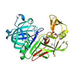
 | | Q8A Hfq from Pseudomonas aeruginosa | | 分子名称: | POTASSIUM ION, Protein hfq, SODIUM ION, ... | | 著者 | Murina, V.N, Filimonov, V.V, Melnik, B.S, Uhlein, M, Mueller, U, Weiss, M, Nikulin, A.D. | | 登録日 | 2013-09-09 | | 公開日 | 2014-07-09 | | 最終更新日 | 2023-09-20 | | 実験手法 | X-RAY DIFFRACTION (2.156 Å) | | 主引用文献 | Effect of conserved intersubunit amino Acid substitutions on hfq protein structure and stability.
Biochemistry Mosc., 79, 2014
|
|
9RCZ
 | |
3NAX
 | | PDK1 in complex with inhibitor MP7 | | 分子名称: | 1-(3,4-difluorobenzyl)-2-oxo-N-{(1R)-2-[(2-oxo-2,3-dihydro-1H-benzimidazol-5-yl)oxy]-1-phenylethyl}-1,2-dihydropyridine-3-carboxamide, 3-phosphoinositide-dependent protein kinase 1 | | 著者 | Yan, Y, Munshi, S.K, Allison, T. | | 登録日 | 2010-06-02 | | 公開日 | 2010-11-24 | | 最終更新日 | 2024-02-21 | | 実験手法 | X-RAY DIFFRACTION (1.75 Å) | | 主引用文献 | Genetic and pharmacological inhibition of PDK1 in cancer cells: characterization of a selective allosteric kinase inhibitor.
J.Biol.Chem., 286, 2011
|
|
3NAY
 | | PDK1 in complex with inhibitor MP6 | | 分子名称: | 3-phosphoinositide-dependent protein kinase 1, 4-(2-cyclopropylethylidene)-9-(1H-pyrazol-4-yl)-6-{[(1R)-1,2,2-trimethylpropyl]amino}benzo[c][1,6]naphthyridin-1(4H)-one | | 著者 | Yan, Y, Munshi, S.K, Allison, T. | | 登録日 | 2010-06-02 | | 公開日 | 2010-11-24 | | 最終更新日 | 2024-11-06 | | 実験手法 | X-RAY DIFFRACTION (2.6 Å) | | 主引用文献 | Selective inhibition of PDK1 using an allosteric kinase inhibitor and RNAi impairs cancer cell migration and anchorage-independent growth of primary tumor lines
J.Biol.Chem., 2010
|
|
5QBC
 | |
5QBL
 | | Crystal structure of Endothiapepsin-NAT17-347147 complex | | 分子名称: | (1S,2S,3S,4R,5R)-2-({[5-(hydroxymethyl)furan-2-yl]methyl}amino)-4-(morpholin-4-yl)-6,8-dioxabicyclo[3.2.1]octan-3-ol, 1,2-ETHANEDIOL, ACETATE ION, ... | | 著者 | Huschmann, F. | | 登録日 | 2017-08-04 | | 公開日 | 2020-04-22 | | 最終更新日 | 2024-10-30 | | 実験手法 | X-RAY DIFFRACTION (1.619 Å) | | 主引用文献 | Crystal structure of Endothiapepsin
To be published
|
|
5QBB
 | |
5QBJ
 | |
5QBR
 | | Crystal structure of Endothiapepsin | | 分子名称: | (3R,5R)-5-[3-(3-fluoro-4-methylphenyl)-1,2,4-oxadiazol-5-yl]pyrrolidin-3-ol, ACETATE ION, DIMETHYL SULFOXIDE, ... | | 著者 | Huschmann, F. | | 登録日 | 2017-08-04 | | 公開日 | 2020-04-22 | | 最終更新日 | 2024-11-06 | | 実験手法 | X-RAY DIFFRACTION (1.418 Å) | | 主引用文献 | Crystal structure of Endothiapepsin
To be published
|
|
5QB8
 | | Crystal structure of Endothiapepsin-FRG080 complex | | 分子名称: | (1S,2S,3S,4R,5R)-2-amino-4-[(2S)-2-(hydroxymethyl)pyrrolidin-1-yl]-6,8-dioxabicyclo[3.2.1]octan-3-ol, ACETATE ION, Endothiapepsin, ... | | 著者 | Huschmann, F. | | 登録日 | 2017-08-04 | | 公開日 | 2020-04-22 | | 最終更新日 | 2024-10-30 | | 実験手法 | X-RAY DIFFRACTION (1.24 Å) | | 主引用文献 | Crystal structure of Endothiapepsin
To be published
|
|
5QBP
 | |
5QBD
 | |
5QBN
 | | Crystal structure of Endothiapepsin-NAT14-350193 complex | | 分子名称: | 2-{(3R,4S)-3-[(5-{[benzyl(methyl)amino]methyl}-1,2-oxazol-3-yl)methyl]piperidin-4-yl}-N-(1,3-thiazol-2-yl)acetamide, ACETATE ION, DIMETHYL SULFOXIDE, ... | | 著者 | Huschmann, F. | | 登録日 | 2017-08-04 | | 公開日 | 2020-04-22 | | 最終更新日 | 2024-10-30 | | 実験手法 | X-RAY DIFFRACTION (1.403 Å) | | 主引用文献 | Crystal structure of Endothiapepsin
To be published
|
|
5QB7
 | |
5QBG
 | |
5QBQ
 | | Crystal structure of Endothiapepsin | | 分子名称: | 4-[2-({methyl[(pyridin-3-yl)methyl]amino}methyl)-1,3-thiazol-4-yl]piperidin-4-ol, ACETATE ION, Endothiapepsin, ... | | 著者 | Huschmann, F. | | 登録日 | 2017-08-04 | | 公開日 | 2020-04-22 | | 最終更新日 | 2024-10-30 | | 実験手法 | X-RAY DIFFRACTION (1.697 Å) | | 主引用文献 | Crystal structure of Endothiapepsin
To be published
|
|
5QB9
 | |
5QBK
 | |
5QBA
 | |
5QBM
 | |


