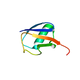
2MTV
 
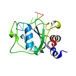 | | Solution Structure of the YTH Domain of YT521-B in complex with N6-Methyladenosine containing RNA | | 分子名称: | RNA_(5'-R(*UP*GP*(6MZ)P*CP*AP*C)-3'), YTH domain-containing protein 1 | | 著者 | Theler, D, Dominguez, C, Blatter, M, Boudet, J, Allain, F.H.-T. | | 登録日 | 2014-09-01 | | 公開日 | 2014-11-26 | | 最終更新日 | 2024-05-01 | | 実験手法 | SOLUTION NMR | | 主引用文献 | Solution structure of the YTH domain in complex with N6-methyladenosine RNA: a reader of methylated RNA.
Nucleic Acids Res., 42, 2014
|
|
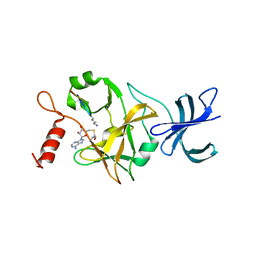
2J80
 | | Structure of Citrate-bound Periplasmic Domain of Sensor Histidine Kinase CitA | | 分子名称: | CITRATE ANION, GLYCEROL, SENSOR KINASE CITA, ... | | 著者 | Sevvana, M, Vijayan, V, Zweckstetter, M, Reinelt, S, Madden, D.R, Sheldrick, G.M, Bott, M, Griesinger, C, Becker, S. | | 登録日 | 2006-10-18 | | 公開日 | 2007-10-23 | | 最終更新日 | 2019-05-08 | | 実験手法 | X-RAY DIFFRACTION (1.6 Å) | | 主引用文献 | A Ligand-Induced Switch in the Periplasmic Domain of Sensor Histidine Kinase Cita.
J.Mol.Biol., 377, 2008
|
|
1HDF
 | | Evolution of the eye lens beta-gamma-crystallin domain fold | | 分子名称: | CALCIUM ION, SPHERULIN 3A | | 著者 | Clout, N.J, Kretschmar, M, Jaenicke, R, Slingsby, C. | | 登録日 | 2000-11-13 | | 公開日 | 2000-12-28 | | 最終更新日 | 2011-07-13 | | 実験手法 | X-RAY DIFFRACTION (2.35 Å) | | 主引用文献 | Crystal Structure of the Calcium-Loaded Spherulin 3A Dimer Sheds Light on the Evolution of the Eye Lens Betagamma-Crystallin Domain Fold
Structure, 9, 2001
|
|
1H3I
 | | Crystal structure of the Histone Methyltransferase SET7/9 | | 分子名称: | HISTONE H3 LYSINE 4 SPECIFIC METHYLTRANSFERASE, MAGNESIUM ION | | 著者 | Wilson, J.R, Jing, C, Walker, P.A, Martin, S.R, Howell, S.A, Blackburn, G.M, Gamblin, S.J, Xiao, B. | | 登録日 | 2002-09-04 | | 公開日 | 2002-11-11 | | 最終更新日 | 2024-05-08 | | 実験手法 | X-RAY DIFFRACTION (2.1 Å) | | 主引用文献 | Crystal Structure and Functional Analysis of the Histone Methyltransferase Set7/9
Cell(Cambridge,Mass.), 111, 2002
|
|
2MWH
 | | NMR solution structure of ligand-free OAA | | 分子名称: | Anti-HIV lectin OAA | | 著者 | Lee, D, Carneiro, M.G, Koharudin, L.M, Griesinger, C, Gronenborn, A.M, Ban, D, Sabo, T, Trigo-Mourino, P, Mazur, A. | | 登録日 | 2014-11-10 | | 公開日 | 2015-04-22 | | 最終更新日 | 2024-05-15 | | 実験手法 | SOLUTION NMR | | 主引用文献 | Sampling of Glycan-Bound Conformers by the Anti-HIV Lectin Oscillatoria agardhii agglutinin in the Absence of Sugar.
Angew.Chem.Int.Ed.Engl., 54, 2015
|
|
1GAM
 | | GAMMA B CRYSTALLIN TRUNCATED C-TERMINAL DOMAIN | | 分子名称: | GAMMA B CRYSTALLIN | | 著者 | Norledge, B.V, Mayr, E.-M, Glockshuber, R, Bateman, O.A, Slingsby, C, Jaenicke, R, Driessen, H.P.C. | | 登録日 | 1996-02-02 | | 公開日 | 1996-07-11 | | 最終更新日 | 2024-02-07 | | 実験手法 | X-RAY DIFFRACTION (2.6 Å) | | 主引用文献 | The X-ray structures of two mutant crystallin domains shed light on the evolution of multi-domain proteins.
Nat.Struct.Biol., 3, 1996
|
|
7DEY
 | |
2O20
 | | Crystal structure of transcription regulator CcpA of Lactococcus lactis | | 分子名称: | CHLORIDE ION, Catabolite control protein A, SULFATE ION | | 著者 | Loll, B, Kowalczyk, M, Alings, C, Chieduch, A, Bardowski, J, Saenger, W, Biesiadka, J. | | 登録日 | 2006-11-29 | | 公開日 | 2007-03-27 | | 最終更新日 | 2023-08-30 | | 実験手法 | X-RAY DIFFRACTION (1.9 Å) | | 主引用文献 | Structure of the transcription regulator CcpA from Lactococcus lactis
Acta Crystallogr.,Sect.D, 63, 2007
|
|
1OJG
 | | Sensory domain of the membraneous two-component fumarate sensor DcuS of E. coli | | 分子名称: | SENSOR PROTEIN DCUS | | 著者 | Pappalardo, L, Janausch, I.G, Vijayan, V, Zientz, E, Junker, J, Peti, W, Zweckstetter, M, Unden, G, Griesinger, C. | | 登録日 | 2003-07-10 | | 公開日 | 2003-08-15 | | 最終更新日 | 2024-05-15 | | 実験手法 | SOLUTION NMR | | 主引用文献 | The NMR structure of the sensory domain of the membranous two-component fumarate sensor (histidine protein kinase) DcuS of Escherichia coli.
J. Biol. Chem., 278, 2003
|
|
2Z57
 | | Crystal structure of G56E-propeptide:S324A-subtilisin complex | | 分子名称: | CALCIUM ION, Tk-subtilisin, ZINC ION | | 著者 | Pulido, M.A, Tanaka, S, Sringiew, C, You, D.J, Matsumura, H, Koga, Y, Takano, K, Kanaya, S. | | 登録日 | 2007-06-29 | | 公開日 | 2008-01-01 | | 最終更新日 | 2023-11-01 | | 実験手法 | X-RAY DIFFRACTION (1.8 Å) | | 主引用文献 | Requirement of left-handed glycine residue for high stability of the Tk-subtilisin propeptide as revealed by mutational and crystallographic analyses
J.Mol.Biol., 374, 2007
|
|
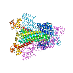
1GV3
 | | The 2.0 Angstrom resolution structure of the catalytic portion of a cyanobacterial membrane-bound manganese superoxide dismutase | | 分子名称: | MANGANESE (II) ION, MANGANESE SUPEROXIDE DISMUTASE | | 著者 | Atzenhofer, W, Regelsberger, G, Jacob, U, Huber, R, Peschek, G.A, Obinger, C. | | 登録日 | 2002-02-05 | | 公開日 | 2002-08-08 | | 最終更新日 | 2023-12-13 | | 実験手法 | X-RAY DIFFRACTION (2 Å) | | 主引用文献 | The 2.0A Resolution Structure of the Catalytic Portion of a Cyanobacterial Membrane-Bound Manganese Superoxide Dismutase
J.Mol.Biol., 321, 2002
|
|
1OJ5
 | | Crystal structure of the Nco-A1 PAS-B domain bound to the STAT6 transactivation domain LXXLL motif | | 分子名称: | IODIDE ION, SIGNAL TRANSDUCER AND ACTIVATOR OF TRANSCRIPTION 6, STEROID RECEPTOR COACTIVATOR 1A | | 著者 | Razeto, A, Ramakrishnan, V, Giller, K, Lakomek, N, Carlomagno, T, Griesinger, C, Lodrini, M, Litterst, C.M, Pftizner, E, Becker, S. | | 登録日 | 2003-07-02 | | 公開日 | 2004-02-12 | | 最終更新日 | 2024-05-08 | | 実験手法 | X-RAY DIFFRACTION (2.21 Å) | | 主引用文献 | Structure of the Ncoa-1/Src-1 Pas-B Domain Bound to the Lxxll Motif of the Stat6 Transactivation Domain
J.Mol.Biol., 336, 2004
|
|
1QRR
 | | CRYSTAL STRUCTURE OF SQD1 PROTEIN COMPLEX WITH NAD AND UDP-GLUCOSE | | 分子名称: | NICOTINAMIDE-ADENINE-DINUCLEOTIDE, SULFATE ION, URIDINE-5'-DIPHOSPHATE-GLUCOSE, ... | | 著者 | Mulichak, A.M, Theisen, M.J, Essigmann, B, Benning, C, Garavito, R.M. | | 登録日 | 1999-06-15 | | 公開日 | 1999-11-10 | | 最終更新日 | 2024-02-14 | | 実験手法 | X-RAY DIFFRACTION (1.6 Å) | | 主引用文献 | Crystal structure of SQD1, an enzyme involved in the biosynthesis of the plant sulfolipid headgroup donor UDP-sulfoquinovose.
Proc.Natl.Acad.Sci.USA, 96, 1999
|
|
1FJP
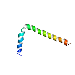 | | NMR Solution Structure of Phospholamban (C41F) | | 分子名称: | CARDIAC PHOSPHOLAMBAN | | 著者 | Lamberth, S, Griesinger, C, Schmid, H, Carafoli, E, Muenchbach, M, Vorherr, T, Krebs, J. | | 登録日 | 2000-08-08 | | 公開日 | 2000-09-06 | | 最終更新日 | 2024-05-22 | | 実験手法 | SOLUTION NMR | | 主引用文献 | NMR Solution Structure of Phospholamban
HELV.CHIM.ACTA, 83, 2000
|
|
1HY0
 | | CRYSTAL STRUCTURE OF WILD TYPE DUCK DELTA 1 CRYSTALLIN (EYE LENS PROTEIN) | | 分子名称: | DELTA CRYSTALLIN I, SULFATE ION | | 著者 | Sampaleanu, L.M, Vallee, F, Slingsby, C, Howell, P.L. | | 登録日 | 2001-01-17 | | 公開日 | 2001-04-21 | | 最終更新日 | 2024-02-07 | | 実験手法 | X-RAY DIFFRACTION (2.2 Å) | | 主引用文献 | Structural studies of duck delta 1 and delta 2 crystallin suggest conformational changes occur during catalysis.
Biochemistry, 40, 2001
|
|
1E7N
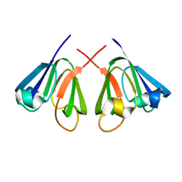 | | The N-terminal domain of beta-B2-crystallin resembles the putative ancestral homodimer | | 分子名称: | BETA-CRYSTALLIN B2 | | 著者 | Clout, N.J, Basak, A, Wieligmann, K, Bateman, O.A, Jaenicke, R, Slingsby, C. | | 登録日 | 2000-08-31 | | 公開日 | 2000-10-05 | | 最終更新日 | 2024-05-08 | | 実験手法 | X-RAY DIFFRACTION (2.35 Å) | | 主引用文献 | The N-Terminal Domain of Betab2-Crystallin Resembles the Putative Ancestral Homodimer.
J.Mol.Biol., 304, 2000
|
|
2KDU
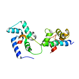 | | Structural basis of the Munc13-1/Ca2+-Calmodulin interaction: A novel 1-26 calmodulin binding motif with a bipartite binding mode | | 分子名称: | CALCIUM ION, Calmodulin, Protein unc-13 homolog A | | 著者 | Rodriguez-Castaneda, F.A, Maestre-Martinez, M, Coudevylle, N, Dimova, K, Jahn, O, Junge, H, Becker, S, Brose, N, Carlomagno, T, Griesinger, C. | | 登録日 | 2009-01-19 | | 公開日 | 2009-12-15 | | 最終更新日 | 2024-05-22 | | 実験手法 | SOLUTION NMR | | 主引用文献 | Modular architecture of Munc13/calmodulin complexes: dual regulation by Ca2+ and possible function in short-term synaptic plasticity.
Embo J., 29, 2010
|
|
1QEW
 | | HUMAN CLASS I HISTOCOMPATIBILITY ANTIGEN (HLA-A 0201) COMPLEX WITH A NONAMERIC PEPTIDE FROM MELANOMA-ASSOCIATED ANTIGEN 3 (RESIDUES 271-279) | | 分子名称: | PROTEIN (BETA-2-MICROGLOBULIN), PROTEIN (HLA CLASS I HISTOCOMPATIBILITY ANTIGEN, B-35 B* 3501 ALPHA CHAIN), ... | | 著者 | Orth, P, Alings, C, Saenger, W, Ziegler, A. | | 登録日 | 1999-04-02 | | 公開日 | 2003-11-18 | | 最終更新日 | 2023-08-16 | | 実験手法 | X-RAY DIFFRACTION (2.2 Å) | | 主引用文献 | HUMAN CLASS I HISTOCOMPATIBILITY ANTIGEN (HLA-A 0201)
COMPLEX WITH A NONAMERIC PEPTIDE FROM MELANOMA-ASSOCIATED
ANTIGEN 3 (RESIDUES 271-279)
To be Published
|
|
1FJK
 | | NMR Solution Structure of Phospholamban (C41F) | | 分子名称: | CARDIAC PHOSPHOLAMBAN | | 著者 | Lamberth, S, Griesinger, C, Schmid, H, Carafoli, E, Muenchbach, M, Vorherr, T, Krebs, J. | | 登録日 | 2000-08-08 | | 公開日 | 2000-09-06 | | 最終更新日 | 2024-05-22 | | 実験手法 | SOLUTION NMR | | 主引用文献 | NMR Solution Structure of Phospholamban
HELV.CHIM.ACTA, 83, 2000
|
|
1HXH
 | | COMAMONAS TESTOSTERONI 3BETA/17BETA HYDROXYSTEROID DEHYDROGENASE | | 分子名称: | 3BETA/17BETA-HYDROXYSTEROID DEHYDROGENASE | | 著者 | Benach, J, Filling, C, Oppermann, U.C.T, Roversi, P, Bricogne, G, Berndt, K.D, Jornvall, H, Ladenstein, R. | | 登録日 | 2001-01-15 | | 公開日 | 2002-12-25 | | 最終更新日 | 2023-08-09 | | 実験手法 | X-RAY DIFFRACTION (1.22 Å) | | 主引用文献 | Structure of Bacterial 3beta/17beta-Hydroxysteroid Dehydrogenase at 1.2 A Resolution: A Model for
Multiple Steroid Recognition
Biochemistry, 41, 2002
|
|
1HY1
 | |
2KOX
 | | NMR residual dipolar couplings identify long range correlated motions in the backbone of the protein ubiquitin | | 分子名称: | Ubiquitin | | 著者 | Fenwick, R.B, Richter, B, Lee, D, Walter, K.F.A, Milovanovic, D, Becker, S, Lakomek, N.A, Griesinger, C, Salvatella, X. | | 登録日 | 2009-10-02 | | 公開日 | 2011-06-08 | | 最終更新日 | 2024-05-01 | | 実験手法 | SOLUTION NMR | | 主引用文献 | Weak Long-Range Correlated Motions in a Surface Patch of Ubiquitin Involved in Molecular Recognition
J.Am.Chem.Soc., 2011
|
|
1I2C
 | | CRYSTAL STRUCTURE OF MUTANT T145A SQD1 PROTEIN COMPLEX WITH NAD AND UDP-GLUCOSE | | 分子名称: | NICOTINAMIDE-ADENINE-DINUCLEOTIDE, SULFATE ION, SULFOLIPID BIOSYNTHESIS PROTEIN SQD1, ... | | 著者 | Theisen, M.J, Sanda, S.L, Ginell, S.L, Benning, C, Garavito, R.M. | | 登録日 | 2001-02-07 | | 公開日 | 2003-07-01 | | 最終更新日 | 2023-08-09 | | 実験手法 | X-RAY DIFFRACTION (1.6 Å) | | 主引用文献 | Characterization of the Active Site of UDP-sulfoquinovose Synthase: Formation of the Sulfonic Acid Product in the Crystalline State.
To be Published
|
|
1O9S
 | | Crystal structure of a ternary complex of the human histone methyltransferase SET7/9 | | 分子名称: | GENE FRAGMENT FOR HISTONE H3, HISTONE-LYSINE N-METHYLTRANSFERASE, H3 LYSINE-4 SPECIFIC, ... | | 著者 | Xiao, B, Jing, C, Wilson, J.R, Walker, P.A, Vasisht, N, Kelly, G, Howell, S, Taylor, I.A, Blackburn, G.M, Gamblin, S.J. | | 登録日 | 2002-12-18 | | 公開日 | 2003-02-06 | | 最終更新日 | 2023-12-13 | | 実験手法 | X-RAY DIFFRACTION (1.75 Å) | | 主引用文献 | Structure and Catalytic Mechanism of the Human Histone Methyltransferase Set7/9
Nature, 421, 2003
|
|
2KXN
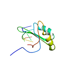 | | NMR structure of human Tra2beta1 RRM in complex with AAGAAC RNA | | 分子名称: | 5'-R(*AP*AP*GP*AP*AP*C)-3', Transformer-2 protein homolog beta | | 著者 | Clery, A, Jayne, S, Benderska, N, Dominguez, C, Stamm, S, Allain, F.H.-T. | | 登録日 | 2010-05-10 | | 公開日 | 2011-03-16 | | 最終更新日 | 2024-05-01 | | 実験手法 | SOLUTION NMR | | 主引用文献 | Molecular basis of purine-rich RNA recognition by the human SR-like protein Tra2-beta1
Nat.Struct.Mol.Biol., 18, 2011
|
|


