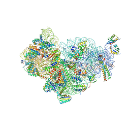
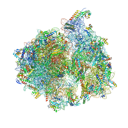
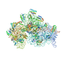
6GB2
 
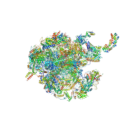 | | Unique features of mammalian mitochondrial translation initiation revealed by cryo-EM. This file contains the 39S ribosomal subunit. | | 分子名称: | 'Mitochondrial ribosomal protein L30, 'Mitochondrial ribosomal protein L55, 'Mitochondrial ribosomal protein L59, ... | | 著者 | Kummer, E, Leibundgut, M, Boehringer, D, Ban, N. | | 登録日 | 2018-04-13 | | 公開日 | 2018-08-08 | | 最終更新日 | 2024-05-15 | | 実験手法 | ELECTRON MICROSCOPY (3.2 Å) | | 主引用文献 | Unique features of mammalian mitochondrial translation initiation revealed by cryo-EM.
Nature, 560, 2018
|
|
7ASD
 | | Structure of native royal jelly filaments | | 分子名称: | (3beta,14beta,17alpha)-ergosta-5,24(28)-dien-3-ol, 2-acetamido-2-deoxy-beta-D-glucopyranose, 2-acetamido-2-deoxy-beta-D-glucopyranose-(1-4)-2-acetamido-2-deoxy-beta-D-glucopyranose, ... | | 著者 | Mattei, S, Ban, A, Picenoni, A, Leibundgut, M, Glockshuber, R, Boehringer, D. | | 登録日 | 2020-10-27 | | 公開日 | 2020-12-30 | | 実験手法 | ELECTRON MICROSCOPY (3.5 Å) | | 主引用文献 | Structure of native glycolipoprotein filaments in honeybee royal jelly.
Nat Commun, 11, 2020
|
|
4A3H
 | | 2',4' DINITROPHENYL-2-DEOXY-2-FLURO-B-D-CELLOBIOSIDE COMPLEX OF THE ENDOGLUCANASE CEL5A FROM BACILLUS AGARADHAERENS AT 1.6 A RESOLUTION | | 分子名称: | 2,4-DINITROPHENYL-2-DEOXY-2-FLUORO-BETA-D-CELLOBIOSIDE, PROTEIN (ENDOGLUCANASE) | | 著者 | Davies, G.J, Brzozowski, A.M, Andersen, K, Schulein, M, Mackenzie, L, Withers, S.G. | | 登録日 | 1998-07-22 | | 公開日 | 1999-07-23 | | 最終更新日 | 2023-12-27 | | 実験手法 | X-RAY DIFFRACTION (1.65 Å) | | 主引用文献 | Snapshots along an enzymatic reaction coordinate: analysis of a retaining beta-glycoside hydrolase.
Biochemistry, 37, 1998
|
|
5NFW
 | | Neutron structure of human transthyretin (TTR) S52P mutant at room temperature to 1.8A resolution (quasi-Laue) | | 分子名称: | Transthyretin | | 著者 | Yee, A.W, Moulin, M, Blakeley, M.P, Cooper, J.B, Haertlein, M, Mitchell, E.P, Forsyth, V.T. | | 登録日 | 2017-03-16 | | 公開日 | 2019-01-02 | | 最終更新日 | 2024-05-01 | | 実験手法 | NEUTRON DIFFRACTION (1.8 Å), X-RAY DIFFRACTION | | 主引用文献 | A molecular mechanism for transthyretin amyloidogenesis.
Nat Commun, 10, 2019
|
|
5NFE
 | | Neutron structure of human transthyretin (TTR) T119M mutant at room temperature to 1.85A resolution | | 分子名称: | Transthyretin | | 著者 | Yee, A.W, Moulin, M, Blakeley, M.P, Ostermann, A, Cooper, J.B, Haertlein, M, Mitchell, E.P, Forsyth, V.T. | | 登録日 | 2017-03-14 | | 公開日 | 2019-01-02 | | 最終更新日 | 2024-05-01 | | 実験手法 | NEUTRON DIFFRACTION (1.853 Å), X-RAY DIFFRACTION | | 主引用文献 | A molecular mechanism for transthyretin amyloidogenesis.
Nat Commun, 10, 2019
|
|
4CE8
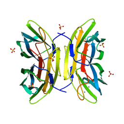 | | Perdeuterated Pseudomonas aeruginosa Lectin II complex with hydrogenated L-Fucose and Calcium | | 分子名称: | CALCIUM ION, FUCOSE-BINDING LECTIN PA-IIL, SULFATE ION, ... | | 著者 | Cuypers, M.G, Mitchell, E.P, Mossou, E, Pokorna, M, Wimmerova, M, Imberty, A, Moulin, M, Haertlein, M, Forsyth, V.T. | | 登録日 | 2013-11-10 | | 公開日 | 2014-11-12 | | 最終更新日 | 2023-12-20 | | 実験手法 | X-RAY DIFFRACTION (0.9 Å) | | 主引用文献 | Perdeuterated Pseudomonas Aeruginosa Lectin II Complex with Hydrogenated L Fucose and Calcium
To be Published
|
|
5OF2
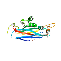 | | The structural versatility of TasA in B. subtilis biofilm formation | | 分子名称: | 1,2-ETHANEDIOL, SULFATE ION, Spore coat-associated protein N | | 著者 | Roske, Y, Diehl, A, Ball, L, Chowdhury, A, Hiller, M, Moliere, N, Kramer, R, Nagaraj, M, Stoeppler, D, Worth, C.L, Schlegel, B, Leidert, M, Cremer, N, Eisenmenger, F, Lopez, D, Schmieder, P, Heinemann, U, Turgay, K, Akbey, U, Oschkinat, H. | | 登録日 | 2017-07-10 | | 公開日 | 2018-03-21 | | 最終更新日 | 2024-01-17 | | 実験手法 | X-RAY DIFFRACTION (1.86 Å) | | 主引用文献 | Structural changes of TasA in biofilm formation ofBacillus subtilis.
Proc. Natl. Acad. Sci. U.S.A., 115, 2018
|
|
5OF1
 | | The structural versatility of TasA in B. subtilis biofilm formation | | 分子名称: | 2-HYDROXYBENZOIC ACID, Spore coat-associated protein N, ethane-1,2-diol | | 著者 | Roske, Y, Diehl, A, Ball, L, Chowdhury, A, Hiller, M, Moliere, N, Kramer, R, Nagaraj, M, Stoeppler, D, Worth, C.L, Schlegel, B, Leidert, M, Cremer, N, Eisenmenger, F, Lopez, D, Schmieder, P, Heinemann, U, Turgay, K, Akbey, U, Oschkinat, H. | | 登録日 | 2017-07-10 | | 公開日 | 2018-03-21 | | 最終更新日 | 2018-04-11 | | 実験手法 | X-RAY DIFFRACTION (1.56 Å) | | 主引用文献 | Structural changes of TasA in biofilm formation ofBacillus subtilis.
Proc. Natl. Acad. Sci. U.S.A., 115, 2018
|
|
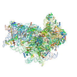
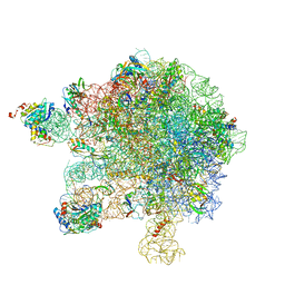
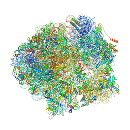
4CE4
 | | 39S large subunit of the porcine mitochondrial ribosome | | 分子名称: | 16S Ribosomal RNA, ICT1, MRPL13, ... | | 著者 | Greber, B.J, Boehringer, D, Leitner, A, Bieri, P, Voigts-Hoffmann, F, Erzberger, J.P, Leibundgut, M, Aebersold, R, Ban, N. | | 登録日 | 2013-11-08 | | 公開日 | 2013-12-18 | | 最終更新日 | 2024-05-08 | | 実験手法 | ELECTRON MICROSCOPY (4.9 Å) | | 主引用文献 | Architecture of the Large Subunit of the Mammalian Mitochondrial Ribosome.
Nature, 505, 2014
|
|
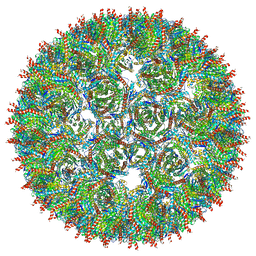
5MQ3
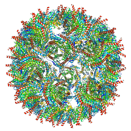 | | Structure of AaLS-neg | | 分子名称: | 6,7-dimethyl-8-ribityllumazine synthase | | 著者 | Sasaki, E, Boehringer, D, Leibundgut, M, Ban, N, Hilvert, D. | | 登録日 | 2016-12-20 | | 公開日 | 2017-03-22 | | 最終更新日 | 2024-05-15 | | 実験手法 | ELECTRON MICROSCOPY (5.4 Å) | | 主引用文献 | Structure and assembly of scalable porous protein cages.
Nat Commun, 8, 2017
|
|
4BTS
 | | THE CRYSTAL STRUCTURE OF THE EUKARYOTIC 40S RIBOSOMAL SUBUNIT IN COMPLEX WITH EIF1 AND EIF1A | | 分子名称: | 18S ribosomal RNA, 40S RIBOSOMAL PROTEIN RACK1, 40S RIBOSOMAL PROTEIN RPS10E, ... | | 著者 | Weisser, M, Voigts-Hoffmann, F, Rabl, J, Leibundgut, M, Ban, N. | | 登録日 | 2013-06-19 | | 公開日 | 2013-07-17 | | 最終更新日 | 2023-12-20 | | 実験手法 | X-RAY DIFFRACTION (3.703 Å) | | 主引用文献 | The crystal structure of the eukaryotic 40S ribosomal subunit in complex with eIF1 and eIF1A.
Nat. Struct. Mol. Biol., 20, 2013
|
|
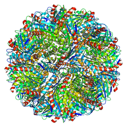
5MQ7
 | | Structure of AaLS-13 | | 分子名称: | 6,7-dimethyl-8-ribityllumazine synthase | | 著者 | Sasaki, E, Boehringer, D, Leibundgut, M, Ban, N, Hilvert, D. | | 登録日 | 2016-12-20 | | 公開日 | 2017-03-22 | | 最終更新日 | 2024-05-15 | | 実験手法 | ELECTRON MICROSCOPY (5.2 Å) | | 主引用文献 | Structure and assembly of scalable porous protein cages.
Nat Commun, 8, 2017
|
|
5MMJ
 | | Structure of the small subunit of the chloroplast ribosome | | 分子名称: | 16S ribosomal RNA, 30S ribosomal protein 2, chloroplastic, ... | | 著者 | Bieri, P, Leibundgut, M, Saurer, M, Boehringer, D, Ban, N. | | 登録日 | 2016-12-10 | | 公開日 | 2017-01-11 | | 最終更新日 | 2024-05-15 | | 実験手法 | ELECTRON MICROSCOPY (3.6 Å) | | 主引用文献 | The complete structure of the chloroplast 70S ribosome in complex with translation factor pY.
EMBO J., 36, 2017
|
|
5OA3
 | | Human 40S-eIF2D-re-initiation complex | | 分子名称: | 18S ribosomal RNA, 40S ribosomal protein S10, 40S ribosomal protein S11, ... | | 著者 | Weisser, M, Schaefer, T, Leibundgut, M, Boehringer, D, Aylett, C.H.S, Ban, N. | | 登録日 | 2017-06-20 | | 公開日 | 2017-08-09 | | 最終更新日 | 2024-05-15 | | 実験手法 | ELECTRON MICROSCOPY (4.2 Å) | | 主引用文献 | Structural and Functional Insights into Human Re-initiation Complexes.
Mol. Cell, 67, 2017
|
|
5MPP
 | | Structure of AaLS-wt | | 分子名称: | 6,7-dimethyl-8-ribityllumazine synthase | | 著者 | Sasaki, E, Boehringer, D, Leibundgut, M, Ban, N, Hilvert, D. | | 登録日 | 2016-12-17 | | 公開日 | 2017-03-22 | | 最終更新日 | 2024-05-15 | | 実験手法 | ELECTRON MICROSCOPY (3.9 Å) | | 主引用文献 | Structure and assembly of scalable porous protein cages.
Nat Commun, 8, 2017
|
|
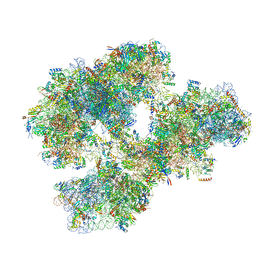
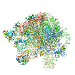
5MMM
 | | Structure of the 70S chloroplast ribosome | | 分子名称: | 16S ribosomal RNA, 23S ribosomal RNA, 30S ribosomal protein 2, ... | | 著者 | Bieri, P, Leibundgut, M, Saurer, M, Boehringer, D, Ban, N. | | 登録日 | 2016-12-11 | | 公開日 | 2017-01-11 | | 最終更新日 | 2024-05-15 | | 実験手法 | ELECTRON MICROSCOPY (3.4 Å) | | 主引用文献 | The complete structure of the chloroplast 70S ribosome in complex with translation factor pY.
EMBO J., 36, 2017
|
|
5MMI
 | | Structure of the large subunit of the chloroplast ribosome | | 分子名称: | 23S ribosomal RNA, 4.5S ribosomal RNA, 50S ribosomal protein 6, ... | | 著者 | Bieri, P, Leibundgut, M, Saurer, M, Boehringer, D, Ban, N. | | 登録日 | 2016-12-10 | | 公開日 | 2017-01-11 | | 最終更新日 | 2024-05-15 | | 実験手法 | ELECTRON MICROSCOPY (3.2 Å) | | 主引用文献 | The complete structure of the chloroplast 70S ribosome in complex with translation factor pY.
EMBO J., 36, 2017
|
|
5OA9
 | | Human translation re-initiation complex containing eIF2D | | 分子名称: | Eukaryotic translation initiation factor 2D | | 著者 | Weisser, M, Schaefer, T, Leibundgut, M, Boehringer, D, Aylett, C.H.S, Ban, N. | | 登録日 | 2017-06-21 | | 公開日 | 2017-07-26 | | 最終更新日 | 2024-05-08 | | 実験手法 | X-RAY DIFFRACTION (1.8 Å) | | 主引用文献 | Structural and Functional Insights into Human Re-initiation Complexes.
Mol. Cell, 67, 2017
|
|
5NCO
 | | Quaternary complex between SRP, SR, and SecYEG bound to the translating ribosome | | 分子名称: | 23S rRNA, 4.5S SRP RNA (Ffs), 50S ribosomal protein L10, ... | | 著者 | Jomaa, A, Hwang Fu, Y, Boerhinger, D, Leibundgut, M, Shan, S.O, Ban, N. | | 登録日 | 2017-03-06 | | 公開日 | 2017-05-24 | | 最終更新日 | 2018-03-28 | | 実験手法 | ELECTRON MICROSCOPY (4.8 Å) | | 主引用文献 | Structure of the quaternary complex between SRP, SR, and translocon bound to the translating ribosome.
Nat Commun, 8, 2017
|
|
5O5J
 | | Structure of the 30S small ribosomal subunit from Mycobacterium smegmatis | | 分子名称: | 16S rRNA, 30S ribosomal protein S10, 30S ribosomal protein S11, ... | | 著者 | Hentschel, J, Burnside, C, Mignot, I, Leibundgut, M, Boehringer, D, Ban, N. | | 登録日 | 2017-06-02 | | 公開日 | 2017-07-12 | | 最終更新日 | 2024-05-15 | | 実験手法 | ELECTRON MICROSCOPY (3.451 Å) | | 主引用文献 | The Complete Structure of the Mycobacterium smegmatis 70S Ribosome.
Cell Rep, 20, 2017
|
|
5O61
 | | The complete structure of the Mycobacterium smegmatis 70S ribosome | | 分子名称: | 16S rRNA, 23S rRNA, 30S ribosomal protein S10, ... | | 著者 | Hentschel, J, Burnside, C, Mignot, I, Leibundgut, M, Boehringer, D, Ban, N. | | 登録日 | 2017-06-03 | | 公開日 | 2017-07-12 | | 最終更新日 | 2024-05-15 | | 実験手法 | ELECTRON MICROSCOPY (3.31 Å) | | 主引用文献 | The Complete Structure of the Mycobacterium smegmatis 70S Ribosome.
Cell Rep, 20, 2017
|
|
5NW3
 | | The cryofrozen atomic resolution X-ray crystal structure of perdeuterated Pyrococcus furiosus Rubredoxin (100K, 0.59A resolution) | | 分子名称: | FE (III) ION, PHOSPHATE ION, POTASSIUM ION, ... | | 著者 | Cuypers, M.G, Mason, S.A, Mossou, E, Haertlein, M, Forsyth, V.T. | | 登録日 | 2017-05-04 | | 公開日 | 2017-06-07 | | 最終更新日 | 2024-05-08 | | 実験手法 | X-RAY DIFFRACTION (0.59 Å) | | 主引用文献 | The cryofrozen atomic resolution X-ray crystal structure of perdeuterated Pyrococcus furiosus Rubredoxin (100K, 0.59A resolution)
To Be Published
|
|
5O60
 | | Structure of the 50S large ribosomal subunit from Mycobacterium smegmatis | | 分子名称: | 23S rRNA, 50S ribosomal protein L10, 50S ribosomal protein L11, ... | | 著者 | Hentschel, J, Burnside, C, Mignot, I, Leibundgut, M, Boehringer, D, Ban, N. | | 登録日 | 2017-06-02 | | 公開日 | 2017-07-12 | | 最終更新日 | 2024-05-15 | | 実験手法 | ELECTRON MICROSCOPY (3.18 Å) | | 主引用文献 | The Complete Structure of the Mycobacterium smegmatis 70S Ribosome.
Cell Rep, 20, 2017
|
|
4AR4
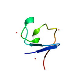 | | Neutron crystallographic structure of the reduced form perdeuterated Pyrococcus furiosus rubredoxin to 1.38 Angstrom resolution. | | 分子名称: | FE (III) ION, Rubredoxin, deuterium(1+), ... | | 著者 | Cuypers, M.G, Mason, S.A, Blakeley, M.P, Mitchell, E.P, Haertlein, M, Forsyth, V.T. | | 登録日 | 2012-04-20 | | 公開日 | 2013-01-16 | | 最終更新日 | 2024-06-19 | | 実験手法 | NEUTRON DIFFRACTION (1.381 Å) | | 主引用文献 | Near-Atomic Resolution Neutron Crystallography on Perdeuterated Pyrococcus Furiosus Rubredoxin: Implication of Hydronium Ions and Protonation State Equilibria in Redox Changes.
Angew.Chem.Int.Ed.Engl., 52, 2013
|
|
4AR3
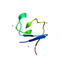 | | Near-atomic resolution neutron crystallography on the oxidised form perdeuterated Pyrococcus furiosus rubredoxin. | | 分子名称: | FE (III) ION, Rubredoxin, deuterium(1+), ... | | 著者 | Cuypers, M.G, Mason, S.A, Blakeley, M.P, Mitchell, E.P, Haertlein, M, Forsyth, V.T. | | 登録日 | 2012-04-20 | | 公開日 | 2013-01-16 | | 最終更新日 | 2024-05-08 | | 実験手法 | NEUTRON DIFFRACTION (1.05 Å) | | 主引用文献 | Near-Atomic Resolution Neutron Crystallography on Perdeuterated Pyrococcus Furiosus Rubredoxin: Implication of Hydronium Ions and Protonation State Equilibria in Redox Changes.
Angew.Chem.Int.Ed.Engl., 52, 2013
|
|


