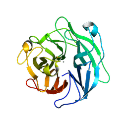
3AJ4
 
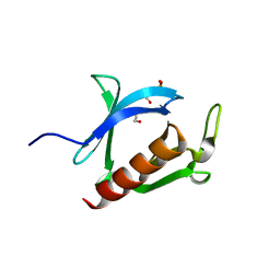 | | Crystal structure of the PH domain of Evectin-2 from human complexed with O-phospho-L-serine | | 分子名称: | 1,2-ETHANEDIOL, PHOSPHOSERINE, Pleckstrin homology domain-containing family B member 2 | | 著者 | Okazaki, S, Kato, R, Wakatsuki, S. | | 登録日 | 2010-05-21 | | 公開日 | 2011-05-25 | | 最終更新日 | 2023-11-01 | | 実験手法 | X-RAY DIFFRACTION (1 Å) | | 主引用文献 | Intracellular phosphatidylserine is essential for retrograde membrane traffic through endosomes
Proc.Natl.Acad.Sci.USA, 108, 2011
|
|
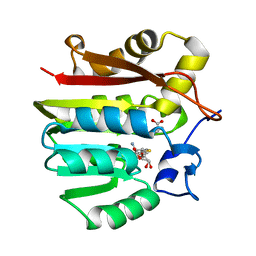
1P1B
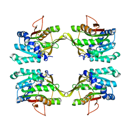
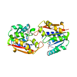 | | Guanidinoacetate methyltransferase | | 分子名称: | Guanidinoacetate N-methyltransferase, S-ADENOSYL-L-HOMOCYSTEINE | | 著者 | Komoto, J, Takusagawa, F. | | 登録日 | 2003-04-12 | | 公開日 | 2003-04-29 | | 最終更新日 | 2024-02-14 | | 実験手法 | X-RAY DIFFRACTION (2.8 Å) | | 主引用文献 | Monoclinic guanidinoacetate methyltransferase and gadolinium ion-binding characteristics.
Acta Crystallogr.,Sect.D, 59, 2003
|
|
4Y0C
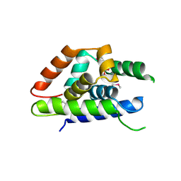 | | The structure of Arabidopsis ClpT2 | | 分子名称: | CHLORIDE ION, Clp protease-related protein At4g12060, chloroplastic, ... | | 著者 | Kimber, M.S, Schultz, L. | | 登録日 | 2015-02-05 | | 公開日 | 2015-05-13 | | 最終更新日 | 2023-11-15 | | 実験手法 | X-RAY DIFFRACTION (1.992 Å) | | 主引用文献 | Structures, Functions, and Interactions of ClpT1 and ClpT2 in the Clp Protease System of Arabidopsis Chloroplasts.
Plant Cell, 27, 2015
|
|
4Y0B
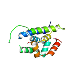 | | The structure of Arabidopsis ClpT1 | | 分子名称: | CHLORIDE ION, Double Clp-N motif protein | | 著者 | Kimber, M.S, Schultz, L. | | 登録日 | 2015-02-05 | | 公開日 | 2015-05-13 | | 最終更新日 | 2020-01-08 | | 実験手法 | X-RAY DIFFRACTION (2.4 Å) | | 主引用文献 | Structures, Functions, and Interactions of ClpT1 and ClpT2 in the Clp Protease System of Arabidopsis Chloroplasts.
Plant Cell, 27, 2015
|
|
1NBI
 | |
1NBH
 | |
1KHH
 | |
1D2C
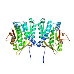 | | METHYLTRANSFERASE | | 分子名称: | PROTEIN (GLYCINE N-METHYLTRANSFERASE) | | 著者 | Huang, Y, Takusagawa, F. | | 登録日 | 1999-09-23 | | 公開日 | 1999-10-06 | | 最終更新日 | 2024-02-07 | | 実験手法 | X-RAY DIFFRACTION (2.5 Å) | | 主引用文献 | Mechanisms for auto-inhibition and forced product release in glycine N-methyltransferase: crystal structures of wild-type, mutant R175K and S-adenosylhomocysteine-bound R175K enzymes.
J.Mol.Biol., 298, 2000
|
|
1IDP
 | |
1WS8
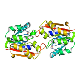
 | | Crystal Structure of Mavicyanin from Cucurbita pepo medullosa (Zucchini) | | 分子名称: | COPPER (II) ION, GLYCEROL, mavicyanin | | 著者 | Xie, Y, Inoue, T, Miyamoto, Y, Matsumura, H, Kunishige, K, Yamaguchi, K, Nojini, M, Suzuki, S, Kai, Y. | | 登録日 | 2004-11-02 | | 公開日 | 2004-11-23 | | 最終更新日 | 2011-07-13 | | 実験手法 | X-RAY DIFFRACTION (1.6 Å) | | 主引用文献 | Structural reorganization of the copper binding site involving Thr15 of mavicyanin from Cucurbita pepo medullosa (zucchini) upon reduction.
J.Biochem.(Tokyo), 137, 2005
|
|
1WLE
 | | Crystal Structure of mammalian mitochondrial seryl-tRNA synthetase complexed with seryl-adenylate | | 分子名称: | SERYL ADENYLATE, Seryl-tRNA synthetase | | 著者 | Chimnaronk, S, Jeppesen, M.G, Suzuki, T, Nyborg, J, Watanabe, K. | | 登録日 | 2004-06-25 | | 公開日 | 2005-09-06 | | 最終更新日 | 2023-10-25 | | 実験手法 | X-RAY DIFFRACTION (1.65 Å) | | 主引用文献 | Dual-mode recognition of noncanonical tRNAs(Ser) by seryl-tRNA synthetase in mammalian mitochondria
Embo J., 24, 2005
|
|
2ZOU
 | | Crystal structure of human F-spondin reeler domain (fragment 2) | | 分子名称: | 1,2-ETHANEDIOL, Spondin-1 | | 著者 | Nagae, M, Nogi, T, Takagi, J. | | 登録日 | 2008-06-07 | | 公開日 | 2008-10-14 | | 最終更新日 | 2023-11-01 | | 実験手法 | X-RAY DIFFRACTION (1.45 Å) | | 主引用文献 | Structure of the F-spondin reeler domain reveals a unique beta-sandwich fold with a deformable disulfide-bonded loop
Acta Crystallogr.,Sect.D, 64, 2008
|
|
3VOO
 | | Cytochrome P450SP alpha (CYP152B1) mutant A245E | | 分子名称: | Fatty acid alpha-hydroxylase, PROTOPORPHYRIN IX CONTAINING FE | | 著者 | Fujishiro, T, Shoji, O, Sugimoto, H, Shiro, Y, Watanabe, Y. | | 登録日 | 2012-01-31 | | 公開日 | 2013-02-06 | | 最終更新日 | 2023-11-08 | | 実験手法 | X-RAY DIFFRACTION (2.34 Å) | | 主引用文献 | A substrate-binding-state mimic of H2O2-dependent cytochrome P450 produced by one-point mutagenesis and peroxygenation of non-native substrates
Catalysis Science And Technology, 6, 2016
|
|
5HZV
 | | Crystal structure of the zona pellucida module of human endoglin/CD105 | | 分子名称: | GLYCEROL, Maltose-binding periplasmic protein,Endoglin, alpha-D-glucopyranose-(1-4)-alpha-D-glucopyranose | | 著者 | Bokhove, M, Saito, T, Jovine, L. | | 登録日 | 2016-02-03 | | 公開日 | 2017-06-07 | | 最終更新日 | 2024-01-10 | | 実験手法 | X-RAY DIFFRACTION (2.7 Å) | | 主引用文献 | Structural Basis of the Human Endoglin-BMP9 Interaction: Insights into BMP Signaling and HHT1.
Cell Rep, 19, 2017
|
|
3VNO
 | | Cytochrome P450SP alpha (CYP152B1) mutant R241E | | 分子名称: | (4S)-2-METHYL-2,4-PENTANEDIOL, Fatty acid alpha-hydroxylase, PROTOPORPHYRIN IX CONTAINING FE | | 著者 | Fujishiro, T, Shoji, O, Sugimoto, H, Shiro, Y, Watanabe, Y. | | 登録日 | 2012-01-17 | | 公開日 | 2013-02-06 | | 最終更新日 | 2023-11-08 | | 実験手法 | X-RAY DIFFRACTION (2.17 Å) | | 主引用文献 | A substrate-binding-state mimic of H2O2-dependent cytochrome P450 produced by one-point mutagenesis and peroxygenation of non-native substrates
Catalysis Science And Technology, 6, 2016
|
|
2ZOT
 | |
3VTJ
 | | Cytochrome P450SP alpha (CYP152B1) mutant A245H | | 分子名称: | Fatty acid alpha-hydroxylase, PROTOPORPHYRIN IX CONTAINING FE | | 著者 | Fujishiro, T, Shoji, O, Sugimoto, H, Shiro, Y, Watanabe, Y. | | 登録日 | 2012-05-30 | | 公開日 | 2013-06-05 | | 最終更新日 | 2023-11-08 | | 実験手法 | X-RAY DIFFRACTION (2.56 Å) | | 主引用文献 | A substrate-binding-state mimic of H2O2-dependent cytochrome P450 produced by one-point mutagenesis and peroxygenation of non-native substrates
Catalysis Science And Technology, 6, 2016
|
|
1WL7
 | | Structure of the thermostable arabinanase | | 分子名称: | CALCIUM ION, arabinanase-TS | | 著者 | Yamaguchi, A, Tada, T, Nakaniwa, T, Kitatani, T. | | 登録日 | 2004-06-21 | | 公開日 | 2005-06-21 | | 最終更新日 | 2023-10-25 | | 実験手法 | X-RAY DIFFRACTION (1.9 Å) | | 主引用文献 | Structural basis for thermostability of endo-1,5-alpha-L-arabinanase from Bacillus thermodenitrificans TS-3.
J.Biochem.(Tokyo), 137, 2005
|
|
1IS1
 | | Crystal structure of ribosome recycling factor from Vibrio parahaemolyticus | | 分子名称: | RIBOSOME RECYCLING FACTOR | | 著者 | Nakano, H, Yamaichi, Y, Uchiyama, S, Yoshida, T, Nishina, K, Kato, H, Ohkubo, T, Honda, T, Yamagata, Y, Kobayashi, Y. | | 登録日 | 2001-11-05 | | 公開日 | 2003-06-17 | | 最終更新日 | 2023-12-27 | | 実験手法 | X-RAY DIFFRACTION (2.2 Å) | | 主引用文献 | Structure and binding mode of a ribosome recycling factor (RRF) from mesophilic bacterium
J.BIOL.CHEM., 278, 2003
|
|
1XCJ
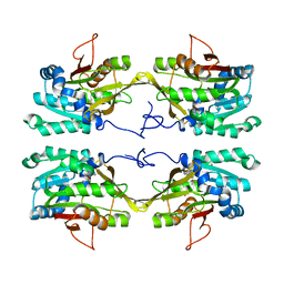
 | | Guanidinoacetate methyltransferase containing S-adenosylhomocysteine and guanidinoacetate | | 分子名称: | GUANIDINO ACETATE, Guanidinoacetate N-methyltransferase, S-ADENOSYL-L-HOMOCYSTEINE | | 著者 | Komoto, J, Yamada, T, Takata, Y, Takusagawa, F. | | 登録日 | 2004-09-02 | | 公開日 | 2004-12-07 | | 最終更新日 | 2024-02-14 | | 実験手法 | X-RAY DIFFRACTION (2 Å) | | 主引用文献 | Catalytic mechanism of guanidinoacetate methyltransferase: crystal structures of guanidinoacetate methyltransferase ternary complexes.
Biochemistry, 43, 2004
|
|
1XCL
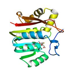 | | Guanidinoacetate methyltransferase containing S-adenosylhomocysteine and guanidine | | 分子名称: | GUANIDINE, Guanidinoacetate N-methyltransferase, S-ADENOSYL-L-HOMOCYSTEINE | | 著者 | Komoto, J, Yamada, T, Takata, Y, Takusagawa, F. | | 登録日 | 2004-09-02 | | 公開日 | 2004-12-07 | | 最終更新日 | 2024-02-14 | | 実験手法 | X-RAY DIFFRACTION (2 Å) | | 主引用文献 | Catalytic mechanism of guanidinoacetate methyltransferase: crystal structures of guanidinoacetate methyltransferase ternary complexes.
Biochemistry, 43, 2004
|
|
2DLC
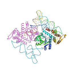 | | Crystal structure of the ternary complex of yeast tyrosyl-tRNA synthetase | | 分子名称: | MAGNESIUM ION, O-(ADENOSINE-5'-O-YL)-N-(L-TYROSYL)PHOSPHORAMIDATE, T-RNA (76-MER), ... | | 著者 | Tsunoda, M, Kusakabe, Y, Tanaka, N, Nakamura, K.T. | | 登録日 | 2006-04-18 | | 公開日 | 2007-06-12 | | 最終更新日 | 2024-03-13 | | 実験手法 | X-RAY DIFFRACTION (2.4 Å) | | 主引用文献 | Structural basis for recognition of cognate tRNA by tyrosyl-tRNA synthetase from three kingdoms.
Nucleic Acids Res., 35, 2007
|
|


