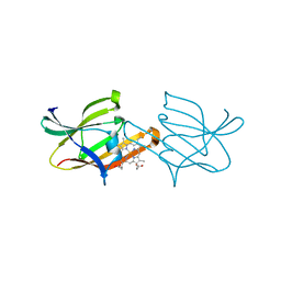
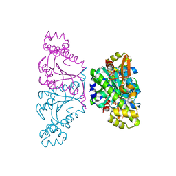
3QUG
 
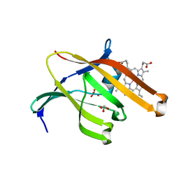 | | Structure of heme transport protein IsdH-NEAT3 from S. aureus in complex with Gallium-porphyrin | | 分子名称: | GLYCEROL, Iron-regulated surface determinant protein H, PROTOPORPHYRIN IX CONTAINING GA, ... | | 著者 | Moriwaki, Y, Caaveiro, J.M.M, Tsumoto, K. | | 登録日 | 2011-02-24 | | 公開日 | 2011-03-30 | | 最終更新日 | 2023-11-01 | | 実験手法 | X-RAY DIFFRACTION (1.7 Å) | | 主引用文献 | Molecular basis of recognition of antibacterial porphyrins by heme-transporter IsdH-NEAT3 of Staphylococcus aureus.
Biochemistry, 50, 2011
|
|
3QUH
 | |
4QCD
 | | Neutron crystal structure of phycocyanobilin:ferredoxin oxidoreductase in complex with biliverdin IXalpha at room temperature. | | 分子名称: | BILIVERDINE IX ALPHA, Phycocyanobilin:ferredoxin oxidoreductase, trideuteriooxidanium | | 著者 | Unno, M, Ishikawa-Suto, K, Ishihara, M, Hagiwara, Y, Sugishima, M, Wada, K, Fukuyama, K. | | 登録日 | 2014-05-10 | | 公開日 | 2015-04-29 | | 最終更新日 | 2024-03-20 | | 実験手法 | NEUTRON DIFFRACTION (1.932 Å), X-RAY DIFFRACTION | | 主引用文献 | Insights into the Proton Transfer Mechanism of a Bilin Reductase PcyA Following Neutron Crystallography.
J. Am. Chem. Soc., 137, 2015
|
|
5X6C
 | | Crystal structure of SepRS-SepCysE from Methanocaldococcus jannaschii | | 分子名称: | ADENOSINE-5'-TRIPHOSPHATE, O-phosphoserine--tRNA(Cys) ligase, SULFATE ION, ... | | 著者 | Chen, M, Kato, K, Yao, M. | | 登録日 | 2017-02-21 | | 公開日 | 2017-12-06 | | 最終更新日 | 2023-11-22 | | 実験手法 | X-RAY DIFFRACTION (3.101 Å) | | 主引用文献 | Structural basis for tRNA-dependent cysteine biosynthesis
Nat Commun, 8, 2017
|
|
5X6B
 | |
4ZM7
 | |
3MBC
 | | Crystal structure of monomeric isocitrate dehydrogenase from Corynebacterium glutamicum in complex with NADP | | 分子名称: | Isocitrate dehydrogenase [NADP], MAGNESIUM ION, NADP NICOTINAMIDE-ADENINE-DINUCLEOTIDE PHOSPHATE | | 著者 | Sidhu, N.S, Aich, S, Sheldrick, G.M, Delbaere, L.T.J. | | 登録日 | 2010-03-25 | | 公開日 | 2011-04-06 | | 最終更新日 | 2023-09-06 | | 実験手法 | X-RAY DIFFRACTION (1.9 Å) | | 主引用文献 | Structure of a highly NADP+-specific isocitrate dehydrogenase.
Acta Crystallogr.,Sect.D, 67, 2011
|
|
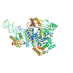
5H7K
 | | Crystal structure of Elongation factor 2 GDP-form | | 分子名称: | Elongation factor 2, GUANOSINE-5'-DIPHOSPHATE | | 著者 | Tanzawa, T, Kato, K, Uchiumi, T, Yao, M. | | 登録日 | 2016-11-18 | | 公開日 | 2018-02-21 | | 最終更新日 | 2024-03-20 | | 実験手法 | X-RAY DIFFRACTION (1.599 Å) | | 主引用文献 | The C-terminal helix of ribosomal P stalk recognizes a hydrophobic groove of elongation factor 2 in a novel fashion
Nucleic Acids Res., 46, 2018
|
|
5H7L
 | | Complex of Elongation factor 2-50S ribosomal protein L12 | | 分子名称: | 50S ribosomal protein L12, Elongation factor 2, PHOSPHOMETHYLPHOSPHONIC ACID GUANYLATE ESTER | | 著者 | Tanzawa, T, Kato, K, Uchiumi, T, Yao, M. | | 登録日 | 2016-11-18 | | 公開日 | 2018-02-21 | | 最終更新日 | 2018-05-02 | | 実験手法 | X-RAY DIFFRACTION (3.1 Å) | | 主引用文献 | The C-terminal helix of ribosomal P stalk recognizes a hydrophobic groove of elongation factor 2 in a novel fashion
Nucleic Acids Res., 46, 2018
|
|
5H7J
 | | Crystal structure of Elongation factor 2 | | 分子名称: | Elongation factor 2, PHOSPHOMETHYLPHOSPHONIC ACID GUANYLATE ESTER | | 著者 | Tanzawa, T, Kato, K, Uchiumi, T, Yao, M. | | 登録日 | 2016-11-18 | | 公開日 | 2018-02-21 | | 最終更新日 | 2018-05-02 | | 実験手法 | X-RAY DIFFRACTION (2.3 Å) | | 主引用文献 | The C-terminal helix of ribosomal P stalk recognizes a hydrophobic groove of elongation factor 2 in a novel fashion
Nucleic Acids Res., 46, 2018
|
|
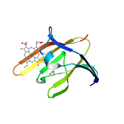
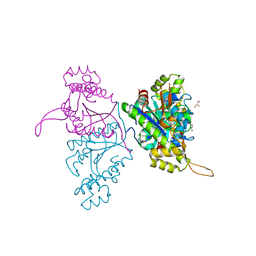
3VTM
 | | Structure of heme transport protein IsdH-NEAT3 from S. aureus in complex with Indium-porphyrin | | 分子名称: | GLYCEROL, Iron-regulated surface determinant protein H, PROTOPORPHYRIN IX CONTAINING INDIUM | | 著者 | Vu, N.T, Caaveiro, J.M.M, Moriwaki, Y, Tsumoto, K. | | 登録日 | 2012-05-31 | | 公開日 | 2013-05-15 | | 最終更新日 | 2023-11-08 | | 実験手法 | X-RAY DIFFRACTION (2.8 Å) | | 主引用文献 | Selective binding of antimicrobial porphyrins to the heme-receptor IsdH-NEAT3 of Staphylococcus aureus
Protein Sci., 22, 2013
|
|
2ROM
 | |
1RDB
 | |
1RDC
 | |
1RDA
 | |
1ROM
 | |
2Z6F
 | |
1V76
 | |
3VUA
 | | Apo IsdH-NEAT3 in space group P3121 at a resolution of 1.85 A | | 分子名称: | ACETATE ION, GLYCEROL, Iron-regulated surface determinant protein H, ... | | 著者 | Vu, N.T, Caaveiro, J.M.M, Moriwaki, Y, Tsumoto, K. | | 登録日 | 2012-06-26 | | 公開日 | 2013-06-26 | | 最終更新日 | 2023-11-08 | | 実験手法 | X-RAY DIFFRACTION (1.85 Å) | | 主引用文献 | Structure of heme transport protein IsdH-NEAT3 from S. aureus in complex with Indium-porphyrin
To be Published
|
|
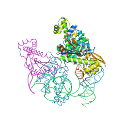
5AXM
 | | Crystal structure of Thg1 like protein (TLP) with tRNA(Phe) | | 分子名称: | MAGNESIUM ION, RNA (75-MER), tRNA(His)-5'-guanylyltransferase (Thg1) like protein | | 著者 | Kimura, S, Suzuki, T, Yu, J, Kato, K, Yao, M. | | 登録日 | 2015-07-31 | | 公開日 | 2016-08-03 | | 最終更新日 | 2023-11-08 | | 実験手法 | X-RAY DIFFRACTION (2.21 Å) | | 主引用文献 | Template-dependent nucleotide addition in the reverse (3'-5') direction by Thg1-like protein
Sci Adv, 2, 2016
|
|
5AXL
 | | Crystal structure of Thg1 like protein (TLP) with GTP | | 分子名称: | GUANOSINE-5'-TRIPHOSPHATE, MAGNESIUM ION, tRNA(His)-5'-guanylyltransferase (Thg1) like protein | | 著者 | Kimura, S, Suzuki, T, Yu, J, Kato, K, Yao, M. | | 登録日 | 2015-07-31 | | 公開日 | 2016-08-03 | | 最終更新日 | 2023-11-08 | | 実験手法 | X-RAY DIFFRACTION (2.998 Å) | | 主引用文献 | Template-dependent nucleotide addition in the reverse (3'-5') direction by Thg1-like protein
Sci Adv, 2, 2016
|
|
5AXK
 | | Crystal structure of Thg1 like protein (TLP) | | 分子名称: | GLYCEROL, tRNA(His)-5'-guanylyltransferase (Thg1) like protein | | 著者 | Kimura, S, Suzuki, T, Yu, J, Kato, K, Yao, M. | | 登録日 | 2015-07-31 | | 公開日 | 2016-08-03 | | 最終更新日 | 2023-11-08 | | 実験手法 | X-RAY DIFFRACTION (2.29 Å) | | 主引用文献 | Template-dependent nucleotide addition in the reverse (3'-5') direction by Thg1-like protein
Sci Adv, 2, 2016
|
|
5AXN
 | | Crystal structure of Thg1 like protein (TLP) with tRNA(Phe) and GDPNP | | 分子名称: | MAGNESIUM ION, PHOSPHOAMINOPHOSPHONIC ACID-GUANYLATE ESTER, RNA (75-MER), ... | | 著者 | Kimura, S, Suzuki, T, Yu, J, Kato, K, Yao, M. | | 登録日 | 2015-07-31 | | 公開日 | 2016-08-03 | | 最終更新日 | 2023-11-08 | | 実験手法 | X-RAY DIFFRACTION (2.703 Å) | | 主引用文献 | Template-dependent nucleotide addition in the reverse (3'-5') direction by Thg1-like protein
Sci Adv, 2, 2016
|
|
3X2L
 | | X-ray structure of PcCel45A apo form at 95K. | | 分子名称: | 2-AMINO-2-HYDROXYMETHYL-PROPANE-1,3-DIOL, 3-methylpentane-1,5-diol, Endoglucanase V-like protein | | 著者 | Nakamura, A, Ishida, T, Ohta, K, Tanaka, H, Inaka, K, Samejima, M, Igarashi, K. | | 登録日 | 2014-12-22 | | 公開日 | 2015-10-14 | | 最終更新日 | 2019-12-18 | | 実験手法 | X-RAY DIFFRACTION (0.83 Å) | | 主引用文献 | "Newton's cradle" proton relay with amide-imidic acid tautomerization in inverting cellulase visualized by neutron crystallography.
Sci Adv, 1, 2015
|
|
2RD3
 | |


