Loading
PDBj
メニューPDBj@FacebookPDBj@X(formerly Twitter)PDBj@BlueSkyPDBj@YouTubewwPDB FoundationwwPDBDonate
RCSB PDBPDBeBMRBAdv. SearchSearch help
Search by PDB author
3BOS
DownloadVisualize
BU of 3bos by Molmil
Crystal structure of a putative dna replication regulator HDA (SAMA_1916) from Shewanella amazonensis sb2b at 1.75 A resolution
分子名称: 1,2-ETHANEDIOL, CHLORIDE ION, CYTIDINE-5'-DIPHOSPHATE, ...
著者Joint Center for Structural Genomics (JCSG)
登録日2007-12-17
公開日2008-01-15
最終更新日2024-11-20
実験手法X-RAY DIFFRACTION (1.75 Å)
主引用文献A structural basis for the regulatory inactivation of DnaA.
J.Mol.Biol., 385, 2009
3D00
DownloadVisualize
BU of 3d00 by Molmil
Crystal structure of a tungsten formylmethanofuran dehydrogenase subunit e (fmde)-like protein (syn_00638) from syntrophus aciditrophicus at 1.90 A resolution
分子名称: CHLORIDE ION, Tungsten formylmethanofuran dehydrogenase subunit E, ZINC ION
著者Joint Center for Structural Genomics (JCSG)
登録日2008-04-30
公開日2008-05-20
最終更新日2024-11-06
実験手法X-RAY DIFFRACTION (1.9 Å)
主引用文献Structures of three members of Pfam PF02663 (FmdE) implicated in microbial methanogenesis reveal a conserved alpha+beta core domain and an auxiliary C-terminal treble-clef zinc finger.
Acta Crystallogr.,Sect.F, 66, 2010
3CGH
DownloadVisualize
BU of 3cgh by Molmil
Crystal structure of a susd homolog (bt_3984) from bacteroides thetaiotaomicron vpi-5482 at 1.70 A resolution
分子名称: ACETATE ION, SusD homolog, ZINC ION
著者Joint Center for Structural Genomics (JCSG)
登録日2008-03-05
公開日2008-03-18
最終更新日2024-10-09
実験手法X-RAY DIFFRACTION (1.7 Å)
主引用文献Structure of BT_3984, a member of the SusD/RagB family of nutrient-binding molecules.
Acta Crystallogr.,Sect.F, 66, 2010
2OOK
DownloadVisualize
BU of 2ook by Molmil
Crystal structure of a protein with unknown function (YP_749275.1) from Shewanella Frigidimarina NCIMB 400 at 1.80 A resolution
分子名称: 1,2-ETHANEDIOL, Hypothetical protein
著者Joint Center for Structural Genomics (JCSG)
登録日2007-01-25
公開日2007-02-06
最終更新日2024-11-13
実験手法X-RAY DIFFRACTION (1.8 Å)
主引用文献Open and closed conformations of two SpoIIAA-like proteins (YP_749275.1 and YP_001095227.1) provide insights into membrane association and ligand binding.
Acta Crystallogr.,Sect.F, 66, 2010
2OOC
DownloadVisualize
BU of 2ooc by Molmil
Crystal structure of Histidine Phosphotransferase ShpA (NP_419930.1) from Caulobacter crescentus at 1.52 A resolution
分子名称: GLYCEROL, Histidine phosphotransferase, TETRAETHYLENE GLYCOL
著者Joint Center for Structural Genomics (JCSG)
登録日2007-01-25
公開日2007-02-06
最終更新日2024-10-30
実験手法X-RAY DIFFRACTION (1.52 Å)
主引用文献Crystal structure of histidine phosphotransfer protein ShpA, an essential regulator of stalk biogenesis in Caulobacter crescentus.
J.Mol.Biol., 390, 2009
3SSF
DownloadVisualize
BU of 3ssf by Molmil
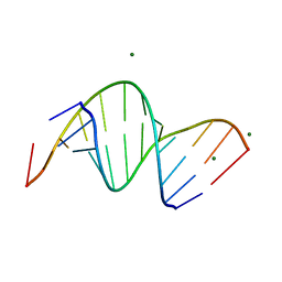
Crystal structure of RNA:DNA dodecamer corresponding to HIV-1 polypurine tract, at 1.6 A resolution.
分子名称: 5'-D(*CP*CP*TP*TP*TP*TP*CP*TP*TP*TP*TP*A)-3', 5'-R(*UP*AP*AP*AP*AP*GP*AP*AP*AP*AP*GP*G)-3', MAGNESIUM ION
著者Drozdzal, P, Michalska, K, Kierzek, R, Lomozik, L, Jaskolski, M.
登録日2011-07-08
公開日2012-02-08
最終更新日2023-09-13
実験手法X-RAY DIFFRACTION (1.6 Å)
主引用文献Structure of an RNA/DNA dodecamer corresponding to the HIV-1 polypurine tract at 1.6 Angstrom resolution
Acta Crystallogr.,Sect.D, 68, 2012
2Q8U
DownloadVisualize
BU of 2q8u by Molmil
CRYSTAL STRUCTURE OF MRE11 FROM THERMOTOGA MARITIMA MSB8 (TM1635) AT 2.20 A RESOLUTION
分子名称: Exonuclease, putative
著者Joint Center for Structural Genomics (JCSG)
登録日2007-06-11
公開日2007-06-26
最終更新日2024-10-30
実験手法X-RAY DIFFRACTION (2.2 Å)
主引用文献Crystal structure of the first eubacterial Mre11 nuclease reveals novel features that may discriminate substrates during DNA repair.
J.Mol.Biol., 397, 2010
4N6H
DownloadVisualize
BU of 4n6h by Molmil
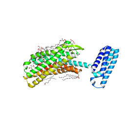
1.8 A Structure of the human delta opioid 7TM receptor (PSI Community Target)
分子名称: (2R)-2,3-dihydroxypropyl (9Z)-octadec-9-enoate, (4bS,8R,8aS,14bR)-7-(cyclopropylmethyl)-5,6,7,8,14,14b-hexahydro-4,8-methano[1]benzofuro[2,3-a]pyrido[4,3-b]carbazole-1,8a(9H)-diol, L(+)-TARTARIC ACID, ...
著者Fenalti, G, Giguere, P.M, Katritch, V, Huang, X.-P, Thompson, A.A, Han, G.W, Cherezov, V, Roth, B.L, Stevens, R.C, GPCR Network (GPCR)
登録日2013-10-12
公開日2013-12-25
最終更新日2024-10-16
実験手法X-RAY DIFFRACTION (1.8 Å)
主引用文献Molecular control of delta-opioid receptor signalling.
Nature, 506, 2014
2PV7
DownloadVisualize
BU of 2pv7 by Molmil
Crystal structure of chorismate mutase / prephenate dehydrogenase (tyrA) (1574749) from Haemophilus influenzae RD at 2.00 A resolution
分子名称: NICOTINAMIDE-ADENINE-DINUCLEOTIDE, T-protein [Includes: Chorismate mutase (EC 5.4.99.5) (CM) and Prephenate dehydrogenase (EC 1.3.1.12) (PDH)], TYROSINE
著者Joint Center for Structural Genomics (JCSG)
登録日2007-05-09
公開日2007-05-22
最終更新日2024-11-20
実験手法X-RAY DIFFRACTION (2 Å)
主引用文献The structure of Haemophilus influenzae prephenate dehydrogenase suggests unique features of bifunctional TyrA enzymes.
Acta Crystallogr.,Sect.F, 66, 2010
2Q3L
DownloadVisualize
BU of 2q3l by Molmil
CRYSTAL STRUCTURE OF AN UNCHARACTERIZED PROTEIN FROM DUF3478 FAMILY WITH A SPOIIAA-LIKE FOLD (SHEW_3102) FROM SHEWANELLA LOIHICA PV-4 AT 2.25 A RESOLUTION
分子名称: (4S)-2-METHYL-2,4-PENTANEDIOL, CHLORIDE ION, SODIUM ION, ...
著者Joint Center for Structural Genomics (JCSG)
登録日2007-05-30
公開日2007-06-12
最終更新日2024-11-20
実験手法X-RAY DIFFRACTION (2.25 Å)
主引用文献Open and closed conformations of two SpoIIAA-like proteins (YP_749275.1 and YP_001095227.1) provide insights into membrane association and ligand binding.
Acta Crystallogr.,Sect.F, 66, 2010
2QTP
DownloadVisualize
BU of 2qtp by Molmil
Crystal structure of a duf1185 family protein (spo0826) from silicibacter pomeroyi dss-3 at 2.10 A resolution
分子名称: Uncharacterized protein
著者Joint Center for Structural Genomics (JCSG)
登録日2007-08-02
公開日2007-08-21
最終更新日2024-10-30
実験手法X-RAY DIFFRACTION (2.1 Å)
主引用文献Structures of the first representatives of Pfam family PF06684 (DUF1185) reveal a novel variant of the Bacillus chorismate mutase fold and suggest a role in amino-acid metabolism.
Acta Crystallogr.,Sect.F, 66, 2010
3HSA
DownloadVisualize
BU of 3hsa by Molmil
Crystal structure of pleckstrin homology domain (YP_926556.1) from SHEWANELLA AMAZONENSIS SB2B at 1.99 A resolution
分子名称: DI(HYDROXYETHYL)ETHER, GLYCEROL, pleckstrin homology domain
著者Joint Center for Structural Genomics (JCSG)
登録日2009-06-10
公開日2009-06-23
最終更新日2023-02-01
実験手法X-RAY DIFFRACTION (1.99 Å)
主引用文献Bacterial pleckstrin homology domains: a prokaryotic origin for the PH domain.
J.Mol.Biol., 396, 2010
3GF8
DownloadVisualize
BU of 3gf8 by Molmil
Crystal structure of putative polysaccharide binding proteins (DUF1812) (NP_809975.1) from BACTEROIDES THETAIOTAOMICRON VPI-5482 at 2.20 A resolution
分子名称: 1,2-ETHANEDIOL, putative polysaccharide binding proteins (DUF1812)
著者Joint Center for Structural Genomics (JCSG)
登録日2009-02-26
公開日2009-03-24
最終更新日2024-11-20
実験手法X-RAY DIFFRACTION (2.2 Å)
主引用文献A conserved fold for fimbrial components revealed by the crystal structure of a putative fimbrial assembly protein (BT1062) from Bacteroides thetaiotaomicron at 2.2 A resolution
Acta Crystallogr.,Sect.F, 66, 2010
1ZX8
DownloadVisualize
BU of 1zx8 by Molmil
CRYSTAL STRUCTURE OF an atypical cyclophilin (peptidylprolyl cis-trans isomerase) (TM1367) FROM THERMOTOGA MARITIMA AT 1.90 A RESOLUTION
分子名称: NICKEL (II) ION, PENTAETHYLENE GLYCOL, hypothetical protein TM1367
著者Joint Center for Structural Genomics (JCSG)
登録日2005-06-07
公開日2005-07-26
最終更新日2024-10-30
実験手法X-RAY DIFFRACTION (1.9 Å)
主引用文献Crystal structure of TM1367 from Thermotoga maritima at 1.90 A resolution reveals an atypical member of the cyclophilin (peptidylprolyl isomerase) fold.
Proteins, 63, 2006
1VK9
DownloadVisualize
BU of 1vk9 by Molmil
CRYSTAL STRUCTURE OF A DUF1893 family protein (TM1506) FROM THERMOTOGA MARITIMA AT 2.70 A RESOLUTION
分子名称: UNKNOWN LIGAND, ZINC ION, conserved hypothetical protein TM1506
著者Joint Center for Structural Genomics (JCSG)
登録日2004-05-06
公開日2004-05-18
最終更新日2024-11-20
実験手法X-RAY DIFFRACTION (2.7 Å)
主引用文献Crystal structure of an ADP-ribosylated protein with a cytidine deaminase-like fold, but unknown function (TM1506), from Thermotoga maritima at 2.70 A resolution.
Proteins, 71, 2008
1VKH
DownloadVisualize
BU of 1vkh by Molmil
CRYSTAL STRUCTURE OF A PUTATIVE SERINE HYDROLASE (YDR428C) FROM SACCHAROMYCES CEREVISIAE AT 1.85 A RESOLUTION
分子名称: CHLORIDE ION, GLYCEROL, putative serine hydrolase
著者Joint Center for Structural Genomics (JCSG)
登録日2004-05-20
公開日2004-06-08
最終更新日2024-10-09
実験手法X-RAY DIFFRACTION (1.85 Å)
主引用文献Crystal structure of an alpha/beta serine hydrolase (YDR428C) from Saccharomyces cerevisiae at 1.85 A resolution
Proteins, 58, 2005
1VR3
DownloadVisualize
BU of 1vr3 by Molmil
Crystal structure of Acireductone dioxygenase (13543033) from Mus musculus at 2.06 A resolution
分子名称: Acireductone dioxygenase, ISOPROPYL ALCOHOL, NICKEL (II) ION, ...
著者Joint Center for Structural Genomics (JCSG)
登録日2005-01-28
公開日2005-02-15
最終更新日2024-11-20
実験手法X-RAY DIFFRACTION (2.06 Å)
主引用文献Crystal structure of acireductone dioxygenase (ARD) from Mus musculus at 2.06 angstrom resolution.
Proteins, 64, 2006
1VKM
DownloadVisualize
BU of 1vkm by Molmil
Crystal structure of an indigoidine synthase a (idga)-like protein (tm1464) from thermotoga maritima msb8 at 1.90 A resolution
分子名称: 1,2-ETHANEDIOL, MANGANESE (II) ION, UNKNOWN LIGAND, ...
著者Joint Center for Structural Genomics (JCSG)
登録日2004-06-09
公開日2004-06-29
最終更新日2024-11-06
実験手法X-RAY DIFFRACTION (1.9 Å)
主引用文献Crystal structure of an indigoidine synthase A (IndA)-like protein (TM1464) from Thermotoga maritima at 1.90 A resolution reveals a new fold.
Proteins, 59, 2005
1ZKG
DownloadVisualize
BU of 1zkg by Molmil
Crystal structure of Transcriptional regulator, TETR family (tm1030) from Thermotoga maritima at 2.30 A resolution
分子名称: UNKNOWN LIGAND, transcriptional regulator, TetR family
著者Joint Center for Structural Genomics (JCSG)
登録日2005-05-02
公開日2005-07-26
最終更新日2024-10-30
実験手法X-RAY DIFFRACTION (2.3 Å)
主引用文献Crystal structure of TM1030 from Thermotoga maritima at 2.3 A resolution reveals molecular details of its transcription repressor function.
Proteins, 68, 2007
1VQ0
DownloadVisualize
BU of 1vq0 by Molmil
Crystal structure of 33 kDa chaperonin (Heat shock protein 33 homolog) (HSP33) (TM1394) from Thermotoga maritima at 2.20 A resolution
分子名称: 1,2-ETHANEDIOL, 33 kDa chaperonin, CHLORIDE ION, ...
著者Joint Center for Structural Genomics (JCSG)
登録日2004-11-30
公開日2004-12-14
最終更新日2023-09-20
実験手法X-RAY DIFFRACTION (2.2 Å)
主引用文献Crystal structure of Hsp33 chaperone (TM1394) from Thermotoga maritima at 2.20 A resolution.
Proteins, 61, 2005
1VKB
DownloadVisualize
BU of 1vkb by Molmil
Crystal structure of an aig2-like protein (a2ld1, ggact, mgc7867) from mus musculus at 1.90 A resolution
分子名称: FORMIC ACID, hypothetical protein
著者Joint Center for Structural Genomics (JCSG)
登録日2004-05-07
公開日2004-05-18
最終更新日2024-10-30
実験手法X-RAY DIFFRACTION (1.9 Å)
主引用文献Crystal structure of a conserved hypothetical protein (gi: 13879369) from Mouse at 1.90 A resolution reveals a new fold.
Proteins, 61, 2005
1VLQ
DownloadVisualize
BU of 1vlq by Molmil
Crystal structure of Acetyl xylan esterase (TM0077) from Thermotoga maritima at 2.10 A resolution
分子名称: GLYCEROL, acetyl xylan esterase
著者Joint Center for Structural Genomics (JCSG)
登録日2004-08-09
公開日2004-08-24
最終更新日2024-10-30
実験手法X-RAY DIFFRACTION (2.1 Å)
主引用文献Functional and structural characterization of a thermostable acetyl esterase from Thermotoga maritima.
Proteins, 80, 2012
1VKY
DownloadVisualize
BU of 1vky by Molmil
Crystal structure of S-adenosylmethionine tRNA ribosyltransferase (TM0574) from Thermotoga maritima at 2.00 A resolution
分子名称: S-adenosylmethionine:tRNA ribosyltransferase-isomerase, UNKNOWN LIGAND
著者Joint Center for Structural Genomics (JCSG)
登録日2004-06-28
公開日2004-08-17
最終更新日2024-05-22
実験手法X-RAY DIFFRACTION (2 Å)
主引用文献Crystal structure of S-adenosylmethionine:tRNA ribosyltransferase-isomerase (QueA) from Thermotoga maritima at 2.0 A resolution reveals a new fold.
Proteins, 59, 2005
3IRB
DownloadVisualize
BU of 3irb by Molmil
Crystal structure of protein with unknown function from DUF35 family (13815350) from SULFOLOBUS SOLFATARICUS at 1.80 A resolution
分子名称: ACETIC ACID, SULFATE ION, ZINC ION, ...
著者Joint Center for Structural Genomics (JCSG)
登録日2009-08-21
公開日2009-09-01
最終更新日2024-11-06
実験手法X-RAY DIFFRACTION (1.8 Å)
主引用文献The structure of SSO2064, the first representative of Pfam family PF01796, reveals a novel two-domain zinc-ribbon OB-fold architecture with a potential acyl-CoA-binding role.
Acta Crystallogr.,Sect.F, 66, 2010
1VQR
DownloadVisualize
BU of 1vqr by Molmil
Crystal structure of a virulence factor (cj0248) from campylobacter jejuni subsp. jejuni at 2.25 A resolution
分子名称: hypothetical protein Cj0248
著者Joint Center for Structural Genomics (JCSG)
登録日2004-12-17
公開日2004-12-28
最終更新日2024-10-09
実験手法X-RAY DIFFRACTION (2.25 Å)
主引用文献Crystal structure of virulence factor CJ0248 from Campylobacter jejuni at 2.25 A resolution reveals a new fold.
Proteins, 62, 2006

238582

件を2025-07-09に公開中

PDB statisticsPDBj update infoContact PDBjnumon