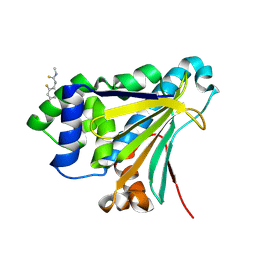
7HAQ
 
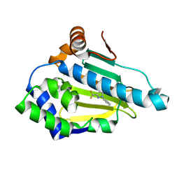 | | PanDDA analysis group deposition -- Crystal structure of HSP90N in complex with Fr13428 | | Descriptor: | 2-phenyl-5-(trifluoromethyl)-1,2-dihydro-3H-pyrazol-3-one, Heat shock protein HSP 90-alpha | | Authors: | Huang, L, Wang, W, Zhu, Z, Li, Q, Li, M, Zhou, H, Xu, Q, Wen, W, Wang, Q, Yu, F. | | Deposit date: | 2024-07-10 | | Release date: | 2025-03-26 | | Method: | X-RAY DIFFRACTION (1.55 Å) | | Cite: | Novel starting points for fragment-based drug design against human heat-shock protein 90 identified using crystallographic fragment screening.
Iucrj, 12, 2025
|
|
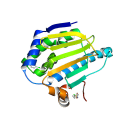
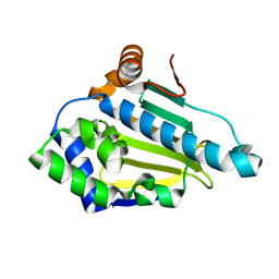
7HBH
 | | PanDDA analysis group deposition -- Crystal structure of HSP90N in complex with Fr13518 | | Descriptor: | (5-tert-butylthiophen-2-yl)(pyrrolidin-1-yl)methanone, Heat shock protein HSP 90-alpha | | Authors: | Huang, L, Wang, W, Zhu, Z, Li, Q, Li, M, Zhou, H, Xu, Q, Wen, W, Wang, Q, Yu, F. | | Deposit date: | 2024-07-10 | | Release date: | 2025-03-26 | | Method: | X-RAY DIFFRACTION (1.8 Å) | | Cite: | Novel starting points for fragment-based drug design against human heat-shock protein 90 identified using crystallographic fragment screening.
Iucrj, 12, 2025
|
|
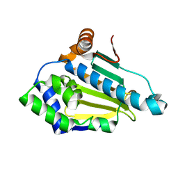
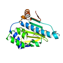
7HB5
 | | PanDDA analysis group deposition -- Crystal structure of HSP90N in complex with Fr13078 | | Descriptor: | Heat shock protein HSP 90-alpha, ~{N}-(4-chlorophenyl)methanesulfonamide | | Authors: | Huang, L, Wang, W, Zhu, Z, Li, Q, Li, M, Zhou, H, Xu, Q, Wen, W, Wang, Q, Yu, F. | | Deposit date: | 2024-07-10 | | Release date: | 2025-03-26 | | Method: | X-RAY DIFFRACTION (1.8 Å) | | Cite: | Novel starting points for fragment-based drug design against human heat-shock protein 90 identified using crystallographic fragment screening.
Iucrj, 12, 2025
|
|
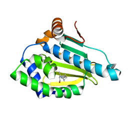
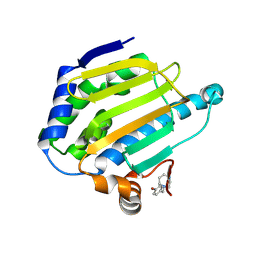
7HBT
 | | PanDDA analysis group deposition -- Crystal structure of HSP90N in complex with PS-3621 | | Descriptor: | Heat shock protein HSP 90-alpha, [3-(4-methylphenyl)-1H-pyrazol-1-yl]acetic acid | | Authors: | Huang, L, Wang, W, Zhu, Z, Li, Q, Li, M, Zhou, H, Xu, Q, Wen, W, Wang, Q, Yu, F. | | Deposit date: | 2024-07-10 | | Release date: | 2025-03-26 | | Method: | X-RAY DIFFRACTION (1.38 Å) | | Cite: | Novel starting points for fragment-based drug design against human heat-shock protein 90 identified using crystallographic fragment screening.
Iucrj, 12, 2025
|
|
7HBZ
 | | PanDDA analysis group deposition -- Crystal structure of HSP90N in complex with 6R-0009 | | Descriptor: | (4R)-5-chloro-3-ethyl[1,2,4]triazolo[4,3-a]pyridine, Heat shock protein HSP 90-alpha | | Authors: | Huang, L, Wang, W, Zhu, Z, Li, Q, Li, M, Zhou, H, Xu, Q, Wen, W, Wang, Q, Yu, F. | | Deposit date: | 2024-07-10 | | Release date: | 2025-03-26 | | Method: | X-RAY DIFFRACTION (1.44 Å) | | Cite: | Novel starting points for fragment-based drug design against human heat-shock protein 90 identified using crystallographic fragment screening.
Iucrj, 12, 2025
|
|
7HBP
 | | PanDDA analysis group deposition -- Crystal structure of HSP90N in complex with Fr13188 | | Descriptor: | Heat shock protein HSP 90-alpha, N-(3,4-difluorophenyl)cyclobutanecarboxamide | | Authors: | Huang, L, Wang, W, Zhu, Z, Li, Q, Li, M, Zhou, H, Xu, Q, Wen, W, Wang, Q, Yu, F. | | Deposit date: | 2024-07-10 | | Release date: | 2025-03-26 | | Method: | X-RAY DIFFRACTION (1.91 Å) | | Cite: | Novel starting points for fragment-based drug design against human heat-shock protein 90 identified using crystallographic fragment screening.
Iucrj, 12, 2025
|
|
7HA9
 | | PanDDA analysis group deposition -- Crystal structure of HSP90N in complex with Fr13551 | | Descriptor: | Heat shock protein HSP 90-alpha, N-[(1R,2S,4R)-bicyclo[2.2.1]heptan-2-yl]-N'-[(2S)-1-hydroxybutan-2-yl]thiourea | | Authors: | Huang, L, Wang, W, Zhu, Z, Li, Q, Li, M, Zhou, H, Xu, Q, Wen, W, Wang, Q, Yu, F. | | Deposit date: | 2024-07-10 | | Release date: | 2025-03-26 | | Method: | X-RAY DIFFRACTION (2 Å) | | Cite: | Novel starting points for fragment-based drug design against human heat-shock protein 90 identified using crystallographic fragment screening.
Iucrj, 12, 2025
|
|
7H9L
 | | PanDDA analysis group deposition -- Crystal structure of HSP90N in complex with Fr14229 | | Descriptor: | (1s,5s)-bicyclo[3.3.1]nonane-1-carboxamide, Heat shock protein HSP 90-alpha | | Authors: | Huang, L, Wang, W, Zhu, Z, Li, Q, Li, M, Zhou, H, Xu, Q, Wen, W, Wang, Q, Yu, F. | | Deposit date: | 2024-07-10 | | Release date: | 2025-03-26 | | Method: | X-RAY DIFFRACTION (1.42 Å) | | Cite: | Novel starting points for fragment-based drug design against human heat-shock protein 90 identified using crystallographic fragment screening.
Iucrj, 12, 2025
|
|
7H9T
 | | PanDDA analysis group deposition -- Crystal structure of HSP90N in complex with Fr12599 | | Descriptor: | (5-methyl-1-phenyl-1H-pyrazol-4-yl)methanol, Heat shock protein HSP 90-alpha | | Authors: | Huang, L, Wang, W, Zhu, Z, Li, Q, Li, M, Zhou, H, Xu, Q, Wen, W, Wang, Q, Yu, F. | | Deposit date: | 2024-07-10 | | Release date: | 2025-03-26 | | Method: | X-RAY DIFFRACTION (2.48 Å) | | Cite: | Novel starting points for fragment-based drug design against human heat-shock protein 90 identified using crystallographic fragment screening.
Iucrj, 12, 2025
|
|
7HA3
 | | PanDDA analysis group deposition -- Crystal structure of HSP90N in complex with Fr14259 | | Descriptor: | 3-ethyl-2-sulfanylidene-1,3-thiazolidin-4-one, Heat shock protein HSP 90-alpha | | Authors: | Huang, L, Wang, W, Zhu, Z, Li, Q, Li, M, Zhou, H, Xu, Q, Wen, W, Wang, Q, Yu, F. | | Deposit date: | 2024-07-10 | | Release date: | 2025-03-26 | | Method: | X-RAY DIFFRACTION (1.98 Å) | | Cite: | Novel starting points for fragment-based drug design against human heat-shock protein 90 identified using crystallographic fragment screening.
Iucrj, 12, 2025
|
|
7HAW
 | | PanDDA analysis group deposition -- Crystal structure of HSP90N in complex with Fr12961 | | Descriptor: | Heat shock protein HSP 90-alpha, N-(2,4-difluorophenyl)-N'-methylthiourea | | Authors: | Huang, L, Wang, W, Zhu, Z, Li, Q, Li, M, Zhou, H, Xu, Q, Wen, W, Wang, Q, Yu, F. | | Deposit date: | 2024-07-10 | | Release date: | 2025-03-26 | | Method: | X-RAY DIFFRACTION (1.65 Å) | | Cite: | Novel starting points for fragment-based drug design against human heat-shock protein 90 identified using crystallographic fragment screening.
Iucrj, 12, 2025
|
|
7HBS
 | | PanDDA analysis group deposition -- Crystal structure of HSP90N in complex with 10X-0806 | | Descriptor: | (5P)-2-chloro-5-(1,3-oxazol-5-yl)pyridine, Heat shock protein HSP 90-alpha | | Authors: | Huang, L, Wang, W, Zhu, Z, Li, Q, Li, M, Zhou, H, Xu, Q, Wen, W, Wang, Q, Yu, F. | | Deposit date: | 2024-07-10 | | Release date: | 2025-03-26 | | Method: | X-RAY DIFFRACTION (1.38 Å) | | Cite: | Novel starting points for fragment-based drug design against human heat-shock protein 90 identified using crystallographic fragment screening.
Iucrj, 12, 2025
|
|
7HAR
 | | PanDDA analysis group deposition -- Crystal structure of HSP90N in complex with Fr12616 | | Descriptor: | 4-(morpholin-4-yl)benzonitrile, Heat shock protein HSP 90-alpha | | Authors: | Huang, L, Wang, W, Zhu, Z, Li, Q, Li, M, Zhou, H, Xu, Q, Wen, W, Wang, Q, Yu, F. | | Deposit date: | 2024-07-10 | | Release date: | 2025-03-26 | | Method: | X-RAY DIFFRACTION (1.77 Å) | | Cite: | Novel starting points for fragment-based drug design against human heat-shock protein 90 identified using crystallographic fragment screening.
Iucrj, 12, 2025
|
|
7HB1
 | | PanDDA analysis group deposition -- Crystal structure of HSP90N in complex with Fr12936 | | Descriptor: | (6-phenoxypyridin-3-yl)methanol, Heat shock protein HSP 90-alpha | | Authors: | Huang, L, Wang, W, Zhu, Z, Li, Q, Li, M, Zhou, H, Xu, Q, Wen, W, Wang, Q, Yu, F. | | Deposit date: | 2024-07-10 | | Release date: | 2025-03-26 | | Method: | X-RAY DIFFRACTION (1.66 Å) | | Cite: | Novel starting points for fragment-based drug design against human heat-shock protein 90 identified using crystallographic fragment screening.
Iucrj, 12, 2025
|
|
7HBF
 | | PanDDA analysis group deposition -- Crystal structure of HSP90N in complex with Fr12938 | | Descriptor: | Heat shock protein HSP 90-alpha, {3-[(pyridin-2-yl)oxy]phenyl}methanol | | Authors: | Huang, L, Wang, W, Zhu, Z, Li, Q, Li, M, Zhou, H, Xu, Q, Wen, W, Wang, Q, Yu, F. | | Deposit date: | 2024-07-10 | | Release date: | 2025-03-26 | | Method: | X-RAY DIFFRACTION (1.97 Å) | | Cite: | Novel starting points for fragment-based drug design against human heat-shock protein 90 identified using crystallographic fragment screening.
Iucrj, 12, 2025
|
|
7HAZ
 | | PanDDA analysis group deposition -- Crystal structure of HSP90N in complex with Fr12919 | | Descriptor: | (2-phenoxyphenyl)methanol, Heat shock protein HSP 90-alpha | | Authors: | Huang, L, Wang, W, Zhu, Z, Li, Q, Li, M, Zhou, H, Xu, Q, Wen, W, Wang, Q, Yu, F. | | Deposit date: | 2024-07-10 | | Release date: | 2025-03-26 | | Method: | X-RAY DIFFRACTION (1.68 Å) | | Cite: | Novel starting points for fragment-based drug design against human heat-shock protein 90 identified using crystallographic fragment screening.
Iucrj, 12, 2025
|
|
7HBJ
 | | PanDDA analysis group deposition -- Crystal structure of HSP90N in complex with Fr13498 | | Descriptor: | 6-(4-chloro-3-methylphenoxy)pyridin-3-amine, Heat shock protein HSP 90-alpha | | Authors: | Huang, L, Wang, W, Zhu, Z, Li, Q, Li, M, Zhou, H, Xu, Q, Wen, W, Wang, Q, Yu, F. | | Deposit date: | 2024-07-10 | | Release date: | 2025-03-26 | | Method: | X-RAY DIFFRACTION (2.65 Å) | | Cite: | Novel starting points for fragment-based drug design against human heat-shock protein 90 identified using crystallographic fragment screening.
Iucrj, 12, 2025
|
|
7HC2
 | | PanDDA analysis group deposition -- Crystal structure of HSP90N in complex with 2X-5009 | | Descriptor: | Heat shock protein HSP 90-alpha, N-(pyridin-4-yl)cyclopropanecarboxamide | | Authors: | Huang, L, Wang, W, Zhu, Z, Li, Q, Li, M, Zhou, H, Xu, Q, Wen, W, Wang, Q, Yu, F. | | Deposit date: | 2024-07-10 | | Release date: | 2025-03-26 | | Method: | X-RAY DIFFRACTION (1.6 Å) | | Cite: | Novel starting points for fragment-based drug design against human heat-shock protein 90 identified using crystallographic fragment screening.
Iucrj, 12, 2025
|
|
7H9Q
 | | PanDDA analysis group deposition -- Crystal structure of HSP90N in complex with Fr13944 | | Descriptor: | 5-fluoro-2-methylbenzamide, Heat shock protein HSP 90-alpha | | Authors: | Huang, L, Wang, W, Zhu, Z, Li, Q, Li, M, Zhou, H, Xu, Q, Wen, W, Wang, Q, Yu, F. | | Deposit date: | 2024-07-10 | | Release date: | 2025-03-26 | | Method: | X-RAY DIFFRACTION (1.51 Å) | | Cite: | Novel starting points for fragment-based drug design against human heat-shock protein 90 identified using crystallographic fragment screening.
Iucrj, 12, 2025
|
|
7H9U
 | | PanDDA analysis group deposition -- Crystal structure of HSP90N in complex with Fr12864 | | Descriptor: | 5-chloranylthiophene-2-sulfonamide, Heat shock protein HSP 90-alpha | | Authors: | Huang, L, Wang, W, Zhu, Z, Li, Q, Li, M, Zhou, H, Xu, Q, Wen, W, Wang, Q, Yu, F. | | Deposit date: | 2024-07-10 | | Release date: | 2025-03-26 | | Method: | X-RAY DIFFRACTION (1.94 Å) | | Cite: | Novel starting points for fragment-based drug design against human heat-shock protein 90 identified using crystallographic fragment screening.
Iucrj, 12, 2025
|
|
7HAI
 | | PanDDA analysis group deposition -- Crystal structure of HSP90N in complex with PS-5525 | | Descriptor: | 2-(3-bromophenyl)-5-methyl-1,3,4-oxadiazole, Heat shock protein HSP 90-alpha | | Authors: | Huang, L, Wang, W, Zhu, Z, Li, Q, Li, M, Zhou, H, Xu, Q, Wen, W, Wang, Q, Yu, F. | | Deposit date: | 2024-07-10 | | Release date: | 2025-03-26 | | Method: | X-RAY DIFFRACTION (1.65 Å) | | Cite: | Novel starting points for fragment-based drug design against human heat-shock protein 90 identified using crystallographic fragment screening.
Iucrj, 12, 2025
|
|
7HAO
 | | PanDDA analysis group deposition -- Crystal structure of HSP90N in complex with Fr13777 | | Descriptor: | (3M)-3-(1H-pyrazol-1-yl)benzonitrile, Heat shock protein HSP 90-alpha | | Authors: | Huang, L, Wang, W, Zhu, Z, Li, Q, Li, M, Zhou, H, Xu, Q, Wen, W, Wang, Q, Yu, F. | | Deposit date: | 2024-07-10 | | Release date: | 2025-03-26 | | Method: | X-RAY DIFFRACTION (1.83 Å) | | Cite: | Novel starting points for fragment-based drug design against human heat-shock protein 90 identified using crystallographic fragment screening.
Iucrj, 12, 2025
|
|
7HA2
 | | PanDDA analysis group deposition -- Crystal structure of HSP90N in complex with Fr14256 | | Descriptor: | 3-amino-4-methylbenzamide, Heat shock protein HSP 90-alpha | | Authors: | Huang, L, Wang, W, Zhu, Z, Li, Q, Li, M, Zhou, H, Xu, Q, Wen, W, Wang, Q, Yu, F. | | Deposit date: | 2024-07-10 | | Release date: | 2025-03-26 | | Method: | X-RAY DIFFRACTION (1.73 Å) | | Cite: | Novel starting points for fragment-based drug design against human heat-shock protein 90 identified using crystallographic fragment screening.
Iucrj, 12, 2025
|
|
7HA5
 | | PanDDA analysis group deposition -- Crystal structure of HSP90N in complex with PS-3550 | | Descriptor: | 1-[(4-bromophenyl)methyl]-1,4-diazepane, Heat shock protein HSP 90-alpha | | Authors: | Huang, L, Wang, W, Zhu, Z, Li, Q, Li, M, Zhou, H, Xu, Q, Wen, W, Wang, Q, Yu, F. | | Deposit date: | 2024-07-10 | | Release date: | 2025-03-26 | | Method: | X-RAY DIFFRACTION (1.53 Å) | | Cite: | Novel starting points for fragment-based drug design against human heat-shock protein 90 identified using crystallographic fragment screening.
Iucrj, 12, 2025
|
|
7HAB
 | | PanDDA analysis group deposition -- Crystal structure of HSP90N in complex with Fr12700 | | Descriptor: | (morpholin-4-yl)(phenyl)methanone, Heat shock protein HSP 90-alpha | | Authors: | Huang, L, Wang, W, Zhu, Z, Li, Q, Li, M, Zhou, H, Xu, Q, Wen, W, Wang, Q, Yu, F. | | Deposit date: | 2024-07-10 | | Release date: | 2025-03-26 | | Method: | X-RAY DIFFRACTION (1.5 Å) | | Cite: | Novel starting points for fragment-based drug design against human heat-shock protein 90 identified using crystallographic fragment screening.
Iucrj, 12, 2025
|
|


