Loading
PDBj
MenuPDBj@FacebookPDBj@TwitterPDBj@YouTubewwPDB FoundationwwPDB
RCSB PDBPDBeBMRBAdv. SearchSearch help
Search by PDB author
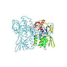
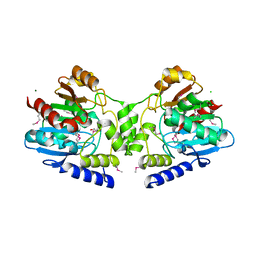
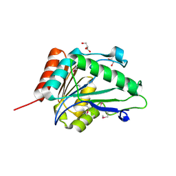
2R11
DownloadVisualize
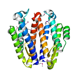
BU of 2r11 by Molmil
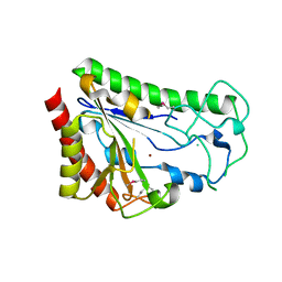
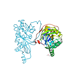
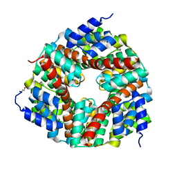
Crystal structure of putative hydrolase (2632844) from Bacillus subtilis at 1.96 A resolution
Descriptor: 1,2-ETHANEDIOL, CHLORIDE ION, Carboxylesterase NP, ...
Authors:Joint Center for Structural Genomics (JCSG)
Deposit date:2007-08-21
Release date:2007-09-04
Last modified:2023-01-25
Method:X-RAY DIFFRACTION (1.96 Å)
Cite:Crystal structure of putative hydrolase (2632844) from Bacillus subtilis at 1.96 A resolution
To be published
2O2X
DownloadVisualize
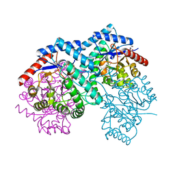
BU of 2o2x by Molmil
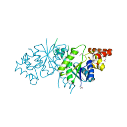
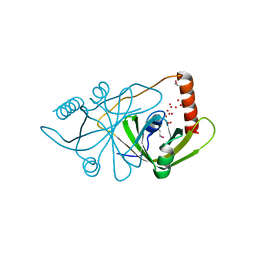
Crystal structure of a putative had-like phosphatase (mll2559) from mesorhizobium loti at 1.50 A resolution
Descriptor: GLYCEROL, hypothetical protein
Authors:Joint Center for Structural Genomics (JCSG)
Deposit date:2006-11-30
Release date:2006-12-19
Last modified:2023-01-25
Method:X-RAY DIFFRACTION (1.5 Å)
Cite:Crystal structure of hypothetical protein (NP_103874.1) from Mesorhizobium loti at 1.50 A resolution
To be published
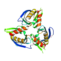
2O4T
DownloadVisualize
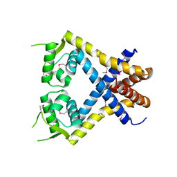
BU of 2o4t by Molmil
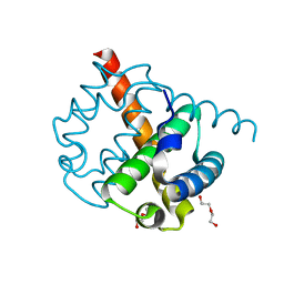
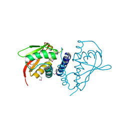
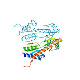
CRYSTAL STRUCTURE OF a protein of the DUF1048 family with a left-handed superhelix fold (BH3976) FROM BACILLUS HALODURANS AT 1.95 A RESOLUTION
Descriptor: BH3976 protein, DI(HYDROXYETHYL)ETHER
Authors:Joint Center for Structural Genomics (JCSG)
Deposit date:2006-12-04
Release date:2006-12-26
Last modified:2023-11-15
Method:X-RAY DIFFRACTION (1.95 Å)
Cite:Crystal structure of BH3976 (10176601) from Bacillus halodurans at 1.95 A resolution
To be published
2PYT
DownloadVisualize
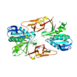
BU of 2pyt by Molmil
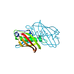
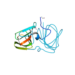
Crystal structure of a putative ethanolamine utilization protein q (eutq, stm2468) from salmonella typhimurium lt2 at 1.90 A resolution
Descriptor: Ethanolamine utilization protein eutQ
Authors:Joint Center for Structural Genomics (JCSG)
Deposit date:2007-05-16
Release date:2007-05-29
Last modified:2023-01-25
Method:X-RAY DIFFRACTION (1.9 Å)
Cite:Crystal structure of Ethanolamine utilization protein eutQ (16421009) from Salmonella typhimurium LT2 at 1.90 A resolution
To be published
2Q7S
DownloadVisualize
BU of 2q7s by Molmil
Crystal structure of N-formylglutamate amidohydrolase (YP_297560.1) from Ralstonia eutropha JMP134 at 2.00 A resolution
Descriptor: N-formylglutamate amidohydrolase, ZINC ION
Authors:Joint Center for Structural Genomics (JCSG)
Deposit date:2007-06-07
Release date:2007-06-19
Last modified:2023-01-25
Method:X-RAY DIFFRACTION (2 Å)
Cite:Crystal structure of N-formylglutamate amidohydrolase (YP_297560.1) from Ralstonia eutropha JMP134 at 2.00 A resolution
To be published
2PG3
DownloadVisualize
BU of 2pg3 by Molmil
Crystal structure of a Queuosine biosynthesis protein queC (ECA1155) from Erwinia carotovora subsp. atroseptica SCRI1043 at 2.40 A resolution
Descriptor: Queuosine biosynthesis protein queC, ZINC ION
Authors:Joint Center for Structural Genomics (JCSG)
Deposit date:2007-04-06
Release date:2007-04-24
Last modified:2023-01-25
Method:X-RAY DIFFRACTION (2.4 Å)
Cite:Crystal structure of hypothetical protein (YP_049261.1) from Erwinia carotovora subsp. atroseptica SCRI1043 at 2.40 A resolution
To be published
2PIM
DownloadVisualize
BU of 2pim by Molmil
CRYSTAL STRUCTURE OF A PUTATIVE THIOESTERASE, PHENYLACETIC ACID DEGRADATION-RELATED PROTEIN (REUT_B4779) FROM RALSTONIA EUTROPHA JMP134 AT 2.20 A RESOLUTION
Descriptor: Phenylacetic acid degradation-related protein
Authors:Joint Center for Structural Genomics (JCSG)
Deposit date:2007-04-13
Release date:2007-05-01
Last modified:2023-01-25
Method:X-RAY DIFFRACTION (2.2 Å)
Cite:Crystal structure of Phenylacetic acid degradation-related protein (YP_298971.1) from Ralstonia eutropha JMP134 at 2.20 A resolution
To be published
2PPV
DownloadVisualize
BU of 2ppv by Molmil
CRYSTAL STRUCTURE OF a protein belonging to the UPF0052 (SE_0549) FROM STAPHYLOCOCCUS EPIDERMIDIS ATCC 12228 AT 2.00 A RESOLUTION
Descriptor: PHOSPHATE ION, Uncharacterized protein
Authors:Joint Center for Structural Genomics (JCSG)
Deposit date:2007-04-30
Release date:2007-05-15
Last modified:2023-01-25
Method:X-RAY DIFFRACTION (2 Å)
Cite:Crystal structure of uncharacterized protein (NP_764104.1) from Staphylococcus epidermidis ATCC 12228 at 2.00 A resolution
To be published
2PN1
DownloadVisualize
BU of 2pn1 by Molmil
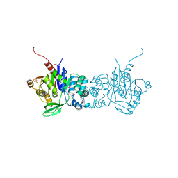
Crystal structure of carbamoylphosphate synthase large subunit (split gene in MJ) (ZP_00538348.1) from Exiguobacterium sp. 255-15 at 2.00 A resolution
Descriptor: Carbamoylphosphate synthase large subunit, GLYCEROL
Authors:Joint Center for Structural Genomics (JCSG)
Deposit date:2007-04-23
Release date:2007-05-08
Last modified:2023-01-25
Method:X-RAY DIFFRACTION (2 Å)
Cite:Crystal structure of carbamoylphosphate synthase large subunit (split gene in MJ) (ZP_00538348.1) from Exiguobacterium sp. 255-15 at 2.00 A resolution
To be published
2PWN
DownloadVisualize
BU of 2pwn by Molmil
Crystal structure of BET3 homolog (13277653) from Mus musculus at 2.04 A resolution
Descriptor: MYRISTIC ACID, Trafficking protein particle complex subunit 3
Authors:Joint Center for Structural Genomics (JCSG)
Deposit date:2007-05-11
Release date:2007-05-29
Last modified:2023-01-25
Method:X-RAY DIFFRACTION (2.04 Å)
Cite:Crystal structure of BET3 homolog (13277653) from Mus musculus at 2.04 A resolution
To be published
2Q0T
DownloadVisualize
BU of 2q0t by Molmil
Crystal structure of a putative gamma-carboxymuconolactone decarboxylase subunit (bxe_b0980) from burkholderia xenovorans lb400 at 1.70 A resolution
Descriptor: Putative gamma-carboxymuconolactone decarboxylase subunit
Authors:Joint Center for Structural Genomics (JCSG)
Deposit date:2007-05-22
Release date:2007-06-12
Last modified:2023-01-25
Method:X-RAY DIFFRACTION (1.7 Å)
Cite:Crystal structure of putative gamma-carboxymuconolactone decarboxylase subunit (YP_554324.1) from Burkholderia xenovorans LB400 at 1.70 A resolution
To be published
2Q9K
DownloadVisualize
BU of 2q9k by Molmil
Crystal structure of a putative oxidoreductase (exig_1997) from exiguobacterium sibiricum 255-15 at 1.59 A resolution
Descriptor: UNKNOWN LIGAND, Uncharacterized protein
Authors:Joint Center for Structural Genomics (JCSG)
Deposit date:2007-06-12
Release date:2007-06-19
Last modified:2023-01-25
Method:X-RAY DIFFRACTION (1.59 Å)
Cite:Crystal structure of conserved uncharacterized protein (ZP_00539648.1) from Exiguobacterium sp. 255-15 at 1.59 A resolution
To be published
2QG3
DownloadVisualize
BU of 2qg3 by Molmil
CRYSTAL STRUCTURE OF A TYW3 METHYLTRANSFERASE-LIKE PROTEIN (AF_2059) FROM ARCHAEOGLOBUS FULGIDUS DSM 4304 AT 1.95 A RESOLUTION
Descriptor: UPF0130 protein AF_2059
Authors:Joint Center for Structural Genomics (JCSG)
Deposit date:2007-06-28
Release date:2007-07-17
Last modified:2023-01-25
Method:X-RAY DIFFRACTION (1.95 Å)
Cite:Crystal structure of uncharacterized protein AF2059 (2648472) from Archaeoglobus fulgidus at 1.95 A resolution
To be published
2OZJ
DownloadVisualize
BU of 2ozj by Molmil
CRYSTAL STRUCTURE OF A CUPIN SUPERFAMILY PROTEIN (DSY2733) FROM DESULFITOBACTERIUM HAFNIENSE DCB-2 AT 1.60 A RESOLUTION
Descriptor: Cupin 2, conserved barrel, GLYCEROL
Authors:Joint Center for Structural Genomics (JCSG)
Deposit date:2007-02-26
Release date:2007-03-20
Last modified:2023-01-25
Method:X-RAY DIFFRACTION (1.6 Å)
Cite:Crystal structure of Cupin 2, conserved barrel (EAT53321.1) from Desulfitobacterium hafniense DCB-2 at 1.60 A resolution
To be published
2P1A
DownloadVisualize
BU of 2p1a by Molmil
Crystal structure of a putative metal-binding protein (bce_2162) from bacillus cereus atcc 10987 at 2.10 A resolution
Descriptor: Hypothetical protein
Authors:Joint Center for Structural Genomics (JCSG)
Deposit date:2007-03-02
Release date:2007-03-20
Last modified:2023-01-25
Method:X-RAY DIFFRACTION (2.1 Å)
Cite:Crystal structure of hypothetical protein (NP_978475.1) from Bacillus cereus ATCC 10987 at 2.10 A resolution
To be published
2QW5
DownloadVisualize
BU of 2qw5 by Molmil
CRYSTAL STRUCTURE OF A PUTATIVE SUGAR PHOSPHATE ISOMERASE/EPIMERASE (AVA4194) FROM ANABAENA VARIABILIS ATCC 29413 AT 1.78 A RESOLUTION
Descriptor: CHLORIDE ION, D-xylose, GLYCEROL, ...
Authors:Joint Center for Structural Genomics (JCSG)
Deposit date:2007-08-09
Release date:2007-09-04
Last modified:2023-01-25
Method:X-RAY DIFFRACTION (1.78 Å)
Cite:Crystal structure of putative sugar phosphate isomerase/epimerase (YP_324688.1) from Anabaena variabilis ATCC 29413 at 1.78 A resolution
To be published
2QWW
DownloadVisualize
BU of 2qww by Molmil
Crystal structure of multiple antibiotic-resistance repressor (MarR) (YP_013417.1) from Listeria monocytogenes 4b F2365 at 2.07 A resolution
Descriptor: Transcriptional regulator, MarR family
Authors:Joint Center for Structural Genomics (JCSG)
Deposit date:2007-08-10
Release date:2007-08-21
Last modified:2023-01-25
Method:X-RAY DIFFRACTION (2.07 Å)
Cite:Crystal structure of multiple antibiotic-resistance repressor (MarR) (YP_013417.1) from Listeria monocytogenes 4b F2365 at 2.07 A resolution
To be published
2QJ8
DownloadVisualize
BU of 2qj8 by Molmil
Crystal structure of an aspartoacylase family protein (mlr6093) from mesorhizobium loti maff303099 at 2.00 A resolution
Descriptor: Mlr6093 protein
Authors:Joint Center for Structural Genomics (JCSG)
Deposit date:2007-07-06
Release date:2007-07-24
Last modified:2023-01-25
Method:X-RAY DIFFRACTION (2 Å)
Cite:Crystal structure of uncharacterized protein mlr6093 (NP_106651.1) from Mesorhizobium loti at 2.00 A resolution
To be published
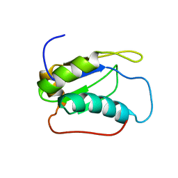
2LQ5
DownloadVisualize
BU of 2lq5 by Molmil
NMR structure of the RNA binding motif 39 (RBM39) from Mus musculus
Descriptor: RNA-binding protein 39
Authors:Serrano, P, Dutta, S.K, Geralt, M, Wuthrich, K, Joint Center for Structural Genomics (JCSG), Partnership for T-Cell Biology (TCELL)
Deposit date:2012-02-23
Release date:2012-03-07
Last modified:2024-05-15
Method:SOLUTION NMR
Cite:NMR structure of the RNA binding motif 39 (RBM39) from Mus musculus
To be Published
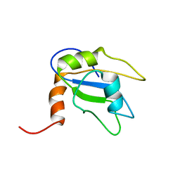
2LMI
DownloadVisualize
BU of 2lmi by Molmil
NMR structure of the protein BC040485 from Homo sapiens
Descriptor: G-rich sequence factor 1
Authors:Dutta, S.K, Serrano, P, Geralt, M, Wuthrich, K, Joint Center for Structural Genomics (JCSG), Partnership for T-Cell Biology (TCELL)
Deposit date:2011-12-01
Release date:2011-12-28
Last modified:2024-05-15
Method:SOLUTION NMR
Cite:NMR structure of the human RNA binding protein BC040485
To be Published
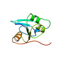
1RDU
DownloadVisualize
BU of 1rdu by Molmil
NMR STRUCTURE OF A PUTATIVE NIFB PROTEIN FROM THERMOTOGA (TM1290), WHICH BELONGS TO THE DUF35 FAMILY
Descriptor: conserved hypothetical protein
Authors:Etezady-Esfarjani, T, Herrmann, T, Peti, W, Klock, H.E, Lesley, S.A, Wuthrich, K, Joint Center for Structural Genomics (JCSG)
Deposit date:2003-11-06
Release date:2004-07-06
Last modified:2024-05-22
Method:SOLUTION NMR
Cite:NMR Structure Determination of the Hypothetical Protein TM1290 from Thermotoga Maritima using Automated NOESY Analysis.
J.Biomol.NMR, 29, 2004
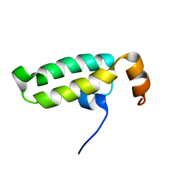
2LO1
DownloadVisualize
BU of 2lo1 by Molmil
NMR structure of the protein BC008182, a DNAJ-like domain from Homo sapiens
Descriptor: DnaJ homolog subfamily A member 1
Authors:Dutta, S.K, Serrano, P, Geralt, M, Wuthrich, K, Joint Center for Structural Genomics (JCSG), Partnership for Stem Cell Biology (STEMCELL)
Deposit date:2012-01-09
Release date:2012-02-15
Last modified:2024-05-15
Method:SOLUTION NMR
Cite:NMR structure of the protein BC008182, DNAJ homolog from Homo sapiens
To be Published
3D1C
DownloadVisualize
BU of 3d1c by Molmil
Crystal structure of Flavin-containing Putative Monooxygenase (NP_373108.1) from STAPHYLOCOCCUS AUREUS MU50 at 2.40 A resolution
Descriptor: FLAVIN-ADENINE DINUCLEOTIDE, Flavin-containing Putative Monooxygenase, SULFATE ION, ...
Authors:Joint Center for Structural Genomics (JCSG)
Deposit date:2008-05-05
Release date:2008-05-20
Last modified:2023-02-01
Method:X-RAY DIFFRACTION (2.4 Å)
Cite:Crystal structure of a Baeyer-Villiger flavin-containing monooxygenase from Staphylococcus aureus MRSA strain MU50.
Proteins, 2014
3CM1
DownloadVisualize
BU of 3cm1 by Molmil
Crystal structure of SsgA-like sporulation-specific cell division protein (YP_290167.1) from Thermobifida fusca YX-ER1 at 2.60 A resolution
Descriptor: SsgA-like sporulation-specific cell division protein
Authors:Joint Center for Structural Genomics (JCSG), Chruszcz, M, Minor, W, Wang, S.
Deposit date:2008-03-20
Release date:2008-04-01
Last modified:2023-02-01
Method:X-RAY DIFFRACTION (2.6 Å)
Cite:Structural and functional characterizations of SsgB, a conserved activator of developmental cell division in morphologically complex actinomycetes.
J.Biol.Chem., 284, 2009
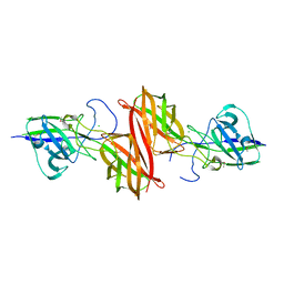
4H40
DownloadVisualize
BU of 4h40 by Molmil
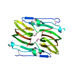
Crystal structure of a putative cell adhesion protein (BF2867) from Bacteroides fragilis NCTC 9343 at 2.57 A resolution
Descriptor: CHLORIDE ION, putative cell adhesion protein
Authors:Joint Center for Structural Genomics (JCSG)
Deposit date:2012-09-14
Release date:2012-10-03
Last modified:2023-02-01
Method:X-RAY DIFFRACTION (2.57 Å)
Cite:A Distinct Type of Pilus from the Human Microbiome.
Cell, 165, 2016

222624

PDB entries from 2024-07-17

PDB statisticsPDBj update infoContact PDBjnumon