Loading
PDBj
MenuPDBj@FacebookPDBj@TwitterPDBj@YouTubewwPDB FoundationwwPDB
RCSB PDBPDBeBMRBAdv. SearchSearch help
Search by PDB author
2DE5
DownloadVisualize
BU of 2de5 by Molmil
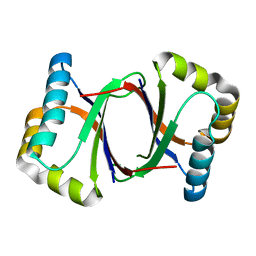
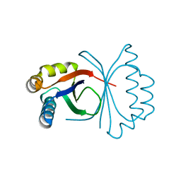
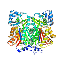
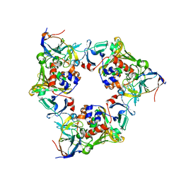
Crystal structure of the electron transfer complex between oxygenase and ferredoxin in carbazole 1,9a-dioxygenase
Descriptor: FE (II) ION, FE2/S2 (INORGANIC) CLUSTER, ferredoxin component of carbazole, ...
Authors:Ashikawa, Y, Nojiri, H.
Deposit date:2006-02-08
Release date:2006-12-05
Last modified:2023-10-25
Method:X-RAY DIFFRACTION (1.9 Å)
Cite:Electron Transfer Complex Formation between Oxygenase and Ferredoxin Components in Rieske Nonheme Iron Oxygenase System
Structure, 14, 2006
2DE6
DownloadVisualize
BU of 2de6 by Molmil
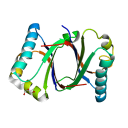
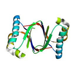
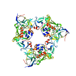
The reduced complex between oxygenase and ferredoxin in carbazole 1,9a-dioxygenase
Descriptor: FE (II) ION, FE2/S2 (INORGANIC) CLUSTER, ferredoxin component of carbazole, ...
Authors:Ashikawa, Y, Nojiri, H.
Deposit date:2006-02-08
Release date:2006-12-05
Last modified:2023-10-25
Method:X-RAY DIFFRACTION (1.8 Å)
Cite:Electron Transfer Complex Formation between Oxygenase and Ferredoxin Components in Rieske Nonheme Iron Oxygenase System
Structure, 14, 2006
5B0D
DownloadVisualize
BU of 5b0d by Molmil
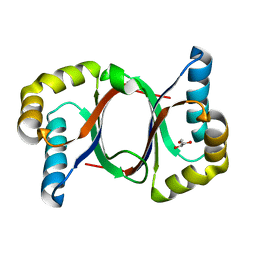
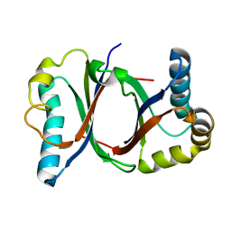
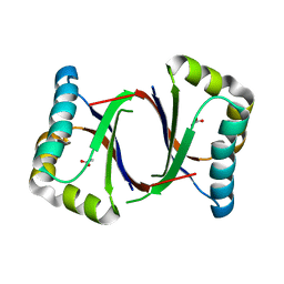
Polyketide cyclase OAC from Cannabis sativa, Y27W mutant
Descriptor: Olivetolic acid cyclase
Authors:Yang, X, Matsui, T, Mori, T, Abe, I, Morita, H.
Deposit date:2015-10-28
Release date:2016-01-27
Last modified:2023-11-08
Method:X-RAY DIFFRACTION (1.801 Å)
Cite:Structural basis for olivetolic acid formation by a polyketide cyclase from Cannabis sativa
Febs J., 283, 2016
5B0E
DownloadVisualize
BU of 5b0e by Molmil
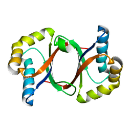
Polyketide cyclase OAC from Cannabis sativa, V59M mutant
Descriptor: GLYCEROL, Olivetolic acid cyclase
Authors:Yang, X, Matsui, T, Mori, T, Abe, I, Morita, H.
Deposit date:2015-10-28
Release date:2016-01-27
Last modified:2023-11-08
Method:X-RAY DIFFRACTION (1.603 Å)
Cite:Structural basis for olivetolic acid formation by a polyketide cyclase from Cannabis sativa
Febs J., 283, 2016
5B0F
DownloadVisualize
BU of 5b0f by Molmil
Polyketide cyclase OAC from Cannabis sativa, Y72F mutant
Descriptor: GLYCEROL, Olivetolic acid cyclase
Authors:Yang, X, Matsui, T, Mori, T, Abe, I, Morita, H.
Deposit date:2015-10-28
Release date:2016-01-27
Last modified:2023-11-08
Method:X-RAY DIFFRACTION (1.6 Å)
Cite:Structural basis for olivetolic acid formation by a polyketide cyclase from Cannabis sativa
Febs J., 283, 2016
5B09
DownloadVisualize
BU of 5b09 by Molmil
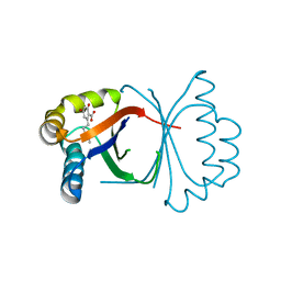
Polyketide cyclase OAC from Cannabis sativa bound with Olivetolic acid
Descriptor: 2,4-bis(oxidanyl)-6-pentyl-benzoic acid, Olivetolic acid cyclase
Authors:Yang, X, Matsui, T, Mori, T, Abe, I, Morita, H.
Deposit date:2015-10-28
Release date:2016-01-27
Last modified:2023-11-08
Method:X-RAY DIFFRACTION (1.7 Å)
Cite:Structural basis for olivetolic acid formation by a polyketide cyclase from Cannabis sativa
Febs J., 283, 2016
5B0G
DownloadVisualize
BU of 5b0g by Molmil
Polyketide cyclase OAC from Cannabis sativa, H78S mutant
Descriptor: Olivetolic acid cyclase
Authors:Yang, X, Matsui, T, Mori, T, Abe, I, Morita, H.
Deposit date:2015-10-28
Release date:2016-01-27
Last modified:2023-11-08
Method:X-RAY DIFFRACTION (1.4 Å)
Cite:Structural basis for olivetolic acid formation by a polyketide cyclase from Cannabis sativa
Febs J., 283, 2016
5B0A
DownloadVisualize
BU of 5b0a by Molmil
Polyketide cyclase OAC from Cannabis sativa, H5Q mutant
Descriptor: Olivetolic acid cyclase
Authors:Yang, X, Matsui, T, Mori, T, Abe, I, Morita, H.
Deposit date:2015-10-28
Release date:2016-01-27
Last modified:2023-11-08
Method:X-RAY DIFFRACTION (2.1 Å)
Cite:Structural basis for olivetolic acid formation by a polyketide cyclase from Cannabis sativa
Febs J., 283, 2016
5B08
DownloadVisualize
BU of 5b08 by Molmil
Polyketide cyclase OAC from Cannabis sativa
Descriptor: Olivetolic acid cyclase
Authors:Yang, X, Matsui, T, Mori, T, Abe, I, Morita, H.
Deposit date:2015-10-28
Release date:2016-01-27
Last modified:2024-03-20
Method:X-RAY DIFFRACTION (1.325 Å)
Cite:Structural basis for olivetolic acid formation by a polyketide cyclase from Cannabis sativa
Febs J., 283, 2016
5B0C
DownloadVisualize
BU of 5b0c by Molmil
Polyketide cyclase OAC from Cannabis sativa, Y27F mutant
Descriptor: Olivetolic acid cyclase
Authors:Yang, X, Matsui, T, Mori, T, Abe, I, Morita, H.
Deposit date:2015-10-28
Release date:2016-01-27
Last modified:2023-11-08
Method:X-RAY DIFFRACTION (1.602 Å)
Cite:Structural basis for olivetolic acid formation by a polyketide cyclase from Cannabis sativa
Febs J., 283, 2016
5B0B
DownloadVisualize
BU of 5b0b by Molmil
Polyketide cyclase OAC from Cannabis sativa, I7F mutant
Descriptor: ACETATE ION, Olivetolic acid cyclase
Authors:Yang, X, Matsui, T, Mori, T, Abe, I, Morita, H.
Deposit date:2015-10-28
Release date:2016-01-27
Last modified:2023-11-08
Method:X-RAY DIFFRACTION (2.187 Å)
Cite:Structural basis for olivetolic acid formation by a polyketide cyclase from Cannabis sativa
Febs J., 283, 2016
2D52
DownloadVisualize
BU of 2d52 by Molmil
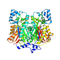
Pentaketide chromone synthase (M207G mutant complexed with Coa)
Descriptor: COENZYME A, pentaketide chromone synthase
Authors:Kohno, T, Morita, H.
Deposit date:2005-10-27
Release date:2006-11-14
Last modified:2024-10-23
Method:X-RAY DIFFRACTION (1.6 Å)
Cite:Structural Insight into Chain-Length Control and Product Specificity of Pentaketide Chromone Synthase from Aloe arborescens
Chem.Biol., 14, 2007
2D51
DownloadVisualize
BU of 2d51 by Molmil
Pentaketide chromone synthase (M207G mutant)
Descriptor: pentaketide chromone synthase
Authors:Kohno, T, Morita, H.
Deposit date:2005-10-27
Release date:2006-11-14
Last modified:2024-10-09
Method:X-RAY DIFFRACTION (1.6 Å)
Cite:Structural Insight into Chain-Length Control and Product Specificity of Pentaketide Chromone Synthase from Aloe arborescens
Chem.Biol., 14, 2007
8PJU
DownloadVisualize
BU of 8pju by Molmil
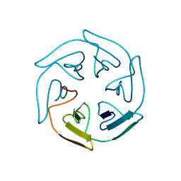
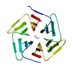
Crystal structure of the computationally designed SAKe6DEtal protein
Descriptor: SAKe6DEtal
Authors:Wouters, S.M.L.
Deposit date:2023-06-23
Release date:2024-07-10
Method:X-RAY DIFFRACTION (1.72 Å)
Cite:Computational design of the SAKe scaffold proteins
To Be Published
8PJQ
DownloadVisualize
BU of 8pjq by Molmil
Crystal structure of the SAKe2BE protein
Descriptor: SAKe2BE
Authors:Wouters, S.M.L.
Deposit date:2023-06-23
Release date:2024-07-10
Method:X-RAY DIFFRACTION (1.15 Å)
Cite:Computational design of the SAKe scaffold proteins
To Be Published
8PKQ
DownloadVisualize
BU of 8pkq by Molmil
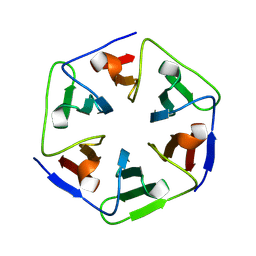
Crystal structure of PknD-34, a repeat fragment from the M. tuberculosis PknD beta-propeller
Descriptor: non-specific serine/threonine protein kinase
Authors:Wouters, S.M.L.
Deposit date:2023-06-27
Release date:2024-07-10
Method:X-RAY DIFFRACTION (1.7 Å)
Cite:Fragment-based assembly of a symmetric protein scaffold
To Be Published
8PJW
DownloadVisualize
BU of 8pjw by Molmil
Crystal structure of the computationally designed SAKe6FE protein
Descriptor: SAKe6FE
Authors:Wouters, S.M.L.
Deposit date:2023-06-23
Release date:2024-07-10
Method:X-RAY DIFFRACTION (1.65 Å)
Cite:Computational design of the SAKe scaffold proteins
To Be Published
8PKS
DownloadVisualize
BU of 8pks by Molmil
Low resolution crystal structure of Keap1-456, a repeat fragment from the human Keap1 beta-propeller
Descriptor: Kelch-like ECH-associated protein 1
Authors:Wouters, S.M.L.
Deposit date:2023-06-27
Release date:2024-07-10
Method:X-RAY DIFFRACTION (2.6 Å)
Cite:Fragment-based assembly of a symmetric protein scaffold
To Be Published
8PJZ
DownloadVisualize
BU of 8pjz by Molmil
Crystal structure of the computationally designed SAKe6EEtal protein
Descriptor: SAKe6EEtal
Authors:Wouters, S.M.L.
Deposit date:2023-06-23
Release date:2024-07-10
Method:X-RAY DIFFRACTION (1.5 Å)
Cite:Computational design of the SAKe scaffold proteins
To Be Published
8PJY
DownloadVisualize
BU of 8pjy by Molmil
Crystal structure of the computationally designed SAKe6EEref protein
Descriptor: SAKe6EEref
Authors:Wouters, S.M.L.
Deposit date:2023-06-23
Release date:2024-07-10
Method:X-RAY DIFFRACTION (1.3 Å)
Cite:Computational design of the SAKe scaffold proteins
To Be Published
8PJV
DownloadVisualize
BU of 8pjv by Molmil
Crystal structure of the computationally designed SAKe6DR protein
Descriptor: SAKe6DR
Authors:Wouters, S.M.L.
Deposit date:2023-06-23
Release date:2024-07-10
Method:X-RAY DIFFRACTION (1.14 Å)
Cite:Computational design of the SAKe scaffold proteins
To Be Published
8PJX
DownloadVisualize
BU of 8pjx by Molmil
Crystal structure of the computationally designed SAKe6FR protein
Descriptor: CALCIUM ION, SAKe6FR
Authors:Wouters, S.M.L.
Deposit date:2023-06-23
Release date:2024-07-10
Method:X-RAY DIFFRACTION (1.7 Å)
Cite:Computational design of the SAKe scaffold proteins
To Be Published
8PJR
DownloadVisualize
BU of 8pjr by Molmil
Crystal structure of the SAKe6AC-LB protein
Descriptor: SAKe6AC-LB
Authors:Wouters, S.M.L.
Deposit date:2023-06-23
Release date:2024-07-10
Method:X-RAY DIFFRACTION (1.71 Å)
Cite:Computational design of the SAKe scaffold protein
To Be Published
8PKR
DownloadVisualize
BU of 8pkr by Molmil
Crystal structure of PknD-345, a repeat fragment from the M. tuberculosis PknD beta-propeller
Descriptor: GLYCEROL, non-specific serine/threonine protein kinase
Authors:Wouters, S.M.L.
Deposit date:2023-06-27
Release date:2024-07-10
Method:X-RAY DIFFRACTION (0.95 Å)
Cite:Computational design of the SAKe scaffold proteins
To Be Published
8PJS
DownloadVisualize
BU of 8pjs by Molmil
Crystal structure of the computationally designed SAKe6CEref protein
Descriptor: SAKeCEref
Authors:Wouters, S.M.L.
Deposit date:2023-06-23
Release date:2024-07-10
Method:X-RAY DIFFRACTION (1.7 Å)
Cite:Computational design of the SAKe scaffold proteins
To Be Published

226707

PDB entries from 2024-10-30

PDB statisticsPDBj update infoContact PDBjnumon