Loading
PDBj
MenuPDBj@FacebookPDBj@TwitterPDBj@YouTubewwPDB FoundationwwPDB
RCSB PDBPDBeBMRBAdv. SearchSearch help
Search by PDB author
5CQX
DownloadVisualize
BU of 5cqx by Molmil
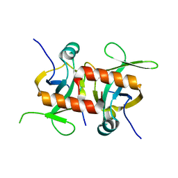
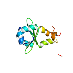
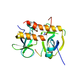
E. coli MazF mutant E24A in complex with MazE residues 68-82 form I
Descriptor: Antitoxin MazE, Endoribonuclease MazF
Authors:Zorzini, V, Loris, R.
Deposit date:2015-07-22
Release date:2016-04-06
Last modified:2024-10-23
Method:X-RAY DIFFRACTION (1.63 Å)
Cite:Substrate Recognition and Activity Regulation of the Escherichia coli mRNA Endonuclease MazF.
J.Biol.Chem., 291, 2016
5CR2
DownloadVisualize
BU of 5cr2 by Molmil
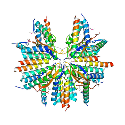
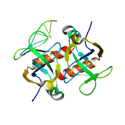
E. coli MazF in complex with single strand DNA substrate analog
Descriptor: Endoribonuclease MazF, ssDNA substrate analog
Authors:Zorzini, V, Loris, R.
Deposit date:2015-07-22
Release date:2016-04-06
Last modified:2024-01-10
Method:X-RAY DIFFRACTION (2.9 Å)
Cite:Substrate Recognition and Activity Regulation of the Escherichia coli mRNA Endonuclease MazF.
J.Biol.Chem., 291, 2016
5CQY
DownloadVisualize
BU of 5cqy by Molmil
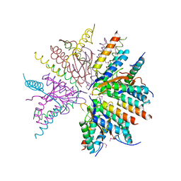
E. coli MazF mutant E24A in complex with MazE residues 68-82 form II
Descriptor: Antitoxin MazE, Endoribonuclease MazF
Authors:Zorzini, V, Loris, R.
Deposit date:2015-07-22
Release date:2016-04-06
Last modified:2024-01-10
Method:X-RAY DIFFRACTION (2.481 Å)
Cite:Substrate Recognition and Activity Regulation of the Escherichia coli mRNA Endonuclease MazF.
J.Biol.Chem., 291, 2016
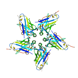
5CW7
DownloadVisualize
BU of 5cw7 by Molmil
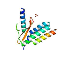
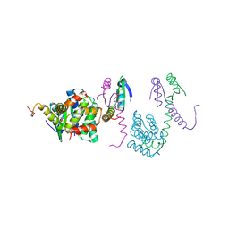
Crystal structure of the PaaA2-ParE2 antitoxin-toxin complex
Descriptor: GLYCEROL, PAAA2, Plasmid stabilization protein ParE
Authors:Sterckx, Y.G.-J, Loris, R.
Deposit date:2015-07-27
Release date:2016-06-01
Method:X-RAY DIFFRACTION (2.827 Å)
Cite:A unique hetero-hexadecameric architecture displayed by the Escherichia coli O157 PaaA2-ParE2 antitoxin-toxin complex.
J.Mol.Biol., 428, 2016
5CZE
DownloadVisualize
BU of 5cze by Molmil
Crystal structure of the PaaA2-ParE2 antitoxin-toxin complex
Descriptor: PaaA2, Plasmid stabilization protein ParE
Authors:Loris, R, Sterckx, Y.G.J.
Deposit date:2015-07-31
Release date:2016-08-24
Last modified:2024-01-10
Method:X-RAY DIFFRACTION (3.82 Å)
Cite:A unique hetero-hexadecameric architecture displayed by the Escherichia coli O157 PaaA2-ParE2 antitoxin-toxin complex.
J.Mol.Biol., 428, 2016
5CZF
DownloadVisualize
BU of 5czf by Molmil
Crystal structure of the PaaA2-ParE2 antitoxin-toxin complex
Descriptor: PaaA2, Plasmid stabilization protein ParE, SULFATE ION
Authors:Loris, R, Sterckx, Y.G.J.
Deposit date:2015-07-31
Release date:2016-06-01
Last modified:2024-01-10
Method:X-RAY DIFFRACTION (2.671 Å)
Cite:A unique hetero-hexadecameric architecture displayed by the Escherichia coli O157 PaaA2-ParE2 antitoxin-toxin complex.
J.Mol.Biol., 428, 2016
6FE4
DownloadVisualize
BU of 6fe4 by Molmil
Crystal structure of the complex between Shiga toxin Stx2 B subunit and neutralising Nb113
Descriptor: Nb113, Shiga-like toxin 2 subunit B
Authors:Bernedo, R, Muyldermans, S, Sterckx, Y.G.J.
Deposit date:2017-12-29
Release date:2018-03-07
Last modified:2024-01-17
Method:X-RAY DIFFRACTION (3 Å)
Cite:Structural Basis for the Specific Neutralization of Stx2a with a Camelid Single Domain Antibody Fragment.
Toxins (Basel), 10, 2018
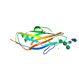
8BXY
DownloadVisualize
BU of 8bxy by Molmil
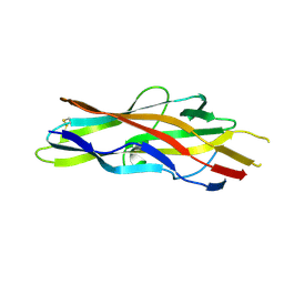
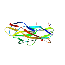
FimH in complex with alpha1,6 core-fucosylated oligomannose-3, crystallized in the trigonal space group
Descriptor: NICKEL (II) ION, SULFATE ION, Type 1 fimbrin D-mannose specific adhesin, ...
Authors:Bridot, C, Bouckaert, J, Krammer, E.-M.
Deposit date:2022-12-11
Release date:2023-04-12
Last modified:2024-10-23
Method:X-RAY DIFFRACTION (1.45 Å)
Cite:Structural insights into a cooperative switch between one and two FimH bacterial adhesins binding pauci- and high-mannose type N-glycan receptors.
J.Biol.Chem., 299, 2023
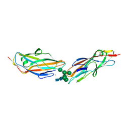
8BY3
DownloadVisualize
BU of 8by3 by Molmil
FimH lectin domain in complex with oligomannose-6
Descriptor: NICKEL (II) ION, SULFATE ION, Type 1 fimbrin D-mannose specific adhesin, ...
Authors:Bouckaert, J, Bourenkov, G.P.
Deposit date:2022-12-11
Release date:2023-04-12
Last modified:2024-10-23
Method:X-RAY DIFFRACTION (3.186 Å)
Cite:Structural insights into a cooperative switch between one and two FimH bacterial adhesins binding pauci- and high-mannose type N-glycan receptors.
J.Biol.Chem., 299, 2023
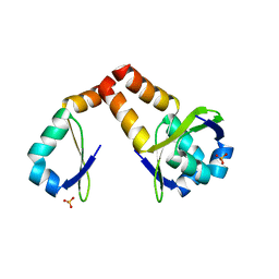
3HS2
DownloadVisualize
BU of 3hs2 by Molmil
Crystal structure of PHD truncated to residue 57 in an orthorhombic space group
Descriptor: Prevent host death protein, SULFATE ION
Authors:Garcia-Pino, A, Loris, R.
Deposit date:2009-06-10
Release date:2010-06-23
Last modified:2023-09-06
Method:X-RAY DIFFRACTION (2.2 Å)
Cite:Allostery and intrinsic disorder mediate transcription regulation by conditional cooperativity.
Cell(Cambridge,Mass.), 142, 2010
3HRY
DownloadVisualize
BU of 3hry by Molmil
Crystal structure of PHD in a trigonal space group and partially disordered
Descriptor: Prevent host death protein
Authors:Garcia-Pino, A, Loris, R.
Deposit date:2009-06-10
Release date:2010-06-23
Last modified:2023-09-06
Method:X-RAY DIFFRACTION (2.25 Å)
Cite:Allostery and intrinsic disorder mediate transcription regulation by conditional cooperativity.
Cell(Cambridge,Mass.), 142, 2010
3K33
DownloadVisualize
BU of 3k33 by Molmil
Crystal structure of the Phd-Doc complex
Descriptor: ACETATE ION, Death on curing protein, Polypeptide of unknown amino acids and source, ...
Authors:Loris, R, Garcia-Pino, A.
Deposit date:2009-10-01
Release date:2010-08-18
Last modified:2023-09-06
Method:X-RAY DIFFRACTION (2.4 Å)
Cite:Allostery and intrinsic disorder mediate transcription regulation by conditional cooperativity.
Cell(Cambridge,Mass.), 142, 2010
3F64
DownloadVisualize
BU of 3f64 by Molmil
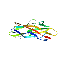
F17a-G lectin domain with bound GlcNAc(beta1-O)paranitrophenyl ligand
Descriptor: 4-nitrophenyl 2-acetamido-2-deoxy-beta-D-glucopyranoside, F17a-G
Authors:Buts, L, De Boer, A, Olsson, J.D.M, Jonckheere, W, De Kerpel, M, De Genst, E, Guerardel, Y, Willaert, R, Wyns, L, Wuhrer, M, Oscarson, S, De Greve, H, Bouckaert, J.
Deposit date:2008-11-05
Release date:2009-11-10
Last modified:2023-11-01
Method:X-RAY DIFFRACTION (1.95 Å)
Cite:Structural Sampling of Glycan Interaction Profiles Reveals Mucosal Receptors for Fimbrial Adhesins of Enterotoxigenic Escherichia coli.
Biology (Basel), 2, 2013
3F6J
DownloadVisualize
BU of 3f6j by Molmil
F17a-G lectin domain with bound GlcNAc(beta1-3)Gal
Descriptor: 2-acetamido-2-deoxy-beta-D-glucopyranose-(1-3)-methyl beta-D-galactopyranoside, F17a-G
Authors:Buts, L, de Boer, A, Olsson, J.D.M, Jonckheere, W, De Kerpel, M, De Genst, E, Guerardel, Y, Willaert, R, Wyns, L, Wuhrer, M, Oscarson, S, De Greve, H, Bouckaert, J.
Deposit date:2008-11-06
Release date:2009-11-17
Last modified:2023-11-01
Method:X-RAY DIFFRACTION (1.75 Å)
Cite:Structural Sampling of Glycan Interaction Profiles Reveals Mucosal Receptors for Fimbrial Adhesins of Enterotoxigenic Escherichia coli
Biology, 2, 2013
3FFO
DownloadVisualize
BU of 3ffo by Molmil
F17b-G lectin domain with bound GlcNAc(beta1-2)man
Descriptor: 2-AMINO-2-HYDROXYMETHYL-PROPANE-1,3-DIOL, 2-acetamido-2-deoxy-beta-D-glucopyranose-(1-2)-beta-D-mannopyranose, Adhesin, ...
Authors:Buts, L, De Boer, A, Olsson, J.D.M, Jonckheere, W, De Kerpel, M, De Genst, E, Guerardel, Y, Willaert, R, Wyns, L, Wuhrer, M, Oscarson, S, De Greve, H, Bouckaert, J.
Deposit date:2008-12-04
Release date:2009-12-22
Last modified:2023-11-01
Method:X-RAY DIFFRACTION (2.1 Å)
Cite:Structural Sampling of Glycan Interaction Profiles Reveals Mucosal Receptors for Fimbrial Adhesins of Enterotoxigenic Escherichia coli.
Biology (Basel), 2, 2013
<1234


226707

PDB entries from 2024-10-30

PDB statisticsPDBj update infoContact PDBjnumon