Loading
PDBj
MenuPDBj@FacebookPDBj@TwitterPDBj@YouTubewwPDB FoundationwwPDB
RCSB PDBPDBeBMRBAdv. SearchSearch help
Search by PDB author
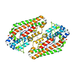
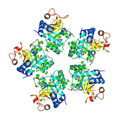
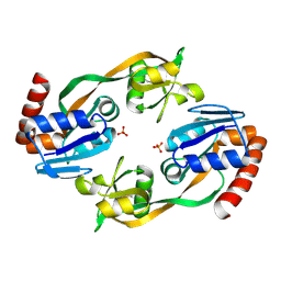
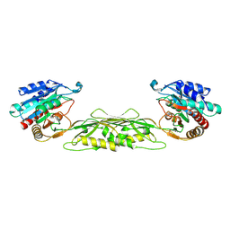
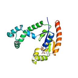
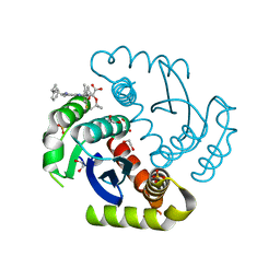
6NCJ
DownloadVisualize
BU of 6ncj by Molmil
Structure of HIV-1 Integrase with potent 5,6,7,8-Tetrahydro-1,6-naphthyridine Derivatives Allosteric Site Inhibitors
Descriptor: (2~{S})-2-[4-(8-fluoranyl-5-methyl-3,4-dihydro-2~{H}-chromen-6-yl)-2-methyl-6-[[(1~{S},2~{R})-2-phenylcyclopropyl]methyl]-7,8-dihydro-5~{H}-1,6-naphthyridin-3-yl]-2-[(2-methylpropan-2-yl)oxy]ethanoic acid, 1,2-ETHANEDIOL, Integrase, ...
Authors:Nolte, R.T.
Deposit date:2018-12-11
Release date:2019-01-16
Last modified:2024-03-13
Method:X-RAY DIFFRACTION (2 Å)
Cite:5,6,7,8-Tetrahydro-1,6-naphthyridine Derivatives as Potent HIV-1-Integrase-Allosteric-Site Inhibitors.
J. Med. Chem., 62, 2019
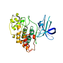
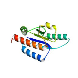
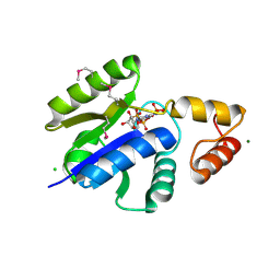
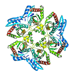
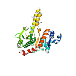
8FUR
DownloadVisualize
BU of 8fur by Molmil
Crystal structure of human IDO1 with compound 11
Descriptor: Indoleamine 2,3-dioxygenase 1, N-(4-methylphenyl)-N'-[(1P,2'P)-4-propoxy-5-propyl-2'-(1H-tetrazol-5-yl)[1,1'-biphenyl]-3-yl]urea
Authors:Critton, D.A, Lewis, H.A.
Deposit date:2023-01-18
Release date:2023-04-26
Last modified:2023-05-03
Method:X-RAY DIFFRACTION (2.285 Å)
Cite:Synthesis and biological evaluation of biaryl alkyl ethers as inhibitors of IDO1.
Bioorg.Med.Chem.Lett., 88, 2023
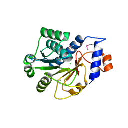
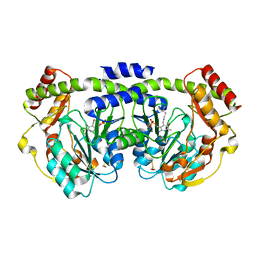
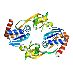
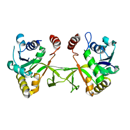
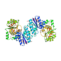
8FF8
DownloadVisualize
BU of 8ff8 by Molmil
CRYSTAL STRUCTURE OF GLYCOGEN SYNTHASE KINASE 3 BETA COMPLEXED WITH 2-[(4-CYANOPHENYL)AMINO]-N-(4-PHENYLPYRIDIN-3-YL)PYRIMIDINE-4-CARBOXAMIDE
Descriptor: 2-(4-cyanoanilino)-N-(4-phenylpyridin-3-yl)pyrimidine-4-carboxamide, Glycogen synthase kinase-3 beta
Authors:Muckelbauer, J.K, Lewis, H.A.
Deposit date:2022-12-08
Release date:2023-06-07
Last modified:2024-05-22
Method:X-RAY DIFFRACTION (2.33 Å)
Cite:Discovery of 2-(Anilino)pyrimidine-4-carboxamides as Highly Potent, Selective, and Orally Active Glycogen Synthase Kinase-3 (GSK-3) Inhibitors.
J.Med.Chem., 66, 2023
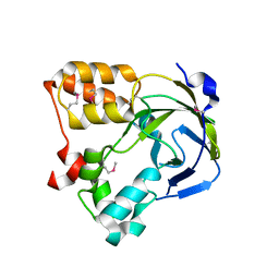
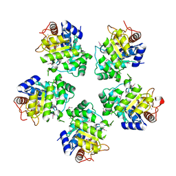
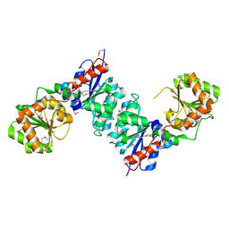
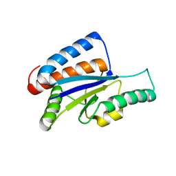
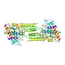
1JZT
DownloadVisualize
BU of 1jzt by Molmil
Crystal structure of yeast ynu0, YNL200c
Descriptor: CHLORIDE ION, Hypothetical 27.5 kDa protein in SPX19-GCR2 intergenic region
Authors:Jiang, J.-S, Manning, N.O, Burley, S.K, New York SGX Research Center for Structural Genomics (NYSGXRC)
Deposit date:2001-09-17
Release date:2001-09-26
Last modified:2021-02-03
Method:X-RAY DIFFRACTION (1.94 Å)
Cite:Crystal Structure of Yeast Hypothetical Protein YNU0_YEAST
To be Published
1O67
DownloadVisualize
BU of 1o67 by Molmil
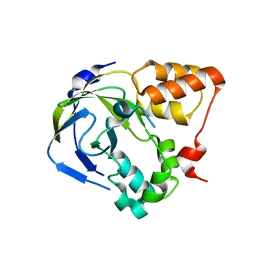
Crystal structure of an hypothetical protein
Descriptor: Hypothetical protein yiiM
Authors:Structural GenomiX
Deposit date:2003-10-23
Release date:2003-11-11
Last modified:2023-12-27
Method:X-RAY DIFFRACTION (2.54 Å)
Cite:Structural analysis of a set of proteins resulting from a bacterial genomics project
Proteins, 60, 2005
1O60
DownloadVisualize
BU of 1o60 by Molmil
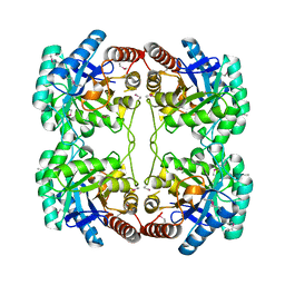
Crystal structure of KDO-8-phosphate synthase
Descriptor: 2-dehydro-3-deoxyphosphooctonate aldolase
Authors:Structural GenomiX
Deposit date:2003-10-23
Release date:2003-11-11
Last modified:2023-12-27
Method:X-RAY DIFFRACTION (1.8 Å)
Cite:Structural analysis of a set of proteins resulting from a bacterial genomics project
Proteins, 60, 2005
1O65
DownloadVisualize
BU of 1o65 by Molmil
Crystal structure of an hypothetical protein
Descriptor: Hypothetical protein yiiM
Authors:Structural GenomiX
Deposit date:2003-10-23
Release date:2003-11-11
Last modified:2023-12-27
Method:X-RAY DIFFRACTION (2.33 Å)
Cite:Structural analysis of a set of proteins resulting from a bacterial genomics project
Proteins, 60, 2005
1O62
DownloadVisualize
BU of 1o62 by Molmil
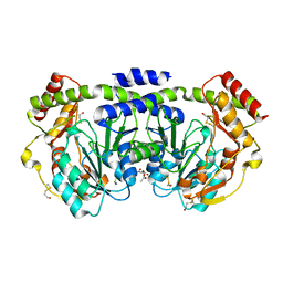
Crystal structure of the apo form of a PLP-dependent enzyme
Descriptor: ACETATE ION, BETA-MERCAPTOETHANOL, aminotransferase
Authors:Structural GenomiX
Deposit date:2003-10-23
Release date:2003-11-11
Last modified:2023-12-27
Method:X-RAY DIFFRACTION (2.1 Å)
Cite:Structural analysis of a set of proteins resulting from a bacterial genomics project.
Proteins, 60, 2005
1O61
DownloadVisualize
BU of 1o61 by Molmil
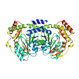
Crystal structure of a PLP-dependent enzyme with PLP
Descriptor: ACETATE ION, PYRIDOXAL-5'-PHOSPHATE, aminotransferase
Authors:Structural GenomiX
Deposit date:2003-10-23
Release date:2003-11-11
Last modified:2023-12-27
Method:X-RAY DIFFRACTION (1.9 Å)
Cite:Structural analysis of a set of proteins resulting from a bacterial genomics project
Proteins, 60, 2005
1O68
DownloadVisualize
BU of 1o68 by Molmil
Crystal structure of 3-methyl-2-oxobutanoate hydroxymethyltransferase
Descriptor: 3-METHYL-2-OXOBUTANOIC ACID, 3-methyl-2-oxobutanoate hydroxymethyltransferase, SODIUM ION
Authors:Structural GenomiX
Deposit date:2003-10-23
Release date:2003-11-11
Last modified:2024-10-30
Method:X-RAY DIFFRACTION (2.1 Å)
Cite:Structural analysis of a set of proteins resulting from a bacterial genomics project
Proteins, 60, 2005
1O6D
DownloadVisualize
BU of 1o6d by Molmil
Crystal structure of a hypothetical protein
Descriptor: Hypothetical UPF0247 protein TM0844
Authors:Structural GenomiX
Deposit date:2003-11-03
Release date:2003-11-25
Last modified:2023-12-27
Method:X-RAY DIFFRACTION (1.66 Å)
Cite:Structural analysis of a set of proteins resulting from a bacterial genomics project
Proteins, 60, 2005
1O69
DownloadVisualize
BU of 1o69 by Molmil
Crystal structure of a PLP-dependent enzyme
Descriptor: (2-AMINO-4-FORMYL-5-HYDROXY-6-METHYLPYRIDIN-3-YL)METHYL DIHYDROGEN PHOSPHATE, BETA-MERCAPTOETHANOL, aminotransferase
Authors:Structural GenomiX
Deposit date:2003-10-23
Release date:2003-11-11
Last modified:2023-12-27
Method:X-RAY DIFFRACTION (1.84 Å)
Cite:Structural analysis of a set of proteins resulting from a bacterial genomics project
Proteins, 60, 2005
1O66
DownloadVisualize
BU of 1o66 by Molmil
Crystal structure of 3-methyl-2-oxobutanoate hydroxymethyltransferase
Descriptor: 3-methyl-2-oxobutanoate hydroxymethyltransferase, GLYCEROL
Authors:Structural GenomiX
Deposit date:2003-10-23
Release date:2003-11-11
Last modified:2024-10-30
Method:X-RAY DIFFRACTION (1.75 Å)
Cite:Structural analysis of a set of proteins resulting from a bacterial genomics project
Proteins, 60, 2005
1O64
DownloadVisualize
BU of 1o64 by Molmil
Crystal structure of an ATP phosphoribosyltransferase
Descriptor: ATP phosphoribosyltransferase, PHOSPHATE ION
Authors:Structural GenomiX
Deposit date:2003-10-23
Release date:2003-11-11
Last modified:2024-10-30
Method:X-RAY DIFFRACTION (2.1 Å)
Cite:Structural analysis of a set of proteins resulting from a bacterial genomics project
Proteins, 60, 2005
1O6B
DownloadVisualize
BU of 1o6b by Molmil
Crystal structure of phosphopantetheine adenylyltransferase with ADP
Descriptor: ADENOSINE-5'-DIPHOSPHATE, CHLORIDE ION, MAGNESIUM ION, ...
Authors:Structural GenomiX
Deposit date:2003-11-03
Release date:2003-11-25
Last modified:2023-12-27
Method:X-RAY DIFFRACTION (2.2 Å)
Cite:Structural analysis of a set of proteins resulting from a bacterial genomics project
Proteins, 60, 2005
1O63
DownloadVisualize
BU of 1o63 by Molmil
Crystal structure of an ATP phosphoribosyltransferase
Descriptor: ATP phosphoribosyltransferase
Authors:Structural GenomiX
Deposit date:2003-10-23
Release date:2003-11-11
Last modified:2023-12-27
Method:X-RAY DIFFRACTION (2 Å)
Cite:Structural analysis of a set of proteins resulting from a bacterial genomics project
Proteins, 60, 2005
1O6C
DownloadVisualize
BU of 1o6c by Molmil
Crystal structure of UDP-N-acetylglucosamine 2-epimerase
Descriptor: UDP-N-acetylglucosamine 2-epimerase
Authors:Structural GenomiX
Deposit date:2003-11-03
Release date:2003-11-25
Last modified:2024-10-30
Method:X-RAY DIFFRACTION (2.9 Å)
Cite:Structural analysis of a set of proteins resulting from a bacterial genomics project
Proteins, 60, 2005
1VGY
DownloadVisualize
BU of 1vgy by Molmil
Crystal structure of succinyl diaminopimelate desuccinylase
Descriptor: succinyl-diaminopimelate desuccinylase
Authors:Structural GenomiX
Deposit date:2003-11-03
Release date:2003-11-25
Last modified:2023-12-27
Method:X-RAY DIFFRACTION (1.9 Å)
Cite:Structural analysis of a set of proteins resulting from a bacterial genomics project
Proteins, 60, 2005
1VHJ
DownloadVisualize
BU of 1vhj by Molmil
Crystal structure of purine nucleoside phosphorylase
Descriptor: purine nucleoside phosphorylase
Authors:Structural GenomiX
Deposit date:2003-12-01
Release date:2003-12-30
Last modified:2023-12-27
Method:X-RAY DIFFRACTION (2.23 Å)
Cite:Structural analysis of a set of proteins resulting from a bacterial genomics project
Proteins, 60, 2005
1VGU
DownloadVisualize
BU of 1vgu by Molmil
Crystal structure of 4-diphosphocytidyl-2C-methyl-D-erythritol synthase
Descriptor: 2-C-methyl-D-erythritol 4-phosphate cytidylyltransferase
Authors:Structural GenomiX
Deposit date:2003-11-03
Release date:2003-11-25
Last modified:2023-12-27
Method:X-RAY DIFFRACTION (2.8 Å)
Cite:Structural analysis of a set of proteins resulting from a bacterial genomics project
Proteins, 60, 2005
1VH0
DownloadVisualize
BU of 1vh0 by Molmil
Crystal structure of a hypothetical protein
Descriptor: Hypothetical UPF0247 protein SAV0024/SA0023
Authors:Structural GenomiX
Deposit date:2003-11-03
Release date:2003-11-25
Last modified:2023-12-27
Method:X-RAY DIFFRACTION (2.31 Å)
Cite:Structural analysis of a set of proteins resulting from a bacterial genomics project
Proteins, 60, 2005
1VHL
DownloadVisualize
BU of 1vhl by Molmil
Crystal structure of dephospho-CoA kinase with adenosine-5'-diphosphate
Descriptor: ACETATE ION, ADENOSINE-5'-DIPHOSPHATE, Dephospho-CoA kinase
Authors:Structural GenomiX
Deposit date:2003-12-01
Release date:2003-12-30
Last modified:2023-12-27
Method:X-RAY DIFFRACTION (1.65 Å)
Cite:Structural analysis of a set of proteins resulting from a bacterial genomics project
Proteins, 60, 2005
1VI1
DownloadVisualize
BU of 1vi1 by Molmil
Crystal structure of a fatty acid/phospholipid synthesis protein
Descriptor: Fatty acid/phospholipid synthesis protein plsX
Authors:Structural GenomiX
Deposit date:2003-12-01
Release date:2003-12-30
Last modified:2024-10-30
Method:X-RAY DIFFRACTION (2.95 Å)
Cite:Structural analysis of a set of proteins resulting from a bacterial genomics project
Proteins, 60, 2005
1VGV
DownloadVisualize
BU of 1vgv by Molmil
Crystal structure of UDP-N-acetylglucosamine_2 epimerase
Descriptor: UDP-N-acetylglucosamine 2-epimerase, URIDINE-DIPHOSPHATE-N-ACETYLGLUCOSAMINE
Authors:Structural GenomiX
Deposit date:2003-11-03
Release date:2003-11-25
Last modified:2023-12-27
Method:X-RAY DIFFRACTION (2.31 Å)
Cite:Structural analysis of a set of proteins resulting from a bacterial genomics project
Proteins, 60, 2005
1VIX
DownloadVisualize
BU of 1vix by Molmil
Crystal structure of a putative peptidase T
Descriptor: Peptidase T, SULFATE ION, ZINC ION
Authors:Structural GenomiX
Deposit date:2003-12-01
Release date:2003-12-30
Last modified:2023-12-27
Method:X-RAY DIFFRACTION (2.5 Å)
Cite:Structural analysis of a set of proteins resulting from a bacterial genomics project
Proteins, 60, 2005

226707

PDB entries from 2024-10-30

PDB statisticsPDBj update infoContact PDBjnumon