3GS9
 
 | |
3GXG
 | |
2QML
 | |
2QPX
 | |
3H3I
 | |
3OWR
 | |
3HBK
 | |
2R3E
 | |
3GYA
 | |
2R11
 | |
3P1V
 | |
3P1T
 | |
3PFE
 | |
2R7H
 | |
2RAA
 | |
3PFO
 | |
2RCD
 | |
2RIJ
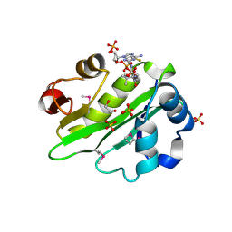
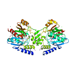
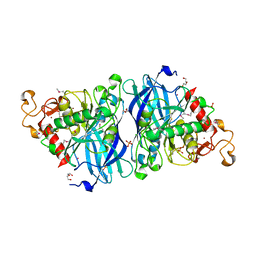
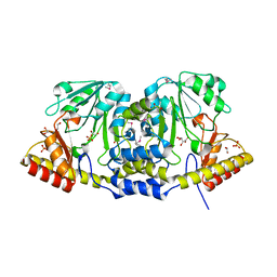
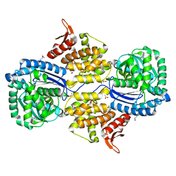
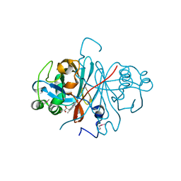
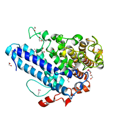
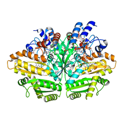
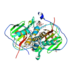
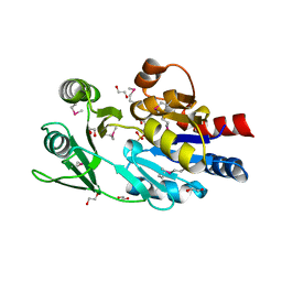
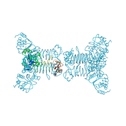 | | Crystal structure of a putative 2,3,4,5-tetrahydropyridine-2-carboxylate n-succinyltransferase (cj1605c, dapd) from campylobacter jejuni at 1.90 A resolution | | Descriptor: | CHLORIDE ION, CITRIC ACID, GLYCEROL, ... | | Authors: | Joint Center for Structural Genomics (JCSG) | | Deposit date: | 2007-10-11 | | Release date: | 2007-10-23 | | Last modified: | 2024-10-30 | | Method: | X-RAY DIFFRACTION (1.9 Å) | | Cite: | Crystal structure of Putative 2,3,4,5-tetrahydropyridine-2-carboxylate N-succinyltransferase (NP_282733.1) from Campylobacter jejuni at 1.90 A resolution
To be published
|
|
3HI0
 | |
2R6V
 | |
3HDX
 | |
3PM9
 | |
3PNX
 | |
3HJ9
 | |
3PGV
 | |


