Loading
PDBj
MenuPDBj@FacebookPDBj@X(formerly Twitter)PDBj@BlueSkyPDBj@YouTubewwPDB FoundationwwPDB
RCSB PDBPDBeBMRBAdv. SearchSearch help
Search by PDB author
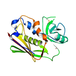
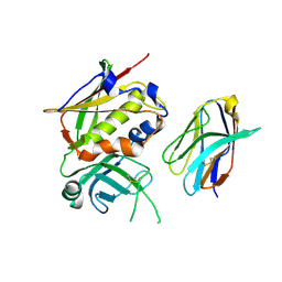
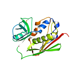
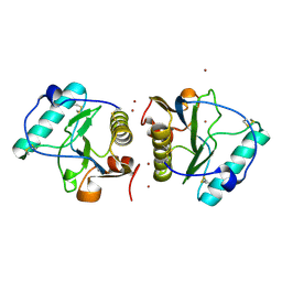
2EAV
DownloadVisualize
BU of 2eav by Molmil
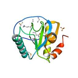
Crystal structure of the C-terminal peptidoglycan-binding domain of human peptidoglycan recognition protein Ibeta
Descriptor: NICKEL (II) ION, Peptidoglycan recognition protein-I-beta
Authors:Cho, S, Mariuzza, R.A.
Deposit date:2007-02-03
Release date:2007-09-18
Last modified:2011-07-13
Method:X-RAY DIFFRACTION (2.2 Å)
Cite:Structural insights into the bactericidal mechanism of human peptidoglycan recognition proteins
Proc.Natl.Acad.Sci.Usa, 104, 2007
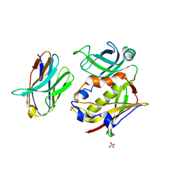
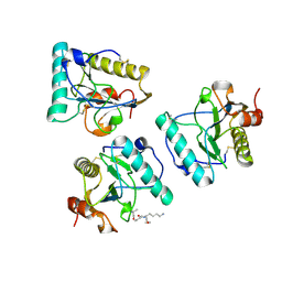
2EAX
DownloadVisualize
BU of 2eax by Molmil
Crystal structure of human PGRP-IBETAC in complex with glycosamyl muramyl pentapeptide
Descriptor: 2-acetamido-2-deoxy-beta-D-glucopyranose-(1-4)-methyl 2-acetamido-3-O-[(1R)-1-carboxyethyl]-2-deoxy-beta-D-glucopyranoside, GLYCOSAMYL MURAMYL PENTAPEPTIDE, Peptidoglycan recognition protein-I-beta
Authors:Cho, S.
Deposit date:2007-02-03
Release date:2007-10-02
Last modified:2024-11-13
Method:X-RAY DIFFRACTION (2.1 Å)
Cite:Structural insights into the bactericidal mechanism of human peptidoglycan recognition proteins
Proc.Natl.Acad.Sci.Usa, 104, 2007
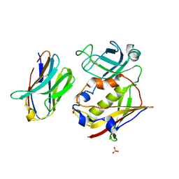
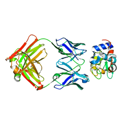
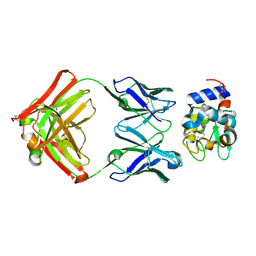
2G9H
DownloadVisualize
BU of 2g9h by Molmil
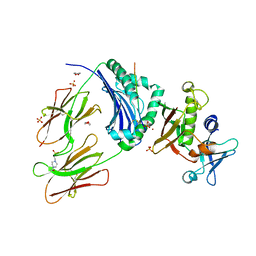
Crystal Structure of Staphylococcal Enterotoxin I (SEI) in Complex with a Human MHC class II Molecule
Descriptor: 1,4-DIETHYLENE DIOXIDE, 4-(2-HYDROXYETHYL)-1-PIPERAZINE ETHANESULFONIC ACID, HLA class II histocompatibility antigen, ...
Authors:Fernandez, M.M, Guan, R, Malchiodi, E.L, Mariuzza, R.A.
Deposit date:2006-03-06
Release date:2006-07-11
Last modified:2023-08-30
Method:X-RAY DIFFRACTION (2 Å)
Cite:Crystal structure of staphylococcal enterotoxin I (SEI) in complex with a human major histocompatibility complex class II molecule.
J.Biol.Chem., 281, 2006
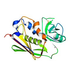
1R1Q
DownloadVisualize
BU of 1r1q by Molmil
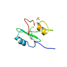
Structural Basis for Differential Recognition of Tyrosine Phosphorylated Sites in the Linker for Activation of T cells (LAT) by the Adaptor Protein Gads
Descriptor: GRB2-related adaptor protein 2, LAT pY191 peptide, SULFATE ION
Authors:Cho, S, Mariuzza, R.A.
Deposit date:2003-09-24
Release date:2004-09-28
Last modified:2024-10-30
Method:X-RAY DIFFRACTION (1.8 Å)
Cite:Structural basis for differential recognition of tyrosine-phosphorylated sites in the linker for activation of T cells (LAT) by the adaptor Gads.
Embo J., 23, 2004
1R1S
DownloadVisualize
BU of 1r1s by Molmil
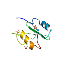
Structural Basis for Differential Recognition of Tyrosine Phosphorylated Sites in the Linker for Activation of T cells (LAT) by the Adaptor Protein Gads
Descriptor: GRB2-related adaptor protein 2, LAT pY226 peptide, SULFATE ION
Authors:Cho, S, Mariuzza, R.A.
Deposit date:2003-09-24
Release date:2004-09-28
Last modified:2024-10-09
Method:X-RAY DIFFRACTION (1.9 Å)
Cite:Structural basis for differential recognition of tyrosine-phosphorylated sites in the linker for activation of T cells (LAT) by the adaptor Gads.
Embo J., 23, 2004
1R1P
DownloadVisualize
BU of 1r1p by Molmil
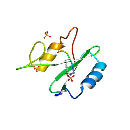
Structural Basis for Differential Recognition of Tyrosine Phosphorylated Sites in the Linker for Activation of T cells (LAT) by the Adaptor Protein Gads
Descriptor: GRB2-related adaptor protein 2, LAT pY171 peptide, SULFATE ION
Authors:Cho, S, Mariuzza, R.A.
Deposit date:2003-09-24
Release date:2004-09-28
Last modified:2024-10-09
Method:X-RAY DIFFRACTION (1.8 Å)
Cite:Structural basis for differential recognition of tyrosine-phosphorylated sites in the linker for activation of T cells (LAT) by the adaptor Gads.
Embo J., 23, 2004
3BVM
DownloadVisualize
BU of 3bvm by Molmil
Manipulating the coupled folding and binding process drives affinity maturation in a protein-protein complex
Descriptor: Enterotoxin type C-3, ZINC ION
Authors:Cho, S.
Deposit date:2008-01-07
Release date:2009-05-12
Last modified:2024-10-30
Method:X-RAY DIFFRACTION (2 Å)
Cite:Manipulating the coupled folding and binding process drives affinity maturation in a protein-protein complex
To be Published
3BYY
DownloadVisualize
BU of 3byy by Molmil
Manipulating the coupled folding and binding process drives affinity maturation in a protein-protein complex
Descriptor: Enterotoxin type C-3, SULFATE ION, T cell receptor beta chain 8.2
Authors:Cho, S, Eric, J.S.
Deposit date:2008-01-16
Release date:2009-05-12
Last modified:2024-10-30
Method:X-RAY DIFFRACTION (2.2 Å)
Cite:Manipulating the coupled folding and binding process drives affinity maturation in a protein-protein complex
To be Published
3BZD
DownloadVisualize
BU of 3bzd by Molmil
Manipulating the coupled folding and binding process drives affinity maturation in a protein-protein complex
Descriptor: Enterotoxin type C-3, SULFATE ION, T cell receptor beta chain 8.2
Authors:Cho, S.
Deposit date:2008-01-17
Release date:2009-05-12
Last modified:2024-10-30
Method:X-RAY DIFFRACTION (2.3 Å)
Cite:Manipulating the coupled folding and binding process drives affinity maturation in a protein-protein complex
To be Published
3BVG
DownloadVisualize
BU of 3bvg by Molmil
Manipulating the coupled folding and binding process drives affinity maturation in a protein-protein complex
Descriptor: Enterotoxin type C-3, ZINC ION
Authors:Cho, S, Eric, J.S.
Deposit date:2008-01-07
Release date:2009-01-27
Last modified:2024-11-06
Method:X-RAY DIFFRACTION (2 Å)
Cite:Assessing energetic contributions to binding from a disordered region in a protein-protein interaction
Biochemistry, 49, 2010
3BYT
DownloadVisualize
BU of 3byt by Molmil
A complex between a variant of staphylococcal enterotoxin C3 and the variable domain of the murine T cell receptor beta chain 8.2
Descriptor: Enterotoxin type C-3, T cell receptor beta chain 8.2
Authors:Cho, S, Eric, J.S.
Deposit date:2008-01-16
Release date:2009-05-12
Last modified:2024-11-13
Method:X-RAY DIFFRACTION (2.3 Å)
Cite:Manipulating the coupled folding and binding process drives affinity maturation in a protein-protein complex
To be Published
3BVZ
DownloadVisualize
BU of 3bvz by Molmil
Manipulating the coupled folding and binding process drives affinity maturation in a protein-protein complex
Descriptor: Enterotoxin type C-3, ZINC ION
Authors:Cho, S.
Deposit date:2008-01-07
Release date:2009-05-12
Last modified:2024-11-06
Method:X-RAY DIFFRACTION (2.3 Å)
Cite:Manipulating the coupled folding and binding process drives affinity maturation in a protein-protein complex
To be Published
1XGP
DownloadVisualize
BU of 1xgp by Molmil
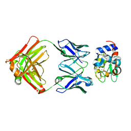
Structure for antibody HyHEL-63 Y33A mutant complexed with hen egg lysozyme
Descriptor: Lysozyme C, antibody kappa heavy chain, antibody kappa light chain
Authors:Li, Y, Mariuzza, R.A.
Deposit date:2004-09-17
Release date:2005-09-06
Last modified:2011-07-13
Method:X-RAY DIFFRACTION (2.1 Å)
Cite:Magnitude of the hydrophobic effect at central versus peripheral sites in protein-protein interfaces
Structure, 13, 2005
1XGQ
DownloadVisualize
BU of 1xgq by Molmil
Structure for antibody HyHEL-63 Y33V mutant complexed with hen egg lysozyme
Descriptor: Lysozyme C, antibody kappa heavy chain, antibody kappa light chain
Authors:Li, Y, Mariuzza, R.A.
Deposit date:2004-09-17
Release date:2005-09-06
Last modified:2024-10-30
Method:X-RAY DIFFRACTION (2.1 Å)
Cite:Magnitude of the hydrophobic effect at central versus peripheral sites in protein-protein interfaces
Structure, 13, 2005
1XGT
DownloadVisualize
BU of 1xgt by Molmil
Structure for antibody HyHEL-63 Y33L mutant complexed with hen egg lysozyme
Descriptor: Lysozyme C, antibody kappa heavy chain, antibody kappa light chain
Authors:Li, Y, Mariuzza, R.A.
Deposit date:2004-09-17
Release date:2005-09-06
Last modified:2024-10-16
Method:X-RAY DIFFRACTION (2.1 Å)
Cite:Magnitude of the hydrophobic effect at central versus peripheral sites in protein-protein interfaces
Structure, 13, 2005
2APH
DownloadVisualize
BU of 2aph by Molmil
Crystal structure of human PGRP-IalphaC in complex with muramyl pentapeptide
Descriptor: N-acetyl-beta-muramic acid, Peptidoglycan recognition protein I-alpha, SULFATE ION, ...
Authors:Guan, R, Roychowdjury, A, Boons, G, Mariuzza, R.A.
Deposit date:2005-08-16
Release date:2006-06-27
Last modified:2024-02-28
Method:X-RAY DIFFRACTION (2.1 Å)
Cite:Crystal structure of human peptidoglycan recognition protein I alpha bound to a muramyl pentapeptide from Gram-positive bacteria.
Protein Sci., 15, 2006
1VBP
DownloadVisualize
BU of 1vbp by Molmil
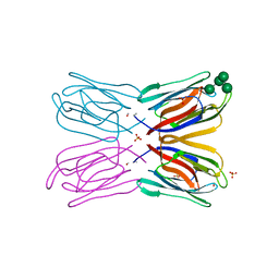
Crystal structure of artocarpin-mannopentose complex
Descriptor: SULFATE ION, alpha-D-mannopyranose-(1-3)-[alpha-D-mannopyranose-(1-6)]alpha-D-mannopyranose, alpha-D-mannopyranose-(1-3)-alpha-D-mannopyranose-(1-6)-[alpha-D-mannopyranose-(1-3)]alpha-D-mannopyranose, ...
Authors:Jeyaprakash, A.A, Srivastav, A, Surolia, A, Vijayan, M.
Deposit date:2004-02-28
Release date:2004-06-15
Last modified:2023-10-25
Method:X-RAY DIFFRACTION (3.5 Å)
Cite:Structural basis for the carbohydrate specificities of artocarpin: variation in the length of a loop as a strategy for generating ligand specificity
J.Mol.Biol., 338, 2004
1VBO
DownloadVisualize
BU of 1vbo by Molmil
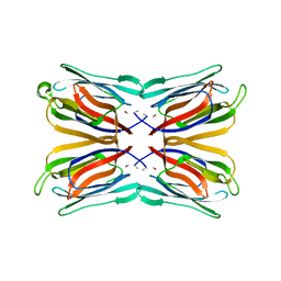
Crystal structure of artocarpin-mannotriose complex
Descriptor: alpha-D-mannopyranose, alpha-D-mannopyranose-(1-3)-[alpha-D-mannopyranose-(1-6)]alpha-D-mannopyranose, artocarpin
Authors:Jeyaprakash, A.A, Srivastav, A, Surolia, A, Vijayan, M.
Deposit date:2004-02-28
Release date:2004-06-15
Last modified:2023-10-25
Method:X-RAY DIFFRACTION (2.35 Å)
Cite:Structural basis for the carbohydrate specificities of artocarpin: variation in the length of a loop as a strategy for generating ligand specificity
J.Mol.Biol., 338, 2004
<12


227344

PDB entries from 2024-11-13

PDB statisticsPDBj update infoContact PDBjnumon