Loading
PDBj
MenuPDBj@FacebookPDBj@TwitterPDBj@YouTubewwPDB FoundationwwPDB
RCSB PDBPDBeBMRBAdv. SearchSearch help
Search by PDB author
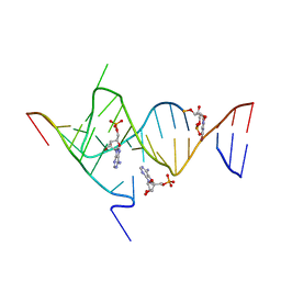
6YLB
DownloadVisualize
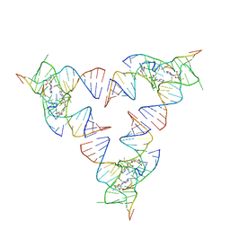
BU of 6ylb by Molmil
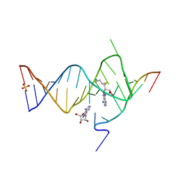
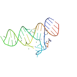
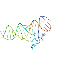
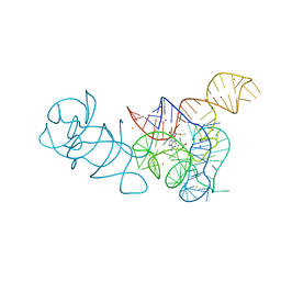
Crystal structure of the SAM-SAH riboswitch with SAM
Descriptor: Chains: A,C,F,I,M,O, Chains: B,D,G,J,N,P, S-ADENOSYLMETHIONINE
Authors:Huang, L, Lilley, D.M.J.
Deposit date:2020-04-07
Release date:2020-07-22
Last modified:2024-01-24
Method:X-RAY DIFFRACTION (2.12 Å)
Cite:Crystal structure and ligand-induced folding of the SAM/SAH riboswitch.
Nucleic Acids Res., 2020
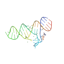
6YML
DownloadVisualize
BU of 6yml by Molmil
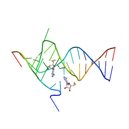
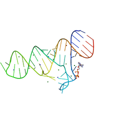
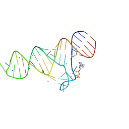
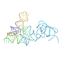
Crystal structure of the SAM-SAH riboswitch with decarboxylated SAH
Descriptor: 5'-S-(3-aminopropyl)-5'-thioadenosine, ADENOSINE MONOPHOSPHATE, Chains: A,C, ...
Authors:Huang, L, Lilley, D.M.J.
Deposit date:2020-04-08
Release date:2020-07-22
Last modified:2024-01-24
Method:X-RAY DIFFRACTION (2.17 Å)
Cite:Crystal structure and ligand-induced folding of the SAM/SAH riboswitch.
Nucleic Acids Res., 48, 2020
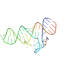
6YMK
DownloadVisualize
BU of 6ymk by Molmil
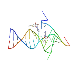
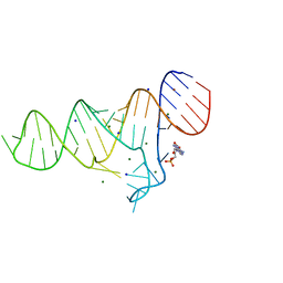
Crystal structure of the SAM-SAH riboswitch with AMP
Descriptor: 5'-DEOXY-5'-METHYLTHIOADENOSINE, Chains: A,C,F,I,M,O, Chains: B,D,G,J,N,P, ...
Authors:Huang, L, Lilley, D.M.J.
Deposit date:2020-04-08
Release date:2020-07-22
Last modified:2024-01-24
Method:X-RAY DIFFRACTION (2.03 Å)
Cite:Crystal structure and ligand-induced folding of the SAM/SAH riboswitch.
Nucleic Acids Res., 48, 2020
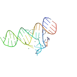
6YMM
DownloadVisualize
BU of 6ymm by Molmil
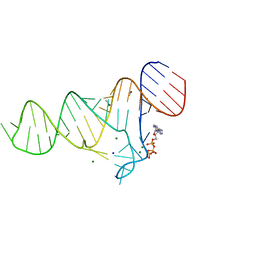
Crystal structure of the SAM-SAH riboswitch with SAM from space group P312
Descriptor: Chains: A, Chains: B,D, S-ADENOSYLMETHIONINE, ...
Authors:Huang, L, Lilley, D.M.J.
Deposit date:2020-04-08
Release date:2020-07-22
Last modified:2024-05-15
Method:X-RAY DIFFRACTION (2.2 Å)
Cite:Crystal structure and ligand-induced folding of the SAM/SAH riboswitch.
Nucleic Acids Res., 48, 2020
6YL5
DownloadVisualize
BU of 6yl5 by Molmil
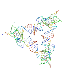
Crystal structure of the SAM-SAH riboswitch with SAH
Descriptor: Chains: A,B,C,D,E,F,G,H,I,J,K,L, MAGNESIUM ION, S-ADENOSYL-L-HOMOCYSTEINE, ...
Authors:Huang, L, Lilley, D.M.J.
Deposit date:2020-04-06
Release date:2020-07-22
Last modified:2024-05-15
Method:X-RAY DIFFRACTION (1.7 Å)
Cite:Crystal structure and ligand-induced folding of the SAM/SAH riboswitch.
Nucleic Acids Res., 2020
6YMI
DownloadVisualize
BU of 6ymi by Molmil
Crystal structure of the SAM-SAH riboswitch with AMP.
Descriptor: 5-BROMOCYTIDINE 5'-(DIHYDROGEN PHOSPHATE), ADENOSINE MONOPHOSPHATE, Chains: A,C,F,I,M,O, ...
Authors:Huang, L, Lilley, D.M.J.
Deposit date:2020-04-08
Release date:2020-07-22
Last modified:2024-02-07
Method:X-RAY DIFFRACTION (2.5 Å)
Cite:Crystal structure and ligand-induced folding of the SAM/SAH riboswitch.
Nucleic Acids Res., 48, 2020
6TF1
DownloadVisualize
BU of 6tf1 by Molmil
Crystal structure of the ADP-binding domain of the NAD+ riboswitch with Adenosine diphosphate (ADP)
Descriptor: ADENOSINE-5'-DIPHOSPHATE, Chains: A, MAGNESIUM ION, ...
Authors:Huang, L, Lilley, D.M.J.
Deposit date:2019-11-12
Release date:2020-09-23
Last modified:2024-01-24
Method:X-RAY DIFFRACTION (2.4 Å)
Cite:Structure and ligand binding of the ADP-binding domain of the NAD+ riboswitch.
Rna, 26, 2020
6TFF
DownloadVisualize
BU of 6tff by Molmil
Crystal structure of the ADP-binding domain of the NAD+ riboswitch with Nicotinamide adenine dinucleotide (NAD+)
Descriptor: BROMIDE ION, Chains: A, MAGNESIUM ION, ...
Authors:Huang, L, Lilley, D.M.J.
Deposit date:2019-11-14
Release date:2020-09-23
Last modified:2024-01-24
Method:X-RAY DIFFRACTION (2.52 Å)
Cite:Structure and ligand binding of the ADP-binding domain of the NAD+ riboswitch.
Rna, 26, 2020
6TF0
DownloadVisualize
BU of 6tf0 by Molmil
Crystal structure of the ADP-binding domain of the NAD+ riboswitch with Nicotinamide adenine dinucleotide, reduced (NADH)
Descriptor: 1,4-DIHYDRONICOTINAMIDE ADENINE DINUCLEOTIDE, Chains: A, MAGNESIUM ION, ...
Authors:Huang, L, Lilley, D.M.J.
Deposit date:2019-11-12
Release date:2020-09-23
Last modified:2024-05-15
Method:X-RAY DIFFRACTION (2.1 Å)
Cite:Structure and ligand binding of the ADP-binding domain of the NAD+ riboswitch.
Rna, 26, 2020
6TF3
DownloadVisualize
BU of 6tf3 by Molmil
Crystal structure of the ADP-binding domain of the NAD+ riboswitch with Cordycepin 5-triphosphate (3-dATP)
Descriptor: 3'-DEOXYADENOSINE-5'-TRIPHOSPHATE, Chains: A, MAGNESIUM ION, ...
Authors:Huang, L, Lilley, D.M.J.
Deposit date:2019-11-13
Release date:2020-09-23
Last modified:2024-01-24
Method:X-RAY DIFFRACTION (2.66 Å)
Cite:Structure and ligand binding of the ADP-binding domain of the NAD+ riboswitch.
Rna, 26, 2020
6TFE
DownloadVisualize
BU of 6tfe by Molmil
Crystal structure of the ADP-binding domain of the NAD+ riboswitch with N6-Methyl-adenosine-5'-triphosphate (m6ATP)
Descriptor: BROMIDE ION, Chains: A, MAGNESIUM ION, ...
Authors:Huang, L, Lilley, D.M.J.
Deposit date:2019-11-14
Release date:2020-09-23
Last modified:2024-05-15
Method:X-RAY DIFFRACTION (2.3 Å)
Cite:Structure and ligand binding of the ADP-binding domain of the NAD+ riboswitch.
Rna, 26, 2020
6TB7
DownloadVisualize
BU of 6tb7 by Molmil
Crystal structure of the ADP-binding domain of the NAD+ riboswitch with Adenosine monophosphate (AMP)
Descriptor: ADENOSINE MONOPHOSPHATE, BROMIDE ION, MAGNESIUM ION, ...
Authors:Huang, L, Lilley, D.M.J.
Deposit date:2019-11-01
Release date:2020-09-30
Last modified:2024-05-15
Method:X-RAY DIFFRACTION (2.53 Å)
Cite:Structure and ligand binding of the ADP-binding domain of the NAD + riboswitch.
Rna, 26, 2020
6TF2
DownloadVisualize
BU of 6tf2 by Molmil
Crystal structure of the ADP-binding domain of the NAD+ riboswitch with Adenosine 5-triphosphate (ATP)
Descriptor: ADENOSINE-5'-TRIPHOSPHATE, BROMIDE ION, Chains: A, ...
Authors:Huang, L, Lilley, D.M.J.
Deposit date:2019-11-12
Release date:2020-09-23
Last modified:2024-05-15
Method:X-RAY DIFFRACTION (2.55 Å)
Cite:Structure and ligand binding of the ADP-binding domain of the NAD+ riboswitch.
Rna, 26, 2020
5LQT
DownloadVisualize
BU of 5lqt by Molmil
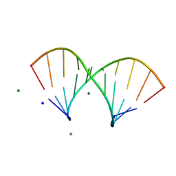
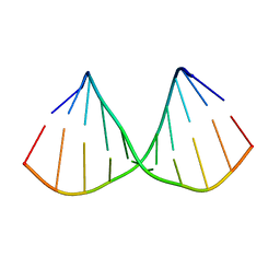
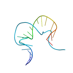
RNA duplex has central consecutive GA pairs flanked by G-C basepairs
Descriptor: CALCIUM ION, MAGNESIUM ION, RNA (5'-R(*GP*(CBV)P*CP*GP*GP*(6MZ)P*CP*GP*GP*C)-3'), ...
Authors:Huang, L, Lilley, D.M.J.
Deposit date:2016-08-17
Release date:2017-06-21
Last modified:2024-05-08
Method:X-RAY DIFFRACTION (1.5 Å)
Cite:Control of box C/D snoRNP assembly by N(6)-methylation of adenine.
EMBO Rep., 18, 2017
6TFG
DownloadVisualize
BU of 6tfg by Molmil
Crystal structure of the ADP-binding domain of the NAD+ riboswitch with Adenosine 3-phosphate 5-phosphosulfate (APPS)
Descriptor: 3'-PHOSPHATE-ADENOSINE-5'-PHOSPHATE SULFATE, Chains: A, MAGNESIUM ION, ...
Authors:Huang, L, Lilley, D.M.J.
Deposit date:2019-11-14
Release date:2020-09-23
Last modified:2024-01-24
Method:X-RAY DIFFRACTION (2.45 Å)
Cite:Structure and ligand binding of the ADP-binding domain of the NAD+ riboswitch.
Rna, 26, 2020
6TFH
DownloadVisualize
BU of 6tfh by Molmil
Crystal structure of the ADP-binding domain of the NAD+ riboswitch with Nicotinamide adenine dinucleotide, reduced (NADH); soaking with Manganese(II) (Mn2+)
Descriptor: Chains: A, MANGANESE (II) ION, NICOTINAMIDE-ADENINE-DINUCLEOTIDE, ...
Authors:Huang, L, Lilley, D.M.J.
Deposit date:2019-11-14
Release date:2020-09-23
Last modified:2024-01-24
Method:X-RAY DIFFRACTION (2.95 Å)
Cite:Structure and ligand binding of the ADP-binding domain of the NAD+ riboswitch.
Rna, 26, 2020
5LR4
DownloadVisualize
BU of 5lr4 by Molmil
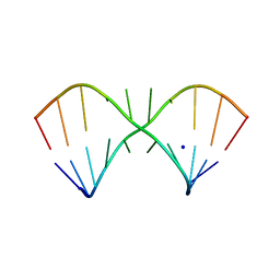
RNA duplex has central consecutive GA pairs flanked by G-U basepairs with a methyl group on the adenine N6
Descriptor: RNA (5'-R(*GP*(CBV)P*CP*GP*GP*(6MZ)P*UP*GP*GP*C)-3'), SODIUM ION
Authors:Huang, L, Lilley, D.M.J.
Deposit date:2016-08-18
Release date:2017-06-21
Last modified:2024-05-08
Method:X-RAY DIFFRACTION (1.8 Å)
Cite:Control of box C/D snoRNP assembly by N(6)-methylation of adenine.
EMBO Rep., 18, 2017
5LQO
DownloadVisualize
BU of 5lqo by Molmil
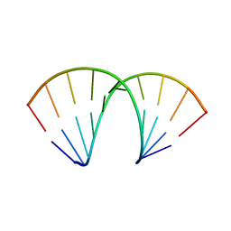
RNA duplex has central consecutive GA pairs flanked by G-C basepairs
Descriptor: RNA (5'-R(*GP*(CBV)P*CP*GP*GP*AP*CP*GP*GP*C)-3')
Authors:Huang, L, Lilley, D.M.J.
Deposit date:2016-08-17
Release date:2017-06-21
Last modified:2024-05-08
Method:X-RAY DIFFRACTION (1.87 Å)
Cite:Control of box C/D snoRNP assembly by N(6)-methylation of adenine.
EMBO Rep., 18, 2017
5LR3
DownloadVisualize
BU of 5lr3 by Molmil
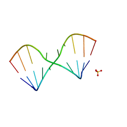
RNA duplex has central consecutive GA pairs flanked by G-U basepairs
Descriptor: RNA (5'-R(*GP*(CBV)P*CP*GP*GP*AP*UP*GP*GP*C)-3'), SULFATE ION
Authors:Huang, L, Lilley, D.M.J.
Deposit date:2016-08-18
Release date:2017-06-21
Last modified:2024-05-08
Method:X-RAY DIFFRACTION (1.65 Å)
Cite:Control of box C/D snoRNP assembly by N(6)-methylation of adenine.
EMBO Rep., 18, 2017
5LR5
DownloadVisualize
BU of 5lr5 by Molmil
N6-methyladenine is accommodated in a conventional A-U basepair
Descriptor: RNA (5'-R(*GP*GP*(6MZ)P*CP*UP*AP*GP*UP*(CBV)P*C)-3'), SULFATE ION
Authors:Huang, L, Lilley, D.M.J.
Deposit date:2016-08-18
Release date:2017-06-21
Last modified:2024-05-08
Method:X-RAY DIFFRACTION (2.4 Å)
Cite:Control of box C/D snoRNP assembly by N(6)-methylation of adenine.
EMBO Rep., 18, 2017
4BW0
DownloadVisualize
BU of 4bw0 by Molmil
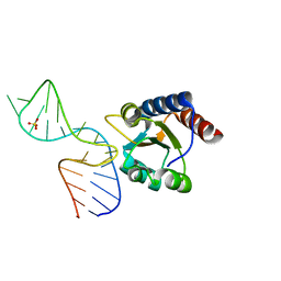
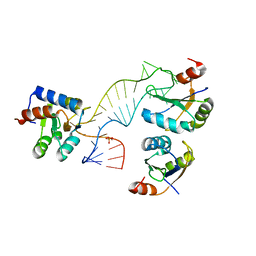
The molecular recognition of kink turn structure by the L7Ae class of proteins
Descriptor: 50S RIBOSOMAL PROTEIN L7AE, HMKT-7, SULFATE ION
Authors:Huang, L, Lilley, D.M.J.
Deposit date:2013-06-29
Release date:2013-11-06
Last modified:2023-12-20
Method:X-RAY DIFFRACTION (2.33 Å)
Cite:The Molecular Recognition of Kink-Turn Structure by the L7Ae Class of Proteins.
RNA, 19, 2013
4C40
DownloadVisualize
BU of 4c40 by Molmil
The molecular recognition of kink turn structure by the L7Ae class of proteins
Descriptor: 5'-(*GP*GP*CP*GP*AP*AP*GP*AP*AP*CP*CP*GP*GP*GP *GP*AP*GP*CP*C)-3'
Authors:Huang, L, Lilley, D.M.J.
Deposit date:2013-08-28
Release date:2013-11-06
Last modified:2023-12-20
Method:X-RAY DIFFRACTION (2.2 Å)
Cite:The Molecular Recognition of Kink-Turn Structure by the L7Ae Class of Proteins.
RNA, 19, 2013
4C4W
DownloadVisualize
BU of 4c4w by Molmil
Structure of a rare, non-standard sequence k-turn bound by L7Ae protein
Descriptor: 50S RIBOSOMAL PROTEIN L7AE, DIHYDROGENPHOSPHATE ION, TSKT-23, ...
Authors:Huang, L, Lilley, D.M.J.
Deposit date:2013-09-09
Release date:2014-02-12
Last modified:2023-12-20
Method:X-RAY DIFFRACTION (2.95 Å)
Cite:Structure of a Rare, Non-Standard Sequence K-Turn Bound by L7Ae Protein
Nucleic Acids Res., 42, 2014
5FKF
DownloadVisualize
BU of 5fkf by Molmil
SAM-I riboswitch bearing the H. marismortui Kt-7 variant 3bn is UC
Descriptor: BARIUM ION, POTASSIUM ION, S-ADENOSYLMETHIONINE, ...
Authors:Huang, L, Lilley, D.M.J.
Deposit date:2015-10-15
Release date:2016-05-25
Last modified:2024-01-10
Method:X-RAY DIFFRACTION (2.8 Å)
Cite:A Critical Base Pair in K-Turns Determines the Conformational Class Adopted, and Correlates with Biological Function.
Nucleic Acids Res., 44, 2016
5FKD
DownloadVisualize
BU of 5fkd by Molmil
SAM-I riboswitch bearing the H. marismortui Kt-7 variant 3bn is UA
Descriptor: BARIUM ION, POTASSIUM ION, S-ADENOSYLMETHIONINE, ...
Authors:Huang, L, Lilley, D.M.J.
Deposit date:2015-10-15
Release date:2016-05-25
Last modified:2024-01-10
Method:X-RAY DIFFRACTION (3 Å)
Cite:A Critical Base Pair in K-Turns Determines the Conformational Class Adopted, and Correlates with Biological Function.
Nucleic Acids Res., 44, 2016

227111

PDB entries from 2024-11-06

PDB statisticsPDBj update infoContact PDBjnumon