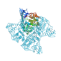
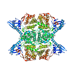
Loading
PDBj
MenuPDBj@FacebookPDBj@X(formerly Twitter)PDBj@BlueSkyPDBj@YouTubewwPDB FoundationwwPDBDonate
RCSB PDBPDBeBMRBAdv. SearchSearch help
Search by PDB author
8C9J
DownloadVisualize
BU of 8c9j by Molmil
Crystal structure of human NQO1 by serial femtosecond crystallography
Descriptor: ACETATE ION, FLAVIN-ADENINE DINUCLEOTIDE, NAD(P)H dehydrogenase [quinone] 1
Authors:Martin-Garcia, J.M, Grieco, A, Ruiz-Fresneda, M.A, Pacheco-Garcia, J.L, Pey, A, Botha, S, Ros, A.
Deposit date:2023-01-23
Release date:2023-06-28
Last modified:2024-02-07
Method:X-RAY DIFFRACTION (2.7 Å)
Cite:Modular droplet injector for sample conservation providing new structural insight for the conformational heterogeneity in the disease-associated NQO1 enzyme.
Lab Chip, 23, 2023
6VWT
DownloadVisualize
BU of 6vwt by Molmil
Transitional unit cell 1 of adenine riboswitch aptamer crystal phase transition upon ligand binding
Descriptor: ADENINE, MAGNESIUM ION, adenine riboswitch aptamer variant
Authors:Stagno, J.R, Wang, Y.-X.
Deposit date:2020-02-20
Release date:2020-11-11
Last modified:2023-10-11
Method:X-RAY DIFFRACTION (3.03 Å)
Cite:Synchronous RNA conformational changes trigger ordered phase transitions in crystals.
Nat Commun, 12, 2021
6NKN
DownloadVisualize
BU of 6nkn by Molmil
Time-resolved SFX structure of the PR intermediate of cytochrome c oxidase at room temperature
Descriptor: (1R)-2-{[{[(2S)-2,3-DIHYDROXYPROPYL]OXY}(HYDROXY)PHOSPHORYL]OXY}-1-[(PALMITOYLOXY)METHYL]ETHYL (11E)-OCTADEC-11-ENOATE, (1S)-2-{[(2-AMINOETHOXY)(HYDROXY)PHOSPHORYL]OXY}-1-[(STEAROYLOXY)METHYL]ETHYL (5E,8E,11E,14E)-ICOSA-5,8,11,14-TETRAENOATE, (7R,17E,20E)-4-HYDROXY-N,N,N-TRIMETHYL-9-OXO-7-[(PALMITOYLOXY)METHYL]-3,5,8-TRIOXA-4-PHOSPHAHEXACOSA-17,20-DIEN-1-AMINIUM 4-OXIDE, ...
Authors:Rousseau, D.L, Yeh, S.-R, Ishigami, I.
Deposit date:2019-01-07
Release date:2019-03-20
Last modified:2023-10-11
Method:X-RAY DIFFRACTION (2.5 Å)
Cite:Snapshot of an oxygen intermediate in the catalytic reaction of cytochromecoxidase.
Proc. Natl. Acad. Sci. U.S.A., 116, 2019
6NMF
DownloadVisualize
BU of 6nmf by Molmil
SFX structure of reduced cytochrome c oxidase at room temperature
Descriptor: (1R)-2-{[{[(2S)-2,3-DIHYDROXYPROPYL]OXY}(HYDROXY)PHOSPHORYL]OXY}-1-[(PALMITOYLOXY)METHYL]ETHYL (11E)-OCTADEC-11-ENOATE, (1S)-2-{[(2-AMINOETHOXY)(HYDROXY)PHOSPHORYL]OXY}-1-[(STEAROYLOXY)METHYL]ETHYL (5E,8E,11E,14E)-ICOSA-5,8,11,14-TETRAENOATE, (7R,17E,20E)-4-HYDROXY-N,N,N-TRIMETHYL-9-OXO-7-[(PALMITOYLOXY)METHYL]-3,5,8-TRIOXA-4-PHOSPHAHEXACOSA-17,20-DIEN-1-AMINIUM 4-OXIDE, ...
Authors:Rousseau, D.L, Yeh, S.-R, Ishigami, I.
Deposit date:2019-01-10
Release date:2019-03-13
Last modified:2023-10-11
Method:X-RAY DIFFRACTION (2.8 Å)
Cite:Snapshot of an oxygen intermediate in the catalytic reaction of cytochromecoxidase.
Proc. Natl. Acad. Sci. U.S.A., 116, 2019
6NMP
DownloadVisualize
BU of 6nmp by Molmil
SFX structure of oxidized cytochrome c oxidase at room temperature
Descriptor: (1R)-2-{[{[(2S)-2,3-DIHYDROXYPROPYL]OXY}(HYDROXY)PHOSPHORYL]OXY}-1-[(PALMITOYLOXY)METHYL]ETHYL (11E)-OCTADEC-11-ENOATE, (1S)-2-{[(2-AMINOETHOXY)(HYDROXY)PHOSPHORYL]OXY}-1-[(STEAROYLOXY)METHYL]ETHYL (5E,8E,11E,14E)-ICOSA-5,8,11,14-TETRAENOATE, (7R,17E,20E)-4-HYDROXY-N,N,N-TRIMETHYL-9-OXO-7-[(PALMITOYLOXY)METHYL]-3,5,8-TRIOXA-4-PHOSPHAHEXACOSA-17,20-DIEN-1-AMINIUM 4-OXIDE, ...
Authors:Rousseau, D.L, Yeh, S.-R, Ishigami, I.
Deposit date:2019-01-11
Release date:2019-03-13
Last modified:2023-10-11
Method:X-RAY DIFFRACTION (2.9 Å)
Cite:Snapshot of an oxygen intermediate in the catalytic reaction of cytochromecoxidase.
Proc. Natl. Acad. Sci. U.S.A., 116, 2019
6U57
DownloadVisualize
BU of 6u57 by Molmil
KDO8PS Structure Determined at the EuXFEL using Segmented Flow Injection
Descriptor: 2-dehydro-3-deoxyphosphooctonate aldolase
Authors:Botha, S, Ros, A.
Deposit date:2019-08-27
Release date:2020-09-02
Last modified:2023-10-11
Method:X-RAY DIFFRACTION (2.8 Å)
Cite:Segmented flow generator for serial crystallography at the European X-ray free electron laser.
Nat Commun, 11, 2020
5HD3
DownloadVisualize
BU of 5hd3 by Molmil
Femtosecond Structural Dynamics Drives the Trans/Cis Isomerization in Photoactive Yellow Protein: Dark structure of photoactive yellow protein
Descriptor: Photoactive yellow protein
Authors:Pande, K, Tenboer, J, Schmidt, M.
Deposit date:2016-01-04
Release date:2016-05-18
Last modified:2023-11-15
Method:X-RAY DIFFRACTION (1.6 Å)
Cite:Femtosecond structural dynamics drives the trans/cis isomerization in photoactive yellow protein.
Science, 352, 2016
5HDC
DownloadVisualize
BU of 5hdc by Molmil
Femtosecond Structural Dynamics Drives the Trans/Cis Isomerization in Photoactive Yellow Protein: 100 fs to 400 fs Structure
Descriptor: Photoactive yellow protein
Authors:Pande, K, Tenboer, J, Schmidt, M.
Deposit date:2016-01-05
Release date:2016-05-25
Last modified:2023-11-15
Method:X-RAY DIFFRACTION (1.6 Å)
Cite:Femtosecond structural dynamics drives the trans/cis isomerization in photoactive yellow protein.
Science, 352, 2016
5HDD
DownloadVisualize
BU of 5hdd by Molmil
Femtosecond Structural Dynamics Drives the Trans/Cis Isomerization in Photoactive Yellow Protein: 800 fs to 1200 fs Structure
Descriptor: Photoactive yellow protein
Authors:Pande, K, Tenboer, J, Schmidt, M.
Deposit date:2016-01-05
Release date:2016-05-25
Last modified:2023-11-15
Method:X-RAY DIFFRACTION (1.6 Å)
Cite:Femtosecond structural dynamics drives the trans/cis isomerization in photoactive yellow protein.
Science, 352, 2016
5HD5
DownloadVisualize
BU of 5hd5 by Molmil
Femtosecond Structural Dynamics Drives the Trans/Cis Isomerization in Photoactive Yellow Protein: 200 ns time delay photo-activated (light) structure
Descriptor: Photoactive yellow protein
Authors:Pande, K, Tenboer, J, Schmidt, M.
Deposit date:2016-01-04
Release date:2016-05-25
Last modified:2023-11-15
Method:X-RAY DIFFRACTION (1.6 Å)
Cite:Femtosecond structural dynamics drives the trans/cis isomerization in photoactive yellow protein.
Science, 352, 2016
5HDS
DownloadVisualize
BU of 5hds by Molmil
Femtosecond Structural Dynamics Drives the Trans/Cis Isomerization in Photoactive Yellow Protein: 3 ps Structure
Descriptor: Photoactive yellow protein
Authors:Pande, K, Tenboer, J, Schmidt, M.
Deposit date:2016-01-05
Release date:2016-05-25
Last modified:2023-11-15
Method:X-RAY DIFFRACTION (1.6 Å)
Cite:Femtosecond structural dynamics drives the trans/cis isomerization in photoactive yellow protein.
Science, 352, 2016
5JM7
DownloadVisualize
BU of 5jm7 by Molmil
The structure of aerobactin synthetase IucA from a hypervirulent pathotype of Klebsiella pneumoniae
Descriptor: Aerobactin synthase IucA, SULFATE ION
Authors:Drake, E.J, Bailey, D.C, Gulick, A.M.
Deposit date:2016-04-28
Release date:2016-06-15
Last modified:2024-03-06
Method:X-RAY DIFFRACTION (2.4 Å)
Cite:Structural and Functional Characterization of Aerobactin Synthetase IucA from a Hypervirulent Pathotype of Klebsiella pneumoniae.
Biochemistry, 55, 2016
5JM8
DownloadVisualize
BU of 5jm8 by Molmil
The structure of ATP-bound aerobactin synthetase IucA from a hypervirulent pathotype of Klebsiella pneumoniae
Descriptor: ADENOSINE-5'-TRIPHOSPHATE, Aerobactin synthase IucA, MAGNESIUM ION
Authors:Bailey, D.C, Drake, E.J, Gulick, A.M.
Deposit date:2016-04-28
Release date:2016-06-15
Last modified:2024-03-06
Method:X-RAY DIFFRACTION (2.2 Å)
Cite:Structural and Functional Characterization of Aerobactin Synthetase IucA from a Hypervirulent Pathotype of Klebsiella pneumoniae.
Biochemistry, 55, 2016
<12


234785

PDB entries from 2025-04-16

PDB statisticsPDBj update infoContact PDBjnumon