Loading
PDBj
MenuPDBj@FacebookPDBj@TwitterPDBj@YouTubewwPDB FoundationwwPDB
RCSB PDBPDBeBMRBAdv. SearchSearch help
Search by PDB author
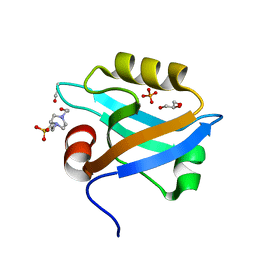
3K82
DownloadVisualize
BU of 3k82 by Molmil
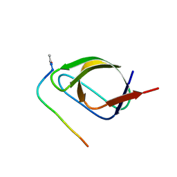
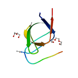
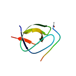
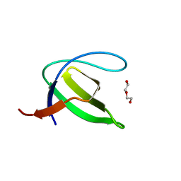
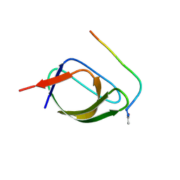
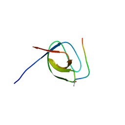
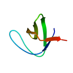
Crystal Structure of the third PDZ domain of PSD-95
Descriptor: 4-(2-HYDROXYETHYL)-1-PIPERAZINE ETHANESULFONIC ACID, Disks large homolog 4, GLYCEROL, ...
Authors:Camara-Artigas, A, Gavira, J.A.
Deposit date:2009-10-13
Release date:2010-04-07
Last modified:2023-11-15
Method:X-RAY DIFFRACTION (1.4 Å)
Cite:Novel conformational aspects of the third PDZ domain of the neuronal post-synaptic density-95 protein revealed from two 1.4A X-ray structures
J.Struct.Biol., 170, 2010
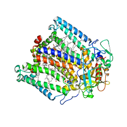
1KBY
DownloadVisualize
BU of 1kby by Molmil
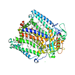
Structure of Photosynthetic Reaction Center with bacteriochlorophyll-bacteriopheophytin heterodimer
Descriptor: BACTERIOCHLOROPHYLL A, BACTERIOPHEOPHYTIN A, CARDIOLIPIN, ...
Authors:Camara-Artigas, A, Magee, C, Goetsch, A, Allen, J.P.
Deposit date:2001-11-07
Release date:2002-11-13
Last modified:2024-02-07
Method:X-RAY DIFFRACTION (2.5 Å)
Cite:The structure of the heterodimer reaction center from Rhodobacter sphaeroides at 2.55 a resolution.
Photosynth.Res., 74, 2002
3V57
DownloadVisualize
BU of 3v57 by Molmil
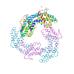
Crystal Structure of the B-phycoerythrin from the red algae Porphyridium Cruentum at pH8
Descriptor: PHYCOERYTHROBILIN, Phycoerythrin alpha subunit, Phycoerythrin beta subunit, ...
Authors:Camara-Artigas, A.
Deposit date:2011-12-16
Release date:2012-10-03
Last modified:2023-09-13
Method:X-RAY DIFFRACTION (1.7 Å)
Cite:pH-dependent structural conformations of B-phycoerythrin from Porphyridium cruentum
Febs J., 279, 2012
3V58
DownloadVisualize
BU of 3v58 by Molmil
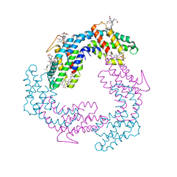
Crystal Structure of the B-phycoerythrin from the red algae Porphyridium Cruentum at pH5
Descriptor: PHYCOERYTHROBILIN, Phycoerythrin alpha subunit, Phycoerythrin beta subunit, ...
Authors:Camara-Artigas, A.
Deposit date:2011-12-16
Release date:2012-10-03
Last modified:2023-09-13
Method:X-RAY DIFFRACTION (1.85 Å)
Cite:pH-dependent structural conformations of B-phycoerythrin from Porphyridium cruentum
Febs J., 279, 2012
4J9E
DownloadVisualize
BU of 4j9e by Molmil
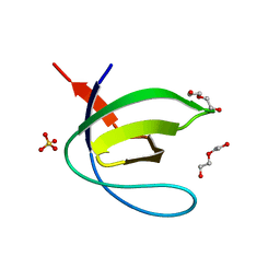
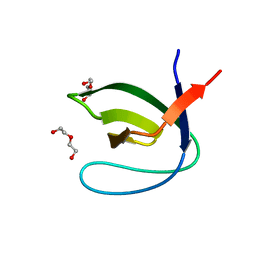
Crystal structure of the N114A mutant of the Abl-SH3 domain complexed with the high affinity peptide P17
Descriptor: P17, SULFATE ION, Tyrosine-protein kinase ABL1
Authors:Camara-Artigas, A.
Deposit date:2013-02-16
Release date:2014-01-29
Last modified:2023-09-20
Method:X-RAY DIFFRACTION (1.4 Å)
Cite:Crystal structure of the N114A mutant of the Abl-SH3 domain complexed with the high affinity peptide P17
To be Published
4J9C
DownloadVisualize
BU of 4j9c by Molmil
Crystal structure of the Abl-SH3 domain H59Q-N96T mutant complexed with the designed high-affinity peptide ligand P17
Descriptor: DI(HYDROXYETHYL)ETHER, P17, TRIETHYLENE GLYCOL, ...
Authors:Camara-Artigas, A.
Deposit date:2013-02-16
Release date:2014-01-29
Last modified:2023-09-20
Method:X-RAY DIFFRACTION (1.051 Å)
Cite:Crystal structure of the Abl-SH3 domain H59Q-N96T mutant complexed with the designed high-affinity peptide ligand P17
To be Published
4J9G
DownloadVisualize
BU of 4j9g by Molmil
Crystal structure of the ABL-SH3 domain complexed with the designed high-affinity peptide ligand P7 at pH7
Descriptor: GLYCEROL, P7, SULFATE ION, ...
Authors:Camara-Artigas, A.
Deposit date:2013-02-16
Release date:2014-01-29
Last modified:2023-09-20
Method:X-RAY DIFFRACTION (1.8 Å)
Cite:Crystal structure of the ABL-SH3 domain complexed with the designed high-affinity peptide ligand P7
To be Published
4JJC
DownloadVisualize
BU of 4jjc by Molmil
Crystal structure of the Abl-SH3 domain at pH5
Descriptor: DI(HYDROXYETHYL)ETHER, Tyrosine-protein kinase ABL1
Authors:Camara-Artigas, A, Martin-Garcia, J.M.
Deposit date:2013-03-07
Release date:2014-03-12
Last modified:2023-11-08
Method:X-RAY DIFFRACTION (1.6 Å)
Cite:Crystal structure of the Abl-SH3 domain at pH5
To be Published
4J9I
DownloadVisualize
BU of 4j9i by Molmil
Crystal structure of the ABL-SH3 domain complexed with the designed high-affinity peptide ligand P17
Descriptor: GLYCEROL, P17, Tyrosine-protein kinase ABL1
Authors:Camara-Artigas, A.
Deposit date:2013-02-16
Release date:2014-01-29
Last modified:2023-09-20
Method:X-RAY DIFFRACTION (2.2 Å)
Cite:Crystal structure of the ABL-SH3 domain complexed with the designed high-affinity peptide ligand P17
To be Published
4J9F
DownloadVisualize
BU of 4j9f by Molmil
Crystal structure of the Abl-SH3 domain complexed with the high affinity peptide P0
Descriptor: GLYCEROL, P0, SULFATE ION, ...
Authors:Camara-Artigas, A.
Deposit date:2013-02-16
Release date:2014-01-29
Last modified:2023-09-20
Method:X-RAY DIFFRACTION (1.094 Å)
Cite:Crystal structure of the Abl-SH3 domain complexed with the high affinity peptide P0
To be Published
4JJB
DownloadVisualize
BU of 4jjb by Molmil
Crystal structure of the Abl-SH3 domain at pH3
Descriptor: DI(HYDROXYETHYL)ETHER, SULFATE ION, Tyrosine-protein kinase ABL1
Authors:Camara-Artigas, A, Martin-Garcia, J.M.
Deposit date:2013-03-07
Release date:2014-03-12
Last modified:2023-11-08
Method:X-RAY DIFFRACTION (1.65 Å)
Cite:Crystal structure of the Abl-SH3 domain at pH3
To be Published
4J9H
DownloadVisualize
BU of 4j9h by Molmil
Crystal structure of the ABL-SH3 domain complexed with the designed high-affinity peptide ligand P7 at pH 8
Descriptor: P7, SULFATE ION, Tyrosine-protein kinase ABL1
Authors:Camara-Artigas, A.
Deposit date:2013-02-16
Release date:2014-01-29
Last modified:2023-09-20
Method:X-RAY DIFFRACTION (1.7 Å)
Cite:Crystal structure of the ABL-SH3 domain complexed with the designed high-affinity peptide ligand P7
To be Published
4JJD
DownloadVisualize
BU of 4jjd by Molmil
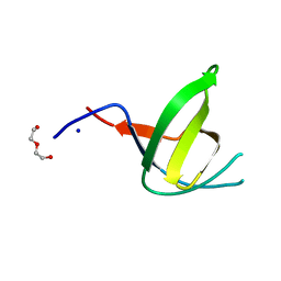
Crystal structure of the N114A Abl-SH3 domain mutant at pH4
Descriptor: DI(HYDROXYETHYL)ETHER, SODIUM ION, Tyrosine-protein kinase ABL1
Authors:Camara-Artigas, A, Martin-Garcia, J.M.
Deposit date:2013-03-07
Release date:2014-03-12
Last modified:2023-11-08
Method:X-RAY DIFFRACTION (1.6 Å)
Cite:Crystal structure of the Abl-SH3 domain at pH5
To be Published
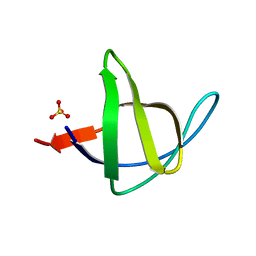
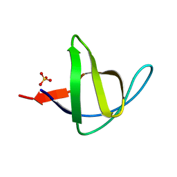
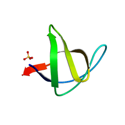
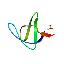
3NGP
DownloadVisualize
BU of 3ngp by Molmil
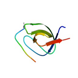
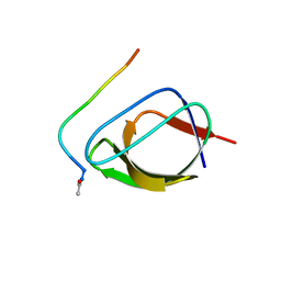
High resolution structure of alpha-spectrin SH3 domain mutant with a redesigned core
Descriptor: Spectrin alpha chain, brain
Authors:Camara-Artigas, A, Gavira, J.A, Martin-Garcia, J.M.
Deposit date:2010-06-12
Release date:2010-09-08
Last modified:2023-09-06
Method:X-RAY DIFFRACTION (1.084 Å)
Cite:High-resolution structure of an alpha-spectrin SH3-domain mutant with a redesigned hydrophobic core.
Acta Crystallogr.,Sect.F, 66, 2010
4J9B
DownloadVisualize
BU of 4j9b by Molmil
Crystal structure of the Abl-SH3 domain H59Q-N96T mutant
Descriptor: DI(HYDROXYETHYL)ETHER, Tyrosine-protein kinase ABL1
Authors:Camara-Artigas, A.
Deposit date:2013-02-16
Release date:2014-01-29
Last modified:2023-09-20
Method:X-RAY DIFFRACTION (1.702 Å)
Cite:Crystal structure of the Abl-SH3 domain H59Q-N96T mutant
To be Published
4J9D
DownloadVisualize
BU of 4j9d by Molmil
Crystal structure of the N114A mutant of the Abl-SH3 domain complexed with the high affinity peptide P0
Descriptor: P0, SULFATE ION, Tyrosine-protein kinase ABL1
Authors:Camara-Artigas, A.
Deposit date:2013-02-16
Release date:2014-01-29
Last modified:2023-09-20
Method:X-RAY DIFFRACTION (1.5 Å)
Cite:Crystal structure of the N114A mutant of the Abl-SH3 domain complexed with the high affinity peptide P0
To be Published
4OMM
DownloadVisualize
BU of 4omm by Molmil
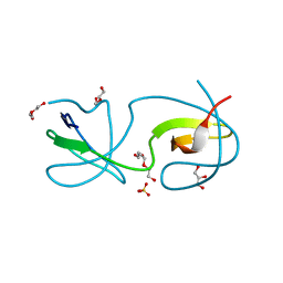
Crystal structure of the intertwined dimer of the c-Src tyrosine kinase SH3 domain mutant N113S
Descriptor: DI(HYDROXYETHYL)ETHER, GLYCEROL, Proto-oncogene tyrosine-protein kinase Src, ...
Authors:Camara-Artigas, A, Bacarizo, J.
Deposit date:2014-01-27
Release date:2015-01-28
Last modified:2023-09-20
Method:X-RAY DIFFRACTION (1.9 Å)
Cite:The role of Gutamine 128 of the SH3 domain of the c-Src tyrosine kinase in 3D domain swapping and amyloid formation
To be Published
6TG7
DownloadVisualize
BU of 6tg7 by Molmil
Crystal structure of the CheY in presence of magnesium
Descriptor: Chemotaxis protein CheY, MAGNESIUM ION
Authors:Camara-Artigas, A, Salinas-Garcia, M.C, Alba-Elena, D.
Deposit date:2019-11-15
Release date:2019-12-04
Last modified:2024-01-24
Method:X-RAY DIFFRACTION (1.65 Å)
Cite:Crystal structure of the CheY in presence of magnesium
To be published
4OMQ
DownloadVisualize
BU of 4omq by Molmil
Crystal structure of the intertwined dimer of the c-Src tyrosine kinase SH3 domain mutant S94A
Descriptor: DI(HYDROXYETHYL)ETHER, Proto-oncogene tyrosine-protein kinase Src, SULFATE ION, ...
Authors:Camara-Artigas, A, Bacarizo, J.
Deposit date:2014-01-27
Release date:2015-01-28
Last modified:2023-09-20
Method:X-RAY DIFFRACTION (2 Å)
Cite:The role of Gutamine 128 of the SH3 domain of the c-Src tyrosine kinase in 3D domain swapping and amyloid formation
To be Published
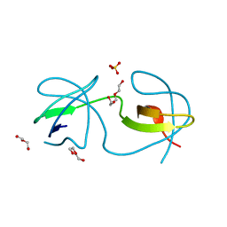
3M0P
DownloadVisualize
BU of 3m0p by Molmil
Crystal Structure of the R21D mutant of alpha-spectrin SH3 domain. Crystal obtained in ammonium sulphate at pH 4.
Descriptor: SULFATE ION, Spectrin alpha chain, brain
Authors:Camara-Artigas, A, Gavira, J.A.
Deposit date:2010-03-03
Release date:2011-01-19
Last modified:2023-09-06
Method:X-RAY DIFFRACTION (2.6 Å)
Cite:Understanding the polymorphic behaviour of a mutant of the alpha-spectrin SH3 domain by means of two 1.1 A structures
Acta Crystallogr.,Sect.D, 2011
3M0S
DownloadVisualize
BU of 3m0s by Molmil
Crystal Structure of the R21D mutant of alpha-spectrin SH3 domain. Crystal obtained in ammonium sulphate at pH 7
Descriptor: SULFATE ION, Spectrin alpha chain, brain
Authors:Camara-Artigas, A, Gavira, J.A.
Deposit date:2010-03-03
Release date:2011-01-19
Last modified:2023-09-06
Method:X-RAY DIFFRACTION (1.6 Å)
Cite:Understanding the polymorphic behaviour of a mutant of the alpha-spectrin SH3 domain by means of two 1.1 A structures
Acta Crystallogr.,Sect.D, 2011
3M0T
DownloadVisualize
BU of 3m0t by Molmil
Crystal Structure of the R21D mutant of alpha-spectrin SH3 domain. Crystal obtained in ammonium sulphate at pH 9.
Descriptor: SULFATE ION, Spectrin alpha chain, brain
Authors:Camara-Artigas, A, Gavira, J.A.
Deposit date:2010-03-03
Release date:2011-01-19
Last modified:2024-02-21
Method:X-RAY DIFFRACTION (1.7 Å)
Cite:Understanding the polymorphic behaviour of a mutant of the alpha-spectrin SH3 domain by means of two 1.1 A structures
Acta Crystallogr.,Sect.D, 2011
3M0Q
DownloadVisualize
BU of 3m0q by Molmil
Crystal Structure of the R21D mutant of alpha-spectrin SH3 domain. Crystal obtained in ammonium sulphate at pH 5.
Descriptor: SULFATE ION, Spectrin alpha chain, brain
Authors:Camara-Artigas, A, Gavira, J.A.
Deposit date:2010-03-03
Release date:2011-01-19
Last modified:2023-09-06
Method:X-RAY DIFFRACTION (1.75 Å)
Cite:Understanding the polymorphic behaviour of a mutant of the alpha-spectrin SH3 domain by means of two 1.1 A structures
Acta Crystallogr.,Sect.D, 2011
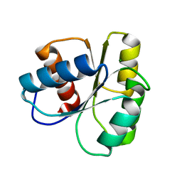
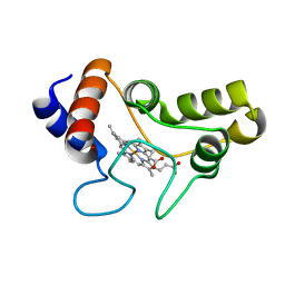
1JDL
DownloadVisualize
BU of 1jdl by Molmil
Structure of cytochrome c2 from Rhodospirillum Centenum
Descriptor: CYTOCHROME C2, ISO-2, PROTOPORPHYRIN IX CONTAINING FE
Authors:Camara-Artigas, A, Williams, J.C, Allen, J.P.
Deposit date:2001-06-14
Release date:2001-11-07
Last modified:2023-08-16
Method:X-RAY DIFFRACTION (1.7 Å)
Cite:Structure of cytochrome c2 from Rhodospirillum centenum.
Acta Crystallogr.,Sect.D, 57, 2001
1JGZ
DownloadVisualize
BU of 1jgz by Molmil
Photosynthetic Reaction Center Mutant With Tyr M 76 Replaced With Lys
Descriptor: BACTERIOCHLOROPHYLL A, BACTERIOPHEOPHYTIN A, CARDIOLIPIN, ...
Authors:Camara-Artigas, A, Magee, C.L, Williams, J.C, Allen, J.P.
Deposit date:2001-06-27
Release date:2001-09-05
Last modified:2023-08-16
Method:X-RAY DIFFRACTION (2.7 Å)
Cite:Individual interactions influence the crystalline order for membrane proteins.
Acta Crystallogr.,Sect.D, 57, 2001

225946

PDB entries from 2024-10-09

PDB statisticsPDBj update infoContact PDBjnumon