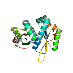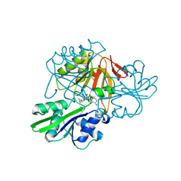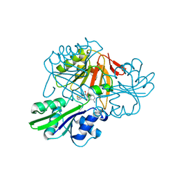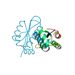
Loading
PDBj
MenuPDBj@FacebookPDBj@TwitterPDBj@YouTubewwPDB FoundationwwPDB
RCSB PDBPDBeBMRBAdv. SearchSearch help
Search by PDB author
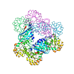
2ED3
DownloadVisualize
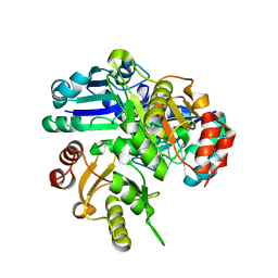
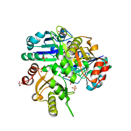
BU of 2ed3 by Molmil
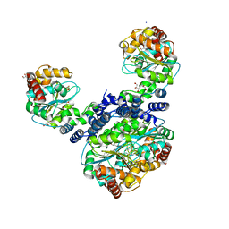
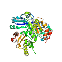
Mutant I127M structure of PH0725 from Pyrococcus horikoshii OT3
Descriptor: S-ADENOSYL-L-HOMOCYSTEINE, diphthine synthase
Authors:Shimizu, K, RIKEN Structural Genomics/Proteomics Initiative (RSGI)
Deposit date:2007-02-14
Release date:2007-08-14
Last modified:2023-10-25
Method:X-RAY DIFFRACTION (2.5 Å)
Cite:Mutant I127M structure of PH0725 from Pyrococcus horikoshii OT3
To be Published
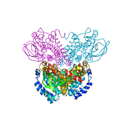
2EEQ
DownloadVisualize
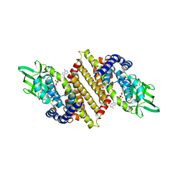
BU of 2eeq by Molmil
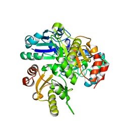
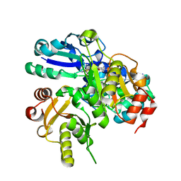
Mutant Y29M structure of PH0725 from Pyrococcus horikoshii OT3
Descriptor: GLYCEROL, S-ADENOSYL-L-HOMOCYSTEINE, SULFATE ION, ...
Authors:Shimizu, K, RIKEN Structural Genomics/Proteomics Initiative (RSGI)
Deposit date:2007-02-16
Release date:2007-08-21
Last modified:2023-10-25
Method:X-RAY DIFFRACTION (2.5 Å)
Cite:Mutant Y29M structure of PH0725 from Pyrococcus horikoshii OT3
To be Published
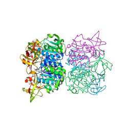
2ELC
DownloadVisualize
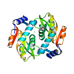
BU of 2elc by Molmil
Crystal structure of TTHA1842 from Thermus thermophilus HB8
Descriptor: Anthranilate phosphoribosyltransferase, GLYCEROL, SODIUM ION
Authors:Shimizu, K, RIKEN Structural Genomics/Proteomics Initiative (RSGI)
Deposit date:2007-03-27
Release date:2007-10-02
Last modified:2023-10-25
Method:X-RAY DIFFRACTION (1.55 Å)
Cite:Crystal structure of TTHA1842 from Thermus thermophilus HB8
To be Published
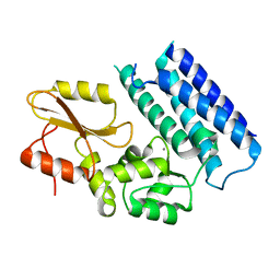
2EMU
DownloadVisualize
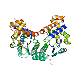
BU of 2emu by Molmil
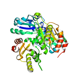
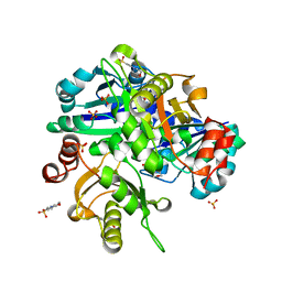
Mutant L21H structure of PH0725 from Pyrococcus horikoshii OT3
Descriptor: S-ADENOSYL-L-HOMOCYSTEINE, SODIUM ION, diphthine synthase
Authors:Shimizu, K, RIKEN Structural Genomics/Proteomics Initiative (RSGI)
Deposit date:2007-03-28
Release date:2007-10-02
Last modified:2023-10-25
Method:X-RAY DIFFRACTION (2.2 Å)
Cite:Mutant L21H structure of PH0725 from Pyrococcus horikoshii OT3
To be Published
2ENU
DownloadVisualize
BU of 2enu by Molmil
Mutant L121M structure of TTHB049 from Thermus thermophilus HB8
Descriptor: Alpha-ribazole-5'-phosphate phosphatase, GLYCEROL
Authors:Shimizu, K, RIKEN Structural Genomics/Proteomics Initiative (RSGI)
Deposit date:2007-03-28
Release date:2007-10-02
Last modified:2023-10-25
Method:X-RAY DIFFRACTION (1.7 Å)
Cite:Mutant L121M structure of TTHB049 from Thermus thermophilus HB8
To be Published
2EMR
DownloadVisualize
BU of 2emr by Molmil
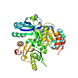
Mutant L65M structure of PH0725 from Pyrococcus horikoshii OT3
Descriptor: Probable diphthine synthase, S-ADENOSYL-L-HOMOCYSTEINE, SODIUM ION
Authors:Shimizu, K, RIKEN Structural Genomics/Proteomics Initiative (RSGI)
Deposit date:2007-03-28
Release date:2007-10-02
Last modified:2023-10-25
Method:X-RAY DIFFRACTION (2.4 Å)
Cite:Mutant L65M structure of PH0725 from Pyrococcus horikoshii OT3
To be Published
2EN5
DownloadVisualize
BU of 2en5 by Molmil
Mutant R262H structure of PH0725 from Pyrococcus horikoshii OT3
Descriptor: GLYCEROL, S-ADENOSYL-L-HOMOCYSTEINE, SODIUM ION, ...
Authors:Shimizu, K, RIKEN Structural Genomics/Proteomics Initiative (RSGI)
Deposit date:2007-03-28
Release date:2007-10-02
Last modified:2023-10-25
Method:X-RAY DIFFRACTION (1.9 Å)
Cite:Mutant R262H structure of PH0725 from Pyrococcus horikoshii OT3
To be Published
2E4N
DownloadVisualize
BU of 2e4n by Molmil
Mutant V251M structure of PH0725 from Pyrococcus horikoshii OT3
Descriptor: S-ADENOSYL-L-HOMOCYSTEINE, SODIUM ION, diphthine synthase
Authors:Shimizu, K, RIKEN Structural Genomics/Proteomics Initiative (RSGI)
Deposit date:2006-12-13
Release date:2007-06-19
Last modified:2023-10-25
Method:X-RAY DIFFRACTION (1.8 Å)
Cite:Mutant V251M structure of PH0725 from Pyrococcus horikoshii OT3
To be Published
2EH2
DownloadVisualize
BU of 2eh2 by Molmil
Mutant V18M structure of PH0725 from Pyrococcus horikoshii OT3
Descriptor: 2-(N-MORPHOLINO)-ETHANESULFONIC ACID, S-ADENOSYL-L-HOMOCYSTEINE, SULFATE ION, ...
Authors:Shimizu, K, RIKEN Structural Genomics/Proteomics Initiative (RSGI)
Deposit date:2007-03-02
Release date:2007-09-04
Last modified:2023-10-25
Method:X-RAY DIFFRACTION (2 Å)
Cite:Mutant V18M structure of PH0725 from Pyrococcus horikoshii OT3
To be Published
2EH5
DownloadVisualize
BU of 2eh5 by Molmil
Mutant L184M structure of PH0725 from Pyrococcus horikoshii OT3
Descriptor: S-ADENOSYL-L-HOMOCYSTEINE, SODIUM ION, diphthine synthase
Authors:Shimizu, K, RIKEN Structural Genomics/Proteomics Initiative (RSGI)
Deposit date:2007-03-04
Release date:2007-09-04
Last modified:2023-10-25
Method:X-RAY DIFFRACTION (2.3 Å)
Cite:Mutant L184M structure of PH0725 from Pyrococcus horikoshii OT3
To be Published
2ELE
DownloadVisualize
BU of 2ele by Molmil
Mutant V18C structure of PH0725 from Pyrococcus horikoshii OT3
Descriptor: S-ADENOSYL-L-HOMOCYSTEINE, SODIUM ION, diphthine synthase
Authors:Shimizu, K, RIKEN Structural Genomics/Proteomics Initiative (RSGI)
Deposit date:2007-03-27
Release date:2007-10-02
Last modified:2023-10-25
Method:X-RAY DIFFRACTION (2.4 Å)
Cite:Mutant V18C structure of PH0725 from Pyrococcus horikoshii OT3
To be Published
2DVL
DownloadVisualize
BU of 2dvl by Molmil
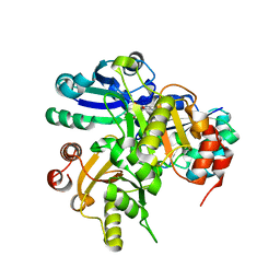
Crystal structure of project TT0160 from Thermus thermophilus HB8
Descriptor: Acyl-CoA dehydrogenase, FLAVIN-ADENINE DINUCLEOTIDE
Authors:Shimizu, K, RIKEN Structural Genomics/Proteomics Initiative (RSGI)
Deposit date:2006-07-31
Release date:2007-01-31
Last modified:2023-10-25
Method:X-RAY DIFFRACTION (2.5 Å)
Cite:Crystal structure of TT0160 from Thermus thermophilus HB8
To be Published
2DY0
DownloadVisualize
BU of 2dy0 by Molmil
Crystal structure of project JW0458 from Escherichia coli
Descriptor: Adenine phosphoribosyltransferase, MAGNESIUM ION
Authors:Shimizu, K, RIKEN Structural Genomics/Proteomics Initiative (RSGI)
Deposit date:2006-09-04
Release date:2007-03-04
Last modified:2023-10-25
Method:X-RAY DIFFRACTION (1.25 Å)
Cite:Crystal structure of JW0458 from Escherichia coli
To be Published
2E18
DownloadVisualize
BU of 2e18 by Molmil
Crystal structure of project PH0182 from Pyrococcus horikoshii OT3
Descriptor: IMIDAZOLE, NH(3)-dependent NAD(+) synthetase, ZINC ION
Authors:Shimizu, K, RIKEN Structural Genomics/Proteomics Initiative (RSGI)
Deposit date:2006-10-18
Release date:2007-04-24
Last modified:2023-10-25
Method:X-RAY DIFFRACTION (2.1 Å)
Cite:Crystal structure of project PH0182 from Pyrococcus horikoshii OT3
To be Published
2ZV3
DownloadVisualize
BU of 2zv3 by Molmil
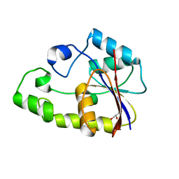
Crystal structure of project MJ0051 from Methanocaldococcus jannaschii DSM 2661
Descriptor: Peptidyl-tRNA hydrolase
Authors:Shimizu, K, RIKEN Structural Genomics/Proteomics Initiative (RSGI)
Deposit date:2008-10-31
Release date:2009-05-05
Last modified:2023-11-01
Method:X-RAY DIFFRACTION (2.1 Å)
Cite:Crystal structure of project MJ0051 from Methanocaldococcus jannaschii DSM 2661
To be published
2ZVC
DownloadVisualize
BU of 2zvc by Molmil
Form 2 structure (C2221) of TT0207 from Thermus thermophilus HB8
Descriptor: Precorrin-3 C17-methyltransferase, S-ADENOSYL-L-HOMOCYSTEINE
Authors:Shimizu, K, RIKEN Structural Genomics/Proteomics Initiative (RSGI)
Deposit date:2008-11-04
Release date:2009-05-05
Last modified:2023-11-01
Method:X-RAY DIFFRACTION (2 Å)
Cite:Form 2 structure (C2221) of TT0207 from Thermus thermophilus HB8
To be published
2ZVB
DownloadVisualize
BU of 2zvb by Molmil
Crystal structure of TT0207 from Thermus thermophilus HB8
Descriptor: Precorrin-3 C17-methyltransferase, S-ADENOSYL-L-HOMOCYSTEINE
Authors:Shimizu, K, RIKEN Structural Genomics/Proteomics Initiative (RSGI)
Deposit date:2008-11-04
Release date:2009-05-05
Last modified:2024-03-13
Method:X-RAY DIFFRACTION (2 Å)
Cite:Crystal structure of TT0207 from Thermus thermophilus HB8
To be published
1WN2
DownloadVisualize
BU of 1wn2 by Molmil
Crystal structure of project ID PH1539 from Pyrococcus horikoshii OT3
Descriptor: Peptidyl-tRNA hydrolase, SODIUM ION
Authors:Shimizu, K, Kunishima, N, RIKEN Structural Genomics/Proteomics Initiative (RSGI)
Deposit date:2004-07-26
Release date:2005-07-19
Last modified:2023-10-25
Method:X-RAY DIFFRACTION (1.2 Å)
Cite:Structure of peptidyl-tRNA hydrolase 2 from Pyrococcus horikoshii OT3: insight into the functional role of its dimeric state.
Acta Crystallogr.,Sect.D, 64, 2008
1X1O
DownloadVisualize
BU of 1x1o by Molmil
Crystal structure of project ID TT0268 from Thermus thermophilus HB8
Descriptor: nicotinate-nucleotide pyrophosphorylase
Authors:Shimizu, K, Kunishima, N, RIKEN Structural Genomics/Proteomics Initiative (RSGI)
Deposit date:2005-04-08
Release date:2006-04-04
Last modified:2023-10-25
Method:X-RAY DIFFRACTION (1.9 Å)
Cite:Crystal structure of project ID TT0268 from Thermus thermophilus HB8
To be Published
1WS9
DownloadVisualize
BU of 1ws9 by Molmil
Crystal structure of project ID TT0172 from Thermus thermophilus HB8
Descriptor: acyl-CoA dehydrogenase
Authors:Shimizu, K, Kunishima, N, RIKEN Structural Genomics/Proteomics Initiative (RSGI)
Deposit date:2004-11-02
Release date:2005-10-18
Last modified:2023-10-25
Method:X-RAY DIFFRACTION (2.3 Å)
Cite:Crystal structure of project ID TT0172 from Thermus thermophilus HB8
To be Published
1WU8
DownloadVisualize
BU of 1wu8 by Molmil
Crystal structure of project PH0463 from Pyrococcus horikoshii OT3
Descriptor: ADENOSINE, hypothetical protein PH0463
Authors:Shimizu, K, Kunishima, N, RIKEN Structural Genomics/Proteomics Initiative (RSGI)
Deposit date:2004-12-02
Release date:2005-11-29
Last modified:2011-07-13
Method:X-RAY DIFFRACTION (2.6 Å)
Cite:Crystal structure of project ID PH0463 from Pyrococcus horikoshii OT3
To be Published
3VRN
DownloadVisualize
BU of 3vrn by Molmil
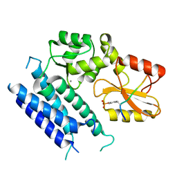
Crystal structure of the tyrosine kinase binding domain of Cbl-c
Descriptor: CALCIUM ION, Signal transduction protein CBL-C
Authors:Takeshita, K, Tezuka, T, Isozaki, Y, Yamashita, E, Suzuki, M, Yamanashi, Y, Yamamoto, T, Nakagawa, A.
Deposit date:2012-04-13
Release date:2013-03-06
Last modified:2023-11-08
Method:X-RAY DIFFRACTION (1.64 Å)
Cite:Structural flexibility regulates phosphopeptide-binding activity of the tyrosine kinase binding domain of Cbl-c
J.Biochem., 152, 2012
3VRR
DownloadVisualize
BU of 3vrr by Molmil
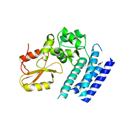
Crystal structure of the tyrosine kinase binding domain of Cbl-c (PL mutant) in complex with phospho-EGFR peptide
Descriptor: CALCIUM ION, Epidermal growth factor receptor, Signal transduction protein CBL-C
Authors:Takeshita, K, Tezuka, T, Isozaki, Y, Yamashita, E, Suzuki, M, Yamanashi, Y, Yamamoto, T, Nakagawa, A.
Deposit date:2012-04-13
Release date:2013-03-06
Last modified:2024-10-30
Method:X-RAY DIFFRACTION (2 Å)
Cite:Structural flexibility regulates phosphopeptide-binding activity of the tyrosine kinase binding domain of Cbl-c.
J.Biochem., 152, 2012
3VRQ
DownloadVisualize
BU of 3vrq by Molmil
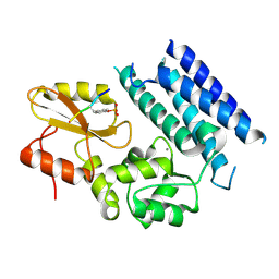
Crystal structure of the tyrosine kinase binding domain of Cbl-c (PL mutant)
Descriptor: CALCIUM ION, Signal transduction protein CBL-C
Authors:Takeshita, K, Tezuka, T, Isozaki, Y, Yamashita, E, Suzuki, M, Yamanashi, Y, Yamamoto, T, Nakagawa, A.
Deposit date:2012-04-13
Release date:2013-03-06
Last modified:2024-03-20
Method:X-RAY DIFFRACTION (2.39 Å)
Cite:Structural flexibility regulates phosphopeptide-binding activity of the tyrosine kinase binding domain of Cbl-c.
J.Biochem., 152, 2012
3VRO
DownloadVisualize
BU of 3vro by Molmil
Crystal structure of the tyrosine kinase binding domain of Cbl-c in complex with phospho-Src peptide
Descriptor: CALCIUM ION, Proto-oncogene tyrosine-protein kinase Src, Signal transduction protein CBL-C
Authors:Takeshita, K, Tezuka, T, Isozaki, Y, Yamashita, E, Suzuki, M, Yamanashi, Y, Yamamoto, T, Nakagawa, A.
Deposit date:2012-04-13
Release date:2013-03-06
Last modified:2024-10-30
Method:X-RAY DIFFRACTION (1.8 Å)
Cite:Structural flexibility regulates phosphopeptide-binding activity of the tyrosine kinase binding domain of Cbl-c.
J.Biochem., 152, 2012

226707

PDB entries from 2024-10-30

PDB statisticsPDBj update infoContact PDBjnumon