






 9VCT
9VCT
Cryo-EM structure of monomeric Suv3-ADP complex
Summary for 9VCT
| Entry DOI | 10.2210/pdb9vct/pdb |
| EMDB information | 64967 |
| Descriptor | ATP-dependent RNA helicase SUPV3L1, mitochondrial, ADENOSINE-5'-DIPHOSPHATE, MAGNESIUM ION (3 entities in total) |
| Functional Keywords | helicase, rna binding protein, hydrolase |
| Biological source | Homo sapiens (human) |
| Total number of polymer chains | 1 |
| Total formula weight | 85902.38 |
| Authors | Patra, M.,Yuan, H.S. (deposition date: 2025-06-06, release date: 2026-05-20, Last modification date: 2026-06-24) |
| Primary citation | Patra, M.,Jain, M.,Li, Y.C.,Chen, Y.P.,Golzarroshan, B.,Yuan, H.S. Asymmetric dimeric assembly of Suv3 helicase facilitates processive RNA unwinding. Nat Commun, 17:-, 2026 Cited by PubMed Abstract: Human Suv3 is a dimeric helicase that collaborates with the exoribonuclease PNPase to mediate RNA decay and surveillance in mitochondria. Despite its pivotal role in maintaining mitochondrial homeostasis, the molecular mechanism underlying Suv3-mediated RNA unwinding has remained elusive. Here, we present near-atomic-resolution cryogenic electron microscopy structures of Suv3 captured in four functional states: the apo form, two binary complexes with ADP and single-stranded RNA (ssRNA), and a ternary complex with ssRNA and an ATP analog (AMP-PNP). These structures reveal an unexpected asymmetric dimeric organization, in which only one of the two protomers engages in the initial binding of ADP, ssRNA, or both ssRNA and AMP-PNP. Complementary biochemical analyses demonstrate that Suv3 dimerization significantly enhances RNA-binding and unwinding efficiency in an ATP-hydrolysis-dependent manner. Together, these findings provide key insights into the dimeric architecture of Suv3 and establish a mechanistic framework for its coordinated function in processive RNA unwinding. PubMed: 41986356DOI: 10.1038/s41467-026-71901-2 PDB entries with the same primary citation |
| Experimental method | ELECTRON MICROSCOPY (3.47 Å) |
Structure validation
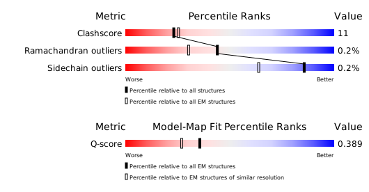
Download full validation report


