






 9EE7
9EE7
Folded domains of Xrs2 from S.cerevisiae
9EE7 の概要
| エントリーDOI | 10.2210/pdb9ee7/pdb |
| 分子名称 | DNA repair protein XRS2 (2 entities in total) |
| 機能のキーワード | dna repair, protein binding |
| 由来する生物種 | Saccharomyces cerevisiae (brewer's yeast) |
| タンパク質・核酸の鎖数 | 1 |
| 化学式量合計 | 37172.78 |
| 構造登録者 | |
| 主引用文献 | Vigneswaran, A.,Shi, K.,Aihara, H.,Evans 3rd, R.L.,Latham, M.P. Crystal structure of the folded domains of Xrs2 from Saccharomyces cerevisiae. Acta Crystallogr.,Sect.F, 81:365-373, 2025 Cited by PubMed Abstract: The MRE11-RAD50-NBS1/Xrs2 (MRN/X) protein complex acts as a first responder in DNA double-strand break repair and telomere-length maintenance, yet the structural architecture of the yeast ortholog Xrs2 has remained unresolved. In this study, we present the first structure of the folded N-terminal region of Xrs2 from Saccharomyces cerevisiae, resolved at 2.38 Å using X-ray crystallography. Like the previously determined crystal structures of Schizosaccharomyces pombe Nbs1, the folded structure of S. cerevisiae Xrs2 adopts an extended three-domain organization at its N-terminus. Electrostatic analysis reveals two distinct charged patches: a positively charged patch on the FHA domain and a negatively charged patch in the cleft between the FHA and BRCT1 domains. This charge segregation is likely to play a role in mediating interactions with various ligands. PubMed: 40767353DOI: 10.1107/S2053230X25006867 主引用文献が同じPDBエントリー |
| 実験手法 | X-RAY DIFFRACTION (2.38 Å) |
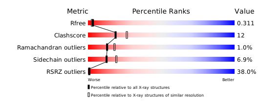
構造検証レポート
検証レポート(詳細版) をダウンロード
をダウンロード


