






 9BN7
9BN7
X-ray crystal structure of TNFa-VNAR C4 complex
Summary for 9BN7
| Entry DOI | 10.2210/pdb9bn7/pdb |
| Descriptor | Tumor necrosis factor, VNARC4, 1,2-ETHANEDIOL, ... (6 entities in total) |
| Functional Keywords | cytokine-immune system complex, cytokine/immune system |
| Biological source | Homo sapiens (human) More |
| Total number of polymer chains | 6 |
| Total formula weight | 92220.11 |
| Authors | Shi, K.,Aihara, H. (deposition date: 2024-05-02, release date: 2025-11-05, Last modification date: 2026-05-20) |
| Primary citation | Ubah, O.C.,Lake, E.W.,Ott, K.L.,Priyanka, S.,Celeda, S.,West, J.L.,Gunaratne, G.S.,Shi, K.,Moeller, N.H.,Porter, A.J.,Aihara, H.,Kosoff, D.,LeBeau, A.M.,Barelle, C.J. The structural basis for the selective antagonism of soluble TNF-alpha by shark variable new antigen receptors. Nat Commun, 17:256-256, 2025 Cited by PubMed Abstract: The pro-inflammatory cytokine tumor necrosis factor-alpha (TNF-α) is synthesized as transmembrane TNF-α (tmTNF-α) where proteolytic processing releases soluble TNF-α (sTNF-α). tmTNF-α can act as either a ligand by activating TNF receptors, or a receptor that transmits reverse (outside-to-inside) signalling after binding to native receptors. All TNF-α therapies bind tmTNF-α and induce reverse signalling which can result in immunosuppression leading to infection. We present crystal structures of two anti-TNF-α Variable New Antigen Receptors (VNARs) in complex with sTNF-α via two distinct epitopes. The VNAR-D1 recognizes an epitope that selectively engages sTNF-α while VNAR-C4 binds an epitope that partially overlaps with other biologic therapies. In activated CD4 T cells, our VNARs do not induce reverse signalling in contrast to currently available therapies. Our findings suggest that neutralization through a unique mechanism may lead to anti-TNF-α agents with an improved safety profile that will benefit high-risk patients. PubMed: 41318641DOI: 10.1038/s41467-025-66967-3 PDB entries with the same primary citation |
| Experimental method | X-RAY DIFFRACTION (1.92 Å) |
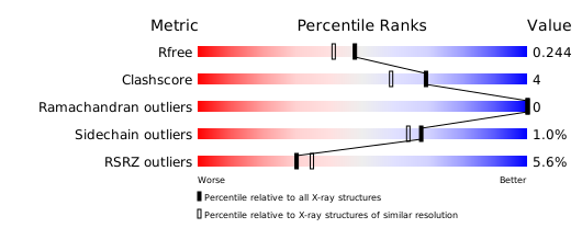
Structure validation
Download full validation report


