






 8ZEF
8ZEF
Crystal structure of Staphylococcus aureus DinG protein in complex with ssDNA and Ca2+
Summary for 8ZEF
| Entry DOI | 10.2210/pdb8zef/pdb |
| Descriptor | 3'-5' exonuclease DinG, DNA (5'-D(P*TP*TP*TP*T)-3'), DNA (5'-D(*TP*TP*TP*TP*TP*TP*T)-3'), ... (4 entities in total) |
| Functional Keywords | exonuclease, hydrolase/dna, dna repair, hydrolase-dna complex |
| Biological source | Staphylococcus aureus (strain NCTC 8325 / PS 47) More |
| Total number of polymer chains | 3 |
| Total formula weight | 107811.86 |
| Authors | |
| Primary citation | Gao, T.,Hao, W.,Gao, J.,Sun, Y.,Sun, Y.,Yang, J.,Cheng, K. Structural and functional investigation of DinG containing a 3'-5' exonuclease domain. Mbio, 16:e0088425-e0088425, 2025 Cited by PubMed Abstract: Damage-inducible gene G (DinG), a bacterial homolog of SF2 helicase, has been extensively studied in . However, the structural and functional characteristics of DinG homologs fused with an N-terminal 3'-5' exonuclease domain, such as DinG (SaDinG), remain unexplored. In this study, we demonstrate that SaDinG possesses 3'-5' exonuclease activity and exhibits 5'-3' helicase activity on diverse DNA substrates, including splayed duplexes, 5'-overhangs, double flaps, bubbles, and gapped duplexes, resolving prior ambiguities about its biochemical functions. Intriguingly, both enzymatic activities were inhibited by elevated ATP concentrations, suggesting a potential ATP-dependent regulatory mechanism . We determined the crystal structures of SaDinG bound to ssDNA at ~3.2 Å resolution and identified key residues essential for its helicase and exonuclease activities through mutational analysis. Phenotypic studies revealed that a SaDinG deletion mutant exhibited heightened sensitivity to DNA crosslinking agents (mitomycin C and formaldehyde) but retained wild-type susceptibility to other DNA-damaging compounds. Complementation with either nuclease-dead or helicase-dead variants failed to restore crosslink resistance, indicating that both activities are indispensable for DNA crosslink repair. These results support a model in which SaDinG functions as a coordinated nuclease-helicase machine specifically adapted for DNA crosslink repair, with its dual enzymatic activities being tightly regulated by physiological ATP concentrations. PubMed: 40586552DOI: 10.1128/mbio.00884-25 PDB entries with the same primary citation |
| Experimental method | X-RAY DIFFRACTION (3.21 Å) |
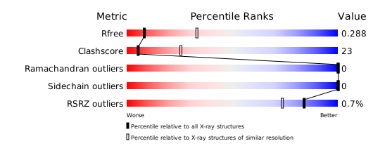
Structure validation
Download full validation report


