






 24TE
24TE
Crystal structure of GH30 Streptomyces avermitilis endo-beta-1,6-galactanase complexed with Gal-beta1,6-Gal
24TE の概要
| エントリーDOI | 10.2210/pdb24te/pdb |
| 分子名称 | Secreted endo-beta-1,6-galactanase, beta-D-galactopyranose-(1-6)-beta-D-galactopyranose, SULFATE ION, ... (6 entities in total) |
| 機能のキーワード | glycoside hydrolase family 30, hydrolase |
| 由来する生物種 | Streptomyces avermitilis MA-4680 = NBRC 14893 |
| タンパク質・核酸の鎖数 | 1 |
| 化学式量合計 | 53457.77 |
| 構造登録者 | |
| 主引用文献 | Fujimoto, Z.,Kishine, N.,Kotake, T.,Kaneko, S. Crystal structure of endo-beta-1,6-galactanase from Streptomyces avermitilis. Acta Crystallogr D Struct Biol, 82:962-971, 2026 Cited by PubMed Abstract: Endo-β-1,6-galactanases hydrolyze β-1,6-linked galactosyl linkages in galactans, yielding β-1,6-linked galacto-oligosaccharides, predominantly galactobiose. Here, we report to our knowledge the first crystal structure of an endo-β-1,6-galactanase, together with its β-1,6-galactobiose-bound complex, revealing the structural basis for substrate recognition by this enzyme. Endo-β-1,6-galactanase from Streptomyces avermitilis (Sa16Gal30A) is a member of glycoside hydrolase family 30 (GH30) subfamily 5. Sa16Gal30A consists of two structural domains: a catalytic (β/α)-barrel domain and an appended β-sandwich domain. The β-1,6-galactobiose complex structure revealed two β-1,6-galactobiose molecules bound within the catalytic domain: one at the catalytic site and another at the distinct surface site distal to the catalytic center. This structure represents the first reported structure of a GH30 subfamily 5 enzyme and provides structural insights into the molecular basis of β-1,6-galactan recognition within the catalytic cleft. Sa16Gal30A possesses three extended regions, loops 2, 4 and 8, in the catalytic domain compared with enzymes from other GH30 subfamilies, and these loops appear to modulate substrate specificity towards β-1,6-galactan by shaping the architecture of the catalytic cleft. In addition, a secondary β-1,6-galactan-binding site was identified at a distal location, which may function as a distal subsite, thereby facilitating the efficient hydrolysis of long β-1,6-galactan chains. PubMed: 42478457DOI: 10.1107/S2059798326006133 主引用文献が同じPDBエントリー |
| 実験手法 | X-RAY DIFFRACTION (2 Å) |
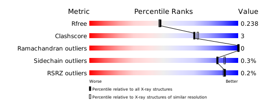
構造検証レポート
検証レポート(詳細版) をダウンロード
をダウンロード


