 Movie
Movie Controller

[English] 日本語
 Yorodumi
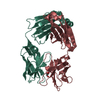
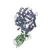
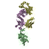
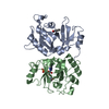
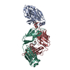
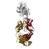
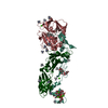
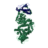
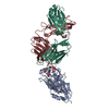
- PDB-6xe1: Structure of SARS-CoV-2 spike protein receptor binding domain in ... -
+ Open data

- Basic information
| Entry | Database: PDB / ID: 6xe1 | ||||||
|---|---|---|---|---|---|---|---|
| Title | Structure of SARS-CoV-2 spike protein receptor binding domain in complex with a potent neutralizing antibody, CV30 Fab | ||||||
 Components Components |
| ||||||
 Keywords Keywords |  VIRAL PROTEIN/IMMUNE SYSTEM / Antibody / COVID-19 / immune system / VIRAL PROTEIN / VIRAL PROTEIN-IMMUNE SYSTEM complex VIRAL PROTEIN/IMMUNE SYSTEM / Antibody / COVID-19 / immune system / VIRAL PROTEIN / VIRAL PROTEIN-IMMUNE SYSTEM complex | ||||||
| Function / homology |  Function and homology information Function and homology informationMaturation of spike protein / viral translation / Translation of Structural Proteins / Virion Assembly and Release / host cell surface / host extracellular space / suppression by virus of host tetherin activity / Induction of Cell-Cell Fusion / structural constituent of virion / host cell endoplasmic reticulum-Golgi intermediate compartment membrane ...Maturation of spike protein / viral translation / Translation of Structural Proteins / Virion Assembly and Release / host cell surface / host extracellular space / suppression by virus of host tetherin activity / Induction of Cell-Cell Fusion / structural constituent of virion / host cell endoplasmic reticulum-Golgi intermediate compartment membrane / entry receptor-mediated virion attachment to host cell / receptor-mediated endocytosis of virus by host cell / Attachment and Entry / membrane fusion / positive regulation of viral entry into host cell / receptor-mediated virion attachment to host cell / receptor ligand activity / host cell surface receptor binding / fusion of virus membrane with host plasma membrane / fusion of virus membrane with host endosome membrane / viral envelope / symbiont-mediated suppression of host type I interferon-mediated signaling pathway / virion attachment to host cell / SARS-CoV-2 activates/modulates innate and adaptive immune responses / host cell plasma membrane / virion membrane / membrane / identical protein binding / plasma membraneSimilarity search - Function | ||||||
| Biological species |  Homo sapiens (human) Homo sapiens (human)  Severe acute respiratory syndrome coronavirus 2 Severe acute respiratory syndrome coronavirus 2 | ||||||
| Method | X-RAY DIFFRACTION / SYNCHROTRON / MOLECULAR REPLACEMENT / Resolution: 2.75 Å | ||||||
 Authors Authors | Hurlburt, N.K. / Pancera, M. | ||||||
 Citation Citation | Journal: Nat Commun / Year: 2020 Title: Structural basis for potent neutralization of SARS-CoV-2 and role of antibody affinity maturation. Authors: Hurlburt, N.K. / Seydoux, E. / Wan, Y.H. / Edara, V.V. / Stuart, A.B. / Feng, J. / Suthar, M.S. / McGuire, A.T. / Stamatatos, L. / Pancera, M. | ||||||
| History |
|
- Structure visualization
| Structure viewer | Molecule: MolmilJmol/JSmol |
|---|
- Downloads & links
-Download
| PDBx/mmCIF format | 6xe1.cif.gz | 140.1 KB | Display | PDBx/mmCIF format |
|---|---|---|---|---|
| PDB format | pdb6xe1.ent.gz | 105.1 KB | Display | PDB format |
| PDBx/mmJSON format | 6xe1.json.gz | Tree view | PDBx/mmJSON format | |
| Others |  Other downloads Other downloads |
-Validation report
| Arichive directory | https://data.pdbj.org/pub/pdb/validation_reports/xe/6xe1ftp://data.pdbj.org/pub/pdb/validation_reports/xe/6xe1 | HTTPS FTP |
|---|
-Related structure data

-Links
 PDBj
PDBj


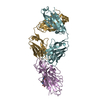
- Assembly
| Deposited unit | 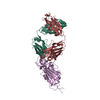
| ||||||||
|---|---|---|---|---|---|---|---|---|---|
| 1 |
| ||||||||
| Unit cell |
|
-Components
| #1: Antibody | Mass: 23491.293 Da / Num. of mol.: 1 Source method: isolated from a genetically manipulated source Source: (gene. exp.) Homo sapiens (human) / Cell line (production host): 293EBNA / Production host: Homo sapiens (human) |
|---|---|
| #2: Antibody | Mass: 23384.883 Da / Num. of mol.: 1 Source method: isolated from a genetically manipulated source Source: (gene. exp.) Homo sapiens (human) / Cell line (production host): 293EBNA / Production host: Homo sapiens (human) |
| #3: Protein | Mass: 30594.381 Da / Num. of mol.: 1 / Fragment: receptor binding domain Source method: isolated from a genetically manipulated source Source: (gene. exp.) Severe acute respiratory syndrome coronavirus 2Gene: S, 2 / Cell line (production host): 293SGlycoDelete / Production host: Homo sapiens (human) / References: UniProt: P0DTC2 |
| #4: Sugar | ChemComp-NAG / N-Acetylglucosamine  Type: D-saccharide, beta linking / Mass: 221.208 Da / Num. of mol.: 1 Type: D-saccharide, beta linking / Mass: 221.208 Da / Num. of mol.: 1Source method: isolated from a genetically manipulated source Formula: C8H15NO6 |
| #5: Water | ChemComp-HOH / Water Mass: 18.015 Da / Num. of mol.: 21 / Source method: isolated from a natural source / Formula: H2O Mass: 18.015 Da / Num. of mol.: 21 / Source method: isolated from a natural source / Formula: H2O |
| Has ligand of interest | N |
-Experimental details
-Experiment
| Experiment | Method: X-RAY DIFFRACTION / Number of used crystals: 1 |
|---|
- Sample preparation
| Crystal | Density Matthews: 3.62 Å3/Da / Density % sol: 66.02 % |
|---|---|
| Crystal grow | Temperature: 298 K / Method: vapor diffusion, hanging drop / pH: 7 / Details: 0.2M ammonium citrate, tribasic, 12% PEG 3350 |
-Data collection
| Diffraction | Mean temperature: 100 K / Serial crystal experiment: N |
|---|---|
| Diffraction source | Source: SYNCHROTRON / Site: APS  / Beamline: 19-ID / Wavelength: 0.9792 Å / Beamline: 19-ID / Wavelength: 0.9792 Å |
| Detector | Type: DECTRIS PILATUS 6M / Detector: PIXEL / Date: May 15, 2020 |
| Radiation | Protocol: SINGLE WAVELENGTH / Monochromatic (M) / Laue (L): M / Scattering type: x-ray |
| Radiation wavelength | Wavelength: 0.9792 Å / Relative weight: 1 |
| Reflection | Resolution: 2.75→48.26 Å / Num. obs: 28897 / % possible obs: 99.93 % / Redundancy: 2 % / CC1/2: 0.999 / Rmerge(I) obs: 0.03068 / Rrim(I) all: 0.04338 / Net I/σ(I): 13.01 |
| Reflection shell | Resolution: 2.75→2.85 Å / Redundancy: 2 % / Rmerge(I) obs: 0.4917 / Mean I/σ(I) obs: 1.45 / Num. unique obs: 2862 / CC1/2: 0.573 / % possible all: 99.97 |
- Processing
| Software |
| ||||||||||||||||||||||||||||||||||||||||||||||||||||||||||||||||||
|---|---|---|---|---|---|---|---|---|---|---|---|---|---|---|---|---|---|---|---|---|---|---|---|---|---|---|---|---|---|---|---|---|---|---|---|---|---|---|---|---|---|---|---|---|---|---|---|---|---|---|---|---|---|---|---|---|---|---|---|---|---|---|---|---|---|---|---|
| Refinement | Method to determine structure: MOLECULAR REPLACEMENT Starting model: 6LZG, 5I1E Resolution: 2.75→48.26 Å / SU ML: 0.41 / Cross valid method: THROUGHOUT / σ(F): 1.35 / Phase error: 27.29 / Stereochemistry target values: ML
| ||||||||||||||||||||||||||||||||||||||||||||||||||||||||||||||||||
| Solvent computation | Shrinkage radii: 0.9 Å / VDW probe radii: 1.11 Å / Solvent model: FLAT BULK SOLVENT MODEL | ||||||||||||||||||||||||||||||||||||||||||||||||||||||||||||||||||
| Displacement parameters | Biso max: 164.59 Å2 / Biso mean: 78.6799 Å2 / Biso min: 42.75 Å2 | ||||||||||||||||||||||||||||||||||||||||||||||||||||||||||||||||||
| Refinement step | Cycle: final / Resolution: 2.75→48.26 Å
| ||||||||||||||||||||||||||||||||||||||||||||||||||||||||||||||||||
| Refine LS restraints |
| ||||||||||||||||||||||||||||||||||||||||||||||||||||||||||||||||||
| LS refinement shell | Refine-ID: X-RAY DIFFRACTION / Rfactor Rfree error: 0 / Total num. of bins used: 10 / % reflection obs: 100 %
|
