 Movie
Movie Controller

[English] 日本語
 Yorodumi
- PDB-4mwf: Structure of Hepatitis C Virus Envelope Glycoprotein E2 core boun... -
+ Open data

- Basic information
| Entry | Database: PDB / ID: 4mwf | |||||||||
|---|---|---|---|---|---|---|---|---|---|---|
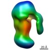
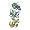
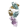
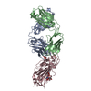
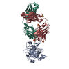
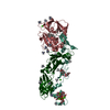
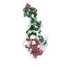
| Title | Structure of Hepatitis C Virus Envelope Glycoprotein E2 core bound to broadly neutralizing antibody AR3C | |||||||||
 Components Components |
| |||||||||
 Keywords Keywords | IMMUNE SYSTEM / Immunoglobulin Fold / HCV E2 | |||||||||
| Function / homology |  Function and homology information Function and homology informationsymbiont-mediated suppression of host JAK-STAT cascade via inhibition of STAT activity / positive regulation of hexokinase activity / translocation of peptides or proteins into host cell cytoplasm / symbiont-mediated perturbation of host cellular process / Toll-like receptor 2 binding / viral capsid assembly / hepacivirin / adhesion receptor-mediated virion attachment to host cell / TBC/RABGAPs / host cell mitochondrial membrane ...symbiont-mediated suppression of host JAK-STAT cascade via inhibition of STAT activity / positive regulation of hexokinase activity / translocation of peptides or proteins into host cell cytoplasm / symbiont-mediated perturbation of host cellular process / Toll-like receptor 2 binding / viral capsid assembly / hepacivirin / adhesion receptor-mediated virion attachment to host cell / TBC/RABGAPs / host cell mitochondrial membrane / host cell lipid droplet / symbiont-mediated transformation of host cell / symbiont-mediated suppression of host TRAF-mediated signal transduction / positive regulation of cytokinesis / symbiont-mediated perturbation of host cell cycle G1/S transition checkpoint / negative regulation of protein secretion / symbiont-mediated suppression of host JAK-STAT cascade via inhibition of STAT1 activity / endoplasmic reticulum-Golgi intermediate compartment membrane / symbiont-mediated suppression of host cytoplasmic pattern recognition receptor signaling pathway via inhibition of MAVS activity / SH3 domain binding / kinase binding / nucleoside-triphosphate phosphatase / viral nucleocapsid / channel activity / monoatomic ion transmembrane transport / clathrin-dependent endocytosis of virus by host cell / entry receptor-mediated virion attachment to host cell / Hydrolases; Acting on peptide bonds (peptidases); Cysteine endopeptidases / RNA helicase activity / host cell perinuclear region of cytoplasm / host cell endoplasmic reticulum membrane / RNA helicase / symbiont-mediated suppression of host type I interferon-mediated signaling pathway / ribonucleoprotein complex / serine-type endopeptidase activity / viral translational frameshifting / symbiont-mediated activation of host autophagy / RNA-directed RNA polymerase / cysteine-type endopeptidase activity / viral RNA genome replication / RNA-directed RNA polymerase activity / fusion of virus membrane with host endosome membrane / viral envelope / host cell nucleus / host cell plasma membrane / virion membrane / structural molecule activity / negative regulation of transcription by RNA polymerase II / ATP hydrolysis activity / proteolysis / RNA binding / zinc ion binding / ATP binding Similarity search - Function | |||||||||
| Biological species |  Homo sapiens (human) Homo sapiens (human) Hepatitis C virus Hepatitis C virus | |||||||||
| Method |  X-RAY DIFFRACTION / SYNCHROTRON / MOLECULAR REPLACEMENT / Resolution: 2.645 Å X-RAY DIFFRACTION / SYNCHROTRON / MOLECULAR REPLACEMENT / Resolution: 2.645 Å | |||||||||
 Authors Authors | Kong, L. / Wilson, I.A. / Law, M. | |||||||||
 Citation Citation | Journal: Science / Year: 2013 Title: Hepatitis C virus E2 envelope glycoprotein core structure. Authors: Leopold Kong / Erick Giang / Travis Nieusma / Rameshwar U Kadam / Kristin E Cogburn / Yuanzi Hua / Xiaoping Dai / Robyn L Stanfield / Dennis R Burton / Andrew B Ward / Ian A Wilson / Mansun Law /  Abstract: Hepatitis C virus (HCV), a Hepacivirus, is a major cause of viral hepatitis, liver cirrhosis, and hepatocellular carcinoma. HCV envelope glycoproteins E1 and E2 mediate fusion and entry into host ...Hepatitis C virus (HCV), a Hepacivirus, is a major cause of viral hepatitis, liver cirrhosis, and hepatocellular carcinoma. HCV envelope glycoproteins E1 and E2 mediate fusion and entry into host cells and are the primary targets of the humoral immune response. The crystal structure of the E2 core bound to broadly neutralizing antibody AR3C at 2.65 angstroms reveals a compact architecture composed of a central immunoglobulin-fold β sandwich flanked by two additional protein layers. The CD81 receptor binding site was identified by electron microscopy and site-directed mutagenesis and overlaps with the AR3C epitope. The x-ray and electron microscopy E2 structures differ markedly from predictions of an extended, three-domain, class II fusion protein fold and therefore provide valuable information for HCV drug and vaccine design. | |||||||||
| History |
|
- Structure visualization
| Structure viewer | Molecule: MolmilJmol/JSmol |
|---|
- Downloads & links
-Download
| PDBx/mmCIF format | 4mwf.cif.gz | 249.9 KB | Display | PDBx/mmCIF format |
|---|---|---|---|---|
| PDB format | pdb4mwf.ent.gz | 198.2 KB | Display | PDB format |
| PDBx/mmJSON format | 4mwf.json.gz | Tree view | PDBx/mmJSON format | |
| Others |  Other downloads Other downloads |
-Validation report
| Arichive directory | https://data.pdbj.org/pub/pdb/validation_reports/mw/4mwfftp://data.pdbj.org/pub/pdb/validation_reports/mw/4mwf | HTTPS FTP |
|---|
-Related structure data


-Links
 PDBj
PDBj


- Assembly
| Deposited unit | 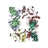
| ||||||||
|---|---|---|---|---|---|---|---|---|---|
| 1 | 
| ||||||||
| 2 | 
| ||||||||
| Unit cell |
|
-Components
-Protein , 1 types, 2 molecules CD
| #3: Protein | Mass: 23286.412 Da / Num. of mol.: 2 / Mutation: N448D, N576D Source method: isolated from a genetically manipulated source Source: (gene. exp.) Hepatitis C virus (isolate H) / Cell line (production host): HEK 293 Freestyle / Production host: Homo sapiens (human) / References: UniProt: P27958 |
|---|
-Antibody , 2 types, 4 molecules AHBL
| #1: Antibody | Mass: 24627.498 Da / Num. of mol.: 2 Source method: isolated from a genetically manipulated source Source: (gene. exp.) Homo sapiens (human) / Production host:  #2: Antibody | Mass: 23266.812 Da / Num. of mol.: 2 Source method: isolated from a genetically manipulated source Source: (gene. exp.) Homo sapiens (human) / Production host: |
|---|
-Sugars , 3 types, 10 molecules 
| #4: Polysaccharide | alpha-D-mannopyranose-(1-2)-alpha-D-mannopyranose-(1-2)-alpha-D-mannopyranose-(1-3)-[alpha-D- ...alpha-D-mannopyranose-(1-2)-alpha-D-mannopyranose-(1-2)-alpha-D-mannopyranose-(1-3)-[alpha-D-mannopyranose-(1-6)-alpha-D-mannopyranose-(1-6)]beta-D-mannopyranose-(1-4)-2-acetamido-2-deoxy-beta-D-glucopyranose-(1-4)-2-acetamido-2-deoxy-beta-D-glucopyranose Source method: isolated from a genetically manipulated source | ||
|---|---|---|---|
| #5: Polysaccharide | Source method: isolated from a genetically manipulated source #6: Sugar | ChemComp-NAG /  Type: D-saccharide, beta linking / Mass: 221.208 Da / Num. of mol.: 7 Type: D-saccharide, beta linking / Mass: 221.208 Da / Num. of mol.: 7Source method: isolated from a genetically manipulated source Formula: C8H15NO6 |
-Details
| Has protein modification | Y |
|---|
-Experimental details
-Experiment
| Experiment | Method: X-RAY DIFFRACTION / Number of used crystals: 5 |
|---|
- Sample preparation
| Crystal | Density Matthews: 2.89 Å3/Da / Density % sol: 57.5 % |
|---|---|
| Crystal grow | Temperature: 293 K / Method: vapor diffusion, sitting drop / pH: 7.5 Details: 20% (w/v) PEG 4000, 10% (v/v) isopropanol, and 0.1 M HEPES pH 7.5, VAPOR DIFFUSION, SITTING DROP, temperature 293K |
-Data collection
| Diffraction | Mean temperature: 100 K |
|---|---|
| Diffraction source | Source: SYNCHROTRON / Site: SSRL / Beamline: BL12-2 / Wavelength: 1.033 Å |
| Detector | Type: MARMOSAIC 325 mm CCD / Detector: CCD / Date: Jan 19, 2013 |
| Radiation | Protocol: SINGLE WAVELENGTH / Monochromatic (M) / Laue (L): M / Scattering type: x-ray |
| Radiation wavelength | Wavelength: 1.033 Å / Relative weight: 1 |
| Reflection | Resolution: 2.65→49.09 Å / Num. obs: 46174 / % possible obs: 93.4 % / Observed criterion σ(F): 0 / Observed criterion σ(I): -3 |
| Reflection shell | Resolution: 2.65→2.7 Å / % possible all: 76.5 |
- Processing
| Software |
| ||||||||||||||||||||||||||||||||||||||||||||||||||||||||||||||||||||||||||||||||||||||||||||||||||||||||||||||||||||||||||||||
|---|---|---|---|---|---|---|---|---|---|---|---|---|---|---|---|---|---|---|---|---|---|---|---|---|---|---|---|---|---|---|---|---|---|---|---|---|---|---|---|---|---|---|---|---|---|---|---|---|---|---|---|---|---|---|---|---|---|---|---|---|---|---|---|---|---|---|---|---|---|---|---|---|---|---|---|---|---|---|---|---|---|---|---|---|---|---|---|---|---|---|---|---|---|---|---|---|---|---|---|---|---|---|---|---|---|---|---|---|---|---|---|---|---|---|---|---|---|---|---|---|---|---|---|---|---|---|---|
| Refinement | Method to determine structure: MOLECULAR REPLACEMENT / Resolution: 2.645→49.078 Å / SU ML: 0.39 / σ(F): 1.35 / Phase error: 31.71 / Stereochemistry target values: ML
| ||||||||||||||||||||||||||||||||||||||||||||||||||||||||||||||||||||||||||||||||||||||||||||||||||||||||||||||||||||||||||||||
| Solvent computation | Shrinkage radii: 0.9 Å / VDW probe radii: 1.11 Å / Solvent model: FLAT BULK SOLVENT MODEL | ||||||||||||||||||||||||||||||||||||||||||||||||||||||||||||||||||||||||||||||||||||||||||||||||||||||||||||||||||||||||||||||
| Refinement step | Cycle: LAST / Resolution: 2.645→49.078 Å
| ||||||||||||||||||||||||||||||||||||||||||||||||||||||||||||||||||||||||||||||||||||||||||||||||||||||||||||||||||||||||||||||
| Refine LS restraints |
| ||||||||||||||||||||||||||||||||||||||||||||||||||||||||||||||||||||||||||||||||||||||||||||||||||||||||||||||||||||||||||||||
| LS refinement shell |
|
