 Movie
Movie Controller


+ Open data

- Basic information
| Entry | Database: EMDB / ID: EMD-5761 | |||||||||
|---|---|---|---|---|---|---|---|---|---|---|
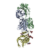
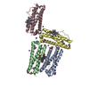
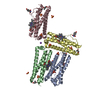
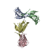
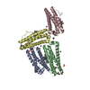
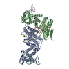
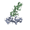
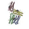
| Title | Hepatitis C Virus E2 Envelope Glycoprotein Core Structure | |||||||||
 Map data Map data | E2 glycoprotein bound to AR2A Fab and CD81, single particle reconstruction | |||||||||
 Sample Sample |
| |||||||||
 Keywords Keywords | hepatitis C / virus / envelope / glycoprotein | |||||||||
| Biological species |  Hepatitis C virus / Hepatitis C virus /  Homo sapiens (human) Homo sapiens (human) | |||||||||
| Method | single particle reconstruction / negative staining / Resolution: 19.0 Å | |||||||||
 Authors Authors | Nieusma T / Kong L / Giang E / Kadam RU / Cogburn KE / Hua Y / Dai X / Stanfield RL / Burton DR / Wilson IA ...Nieusma T / Kong L / Giang E / Kadam RU / Cogburn KE / Hua Y / Dai X / Stanfield RL / Burton DR / Wilson IA / Law M / Ward AB | |||||||||
 Citation Citation | Journal: Science / Year: 2013 Title: Hepatitis C virus E2 envelope glycoprotein core structure. Authors: Leopold Kong / Erick Giang / Travis Nieusma / Rameshwar U Kadam / Kristin E Cogburn / Yuanzi Hua / Xiaoping Dai / Robyn L Stanfield / Dennis R Burton / Andrew B Ward / Ian A Wilson / Mansun Law /  Abstract: Hepatitis C virus (HCV), a Hepacivirus, is a major cause of viral hepatitis, liver cirrhosis, and hepatocellular carcinoma. HCV envelope glycoproteins E1 and E2 mediate fusion and entry into host ...Hepatitis C virus (HCV), a Hepacivirus, is a major cause of viral hepatitis, liver cirrhosis, and hepatocellular carcinoma. HCV envelope glycoproteins E1 and E2 mediate fusion and entry into host cells and are the primary targets of the humoral immune response. The crystal structure of the E2 core bound to broadly neutralizing antibody AR3C at 2.65 angstroms reveals a compact architecture composed of a central immunoglobulin-fold β sandwich flanked by two additional protein layers. The CD81 receptor binding site was identified by electron microscopy and site-directed mutagenesis and overlaps with the AR3C epitope. The x-ray and electron microscopy E2 structures differ markedly from predictions of an extended, three-domain, class II fusion protein fold and therefore provide valuable information for HCV drug and vaccine design. | |||||||||
| History |
|
- Structure visualization
| Movie |
 Movie viewer Movie viewer |
|---|---|
| Structure viewer | EM map: SurfViewMolmilJmol/JSmol |
| Supplemental images |
 UCSF Chimera
- Downloads & links
-EMDB archive
| Map data | emd_5761.map.gz | 3.1 MB | EMDB map data format | |
|---|---|---|---|---|
| Header (meta data) | emd-5761-v30.xmlemd-5761.xml | 11 KB 11 KB | Display Display | EMDB header |
| Images |  emd_5761_1.png emd_5761_1.png | 63.9 KB | ||
| Archive directory |  http://ftp.pdbj.org/pub/emdb/structures/EMD-5761ftp://ftp.pdbj.org/pub/emdb/structures/EMD-5761 http://ftp.pdbj.org/pub/emdb/structures/EMD-5761ftp://ftp.pdbj.org/pub/emdb/structures/EMD-5761 | HTTPS FTP |
-Related structure data


-Links
| EMDB pages | EMDB (EBI/PDBe) / EMDataResource |
|---|
-Map
| File | Download / File: emd_5761.map.gz / Format: CCP4 / Size: 3.3 MB / Type: IMAGE STORED AS FLOATING POINT NUMBER (4 BYTES) | ||||||||||||||||||||||||||||||||||||||||||||||||||||||||||||||||||||
|---|---|---|---|---|---|---|---|---|---|---|---|---|---|---|---|---|---|---|---|---|---|---|---|---|---|---|---|---|---|---|---|---|---|---|---|---|---|---|---|---|---|---|---|---|---|---|---|---|---|---|---|---|---|---|---|---|---|---|---|---|---|---|---|---|---|---|---|---|---|
| Annotation | E2 glycoprotein bound to AR2A Fab and CD81, single particle reconstruction | ||||||||||||||||||||||||||||||||||||||||||||||||||||||||||||||||||||
| Projections & slices | Image control
Images are generated by Spider. | ||||||||||||||||||||||||||||||||||||||||||||||||||||||||||||||||||||
| Voxel size | X=Y=Z: 2.05 Å | ||||||||||||||||||||||||||||||||||||||||||||||||||||||||||||||||||||
| Density |
| ||||||||||||||||||||||||||||||||||||||||||||||||||||||||||||||||||||
| Symmetry | Space group: 1 | ||||||||||||||||||||||||||||||||||||||||||||||||||||||||||||||||||||
| Details | EMDB XML:
CCP4 map header:
| ||||||||||||||||||||||||||||||||||||||||||||||||||||||||||||||||||||
 Z (Sec.)
 Y (Row.)
 X (Col.)


-Supplemental data
- Sample components
-Entire : E2 glycoprotein bound to AR2A Fab and CD81
| Entire | Name: E2 glycoprotein bound to AR2A Fab and CD81 |
|---|---|
| Components |
|
-Supramolecule #1000: E2 glycoprotein bound to AR2A Fab and CD81
| Supramolecule | Name: E2 glycoprotein bound to AR2A Fab and CD81 / type: sample / ID: 1000 / Number unique components: 3 |
|---|
-Macromolecule #1: E2 envelope glycoprotein
| Macromolecule | Name: E2 envelope glycoprotein / type: protein_or_peptide / ID: 1 / Name.synonym: E2 / Number of copies: 1 / Oligomeric state: monomer / Recombinant expression: Yes |
|---|---|
| Source (natural) | Organism: Hepatitis C virus |
| Molecular weight | Theoretical: 46 KDa |
| Recombinant expression | Organism: Homo sapiens (human) / Recombinant cell: HEK 293T / Recombinant plasmid: pCMV-Tag4A-tpa |
-Macromolecule #2: Antigen-binding fragment of antibody AR2A
| Macromolecule | Name: Antigen-binding fragment of antibody AR2A / type: protein_or_peptide / ID: 2 / Number of copies: 1 / Oligomeric state: monomer / Recombinant expression: Yes |
|---|---|
| Source (natural) | Organism: Homo sapiens (human) / synonym: Human |
| Molecular weight | Theoretical: 50 KDa |
| Recombinant expression | Organism:  |
-Macromolecule #3: CD81
| Macromolecule | Name: CD81 / type: protein_or_peptide / ID: 3 / Number of copies: 1 / Oligomeric state: dimer / Recombinant expression: Yes |
|---|---|
| Source (natural) | Organism: Homo sapiens (human) / synonym: Human |
| Recombinant expression | Organism: |
-Experimental details
-Structure determination
| Method | negative staining |
|---|---|
 Processing Processing | single particle reconstruction |
| Aggregation state | particle |
-Sample preparation
| Staining | Type: NEGATIVE / Details: negative stain with uranyl formate |
|---|---|
| Grid | Details: 400 mesh copper |
| Vitrification | Cryogen name: NONE / Instrument: OTHER |
- Electron microscopy
| Microscope | FEI TECNAI SPIRIT |
|---|---|
| Temperature | Average: 298 K |
| Date | May 24, 2013 |
| Image recording | Category: CCD / Film or detector model: TVIPS TEMCAM-F416 (4k x 4k) / Number real images: 319 |
| Tilt angle min | 0 |
| Electron beam | Acceleration voltage: 120 kV / Electron source: LAB6 |
| Electron optics | Illumination mode: FLOOD BEAM / Imaging mode: BRIGHT FIELD / Nominal defocus max: 1.0 µm / Nominal defocus min: 1.0 µm / Nominal magnification: 52000 |
| Sample stage | Specimen holder model: SIDE ENTRY, EUCENTRIC / Tilt angle max: 55 |
| Experimental equipment |  Model: Tecnai Spirit / Image courtesy: FEI Company |
-Image processing
| Details | Particles were selected using automatic (difference-of-Gaussians) picking followed by reference-free classification to eliminate noisy picks or non-target aggregation states. |
|---|---|
| Final reconstruction | Algorithm: OTHER / Resolution.type: BY AUTHOR / Resolution: 19.0 Å / Resolution method: FSC 0.5 CUT-OFF / Software - Name: Appion, Spider, Xmipp, Eman1, Eman2 / Number images used: 32115 |
