 Movie
Movie Controller

[English] 日本語
 Yorodumi
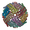
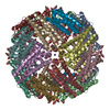
- PDB-6nlg: 1.50 A resolution structure of BfrB (C89S/K96C) from Pseudomonas ... -
+ Open data

- Basic information
| Entry | Database: PDB / ID: 6nlg | ||||||||||||
|---|---|---|---|---|---|---|---|---|---|---|---|---|---|
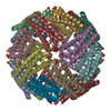
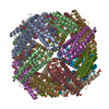
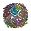
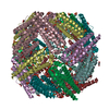
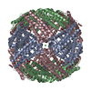
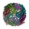
| Title | 1.50 A resolution structure of BfrB (C89S/K96C) from Pseudomonas aeruginosa in complex with a small molecule fragment (analog 1) | ||||||||||||
 Components Components | Bacterioferritin | ||||||||||||
 Keywords Keywords | OXIDOREDUCTASE / ELECTRON TRANSPORT / IRON STORAGE / IRON BINDING / IRON MOBILIZATION / PROTEIN-PROTEIN INTERACTION INHIBITOR | ||||||||||||
| Function / homology |  Function and homology information Function and homology informationiron ion sequestering activity / ferritin complex / ferroxidase / ferroxidase activity / ferric iron binding / iron ion transport / intracellular iron ion homeostasis / iron ion binding / heme binding / cytosol Similarity search - Function | ||||||||||||
| Biological species |   Pseudomonas aeruginosa (bacteria) Pseudomonas aeruginosa (bacteria) | ||||||||||||
| Method |  X-RAY DIFFRACTION / SYNCHROTRON / MOLECULAR REPLACEMENT / molecular replacement / Resolution: 1.5 Å X-RAY DIFFRACTION / SYNCHROTRON / MOLECULAR REPLACEMENT / molecular replacement / Resolution: 1.5 Å | ||||||||||||
 Authors Authors | Lovell, S. / Punchi-Hewage, A. / Battaile, K.P. / Yao, H. / Nammalwar, B. / Gnanasekaran, K.K. / Bunce, R.A. / Reitz, A.B. / Rivera, M. | ||||||||||||
| Funding support |  United States, 3items United States, 3items
| ||||||||||||
 Citation Citation | Journal: J.Am.Chem.Soc. / Year: 2019 Title: Small Molecule Inhibitors of the BfrB-Bfd Interaction Decrease Pseudomonas aeruginosa Fitness and Potentiate Fluoroquinolone Activity. Authors: Punchi Hewage, A.N.D. / Yao, H. / Nammalwar, B. / Gnanasekaran, K.K. / Lovell, S. / Bunce, R.A. / Eshelman, K. / Phaniraj, S.M. / Lee, M.M. / Peterson, B.R. / Battaile, K.P. / Reitz, A.B. / Rivera, M. | ||||||||||||
| History |
|
- Structure visualization
| Structure viewer | Molecule: MolmilJmol/JSmol |
|---|
- Downloads & links
-Download
| PDBx/mmCIF format | 6nlg.cif.gz | 162.6 KB | Display | PDBx/mmCIF format |
|---|---|---|---|---|
| PDB format | pdb6nlg.ent.gz | 127.6 KB | Display | PDB format |
| PDBx/mmJSON format | 6nlg.json.gz | Tree view | PDBx/mmJSON format | |
| Others |  Other downloads Other downloads |
-Validation report
| Arichive directory | https://data.pdbj.org/pub/pdb/validation_reports/nl/6nlgftp://data.pdbj.org/pub/pdb/validation_reports/nl/6nlg | HTTPS FTP |
|---|
-Related structure data
| Related structure data | 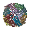 6nlfC  6nliC 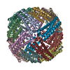 6nljC 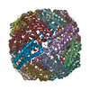 6nlkC  6nllC 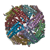 6nlmC 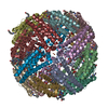 6nlnC 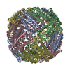 4tofS S: Starting model for refinement C: citing same article ( |
|---|---|
| Similar structure data |


-Links
 PDBj
PDBj


- Assembly
| Deposited unit | 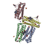
| ||||||||||||||||||||||||
|---|---|---|---|---|---|---|---|---|---|---|---|---|---|---|---|---|---|---|---|---|---|---|---|---|---|
| 1 | x 6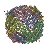
| ||||||||||||||||||||||||
| Unit cell |
| ||||||||||||||||||||||||
| Components on special symmetry positions |
|
-Components
-Protein , 1 types, 4 molecules ABCD
| #1: Protein | Mass: 18538.066 Da / Num. of mol.: 4 / Mutation: C89S, K96C Source method: isolated from a genetically manipulated source Source: (gene. exp.) Pseudomonas aeruginosa (bacteria) / Gene: bfrB, PA3531 / Plasmid: PET11A / Production host: |
|---|
-Non-polymers , 6 types, 569 molecules 


| #2: Chemical |  Mass: 163.130 Da / Num. of mol.: 3 / Source method: obtained synthetically / Formula: C8H5NO3 Mass: 163.130 Da / Num. of mol.: 3 / Source method: obtained synthetically / Formula: C8H5NO3#3: Chemical |  Mass: 616.487 Da / Num. of mol.: 3 / Source method: obtained synthetically / Formula: C34H32FeN4O4 Mass: 616.487 Da / Num. of mol.: 3 / Source method: obtained synthetically / Formula: C34H32FeN4O4#4: Chemical | ChemComp-MPD / (  Mass: 118.174 Da / Num. of mol.: 4 / Source method: obtained synthetically / Formula: C6H14O2 / Comment: precipitant*YM Mass: 118.174 Da / Num. of mol.: 4 / Source method: obtained synthetically / Formula: C6H14O2 / Comment: precipitant*YM#5: Chemical | ChemComp-SO4 /  Mass: 96.063 Da / Num. of mol.: 11 / Source method: obtained synthetically / Formula: SO4 Mass: 96.063 Da / Num. of mol.: 11 / Source method: obtained synthetically / Formula: SO4#6: Chemical | ChemComp-K / |  Mass: 39.098 Da / Num. of mol.: 1 / Source method: obtained synthetically / Formula: K Mass: 39.098 Da / Num. of mol.: 1 / Source method: obtained synthetically / Formula: K#7: Water | ChemComp-HOH / | Mass: 18.015 Da / Num. of mol.: 547 / Source method: isolated from a natural source / Formula: H2O |
|---|
-Details
| Has protein modification | Y |
|---|
-Experimental details
-Experiment
| Experiment | Method: X-RAY DIFFRACTION / Number of used crystals: 1 |
|---|
- Sample preparation
| Crystal | Density Matthews: 3.52 Å3/Da / Density % sol: 65.1 % |
|---|---|
| Crystal grow | Temperature: 291 K / Method: vapor diffusion, sitting drop / pH: 6.5 Details: 35% (v/v) 2-methyl-2,4-pentanediol, 0.1M MES, 0.2M Li2SO4 |
-Data collection
| Diffraction | Mean temperature: 100 K / Serial crystal experiment: N |
|---|---|
| Diffraction source | Source: SYNCHROTRON / Site: APS / Beamline: 17-ID / Wavelength: 1 Å |
| Detector | Type: DECTRIS PILATUS 6M / Detector: PIXEL / Date: Dec 8, 2012 |
| Radiation | Protocol: SINGLE WAVELENGTH / Monochromatic (M) / Laue (L): M / Scattering type: x-ray |
| Radiation wavelength | Wavelength: 1 Å / Relative weight: 1 |
| Reflection | Resolution: 1.5→47.84 Å / Num. obs: 168538 / % possible obs: 99.6 % / Redundancy: 19.9 % / Biso Wilson estimate: 17.26 Å2 / CC1/2: 0.999 / Rmerge(I) obs: 0.106 / Rpim(I) all: 0.024 / Rrim(I) all: 0.108 / Net I/σ(I): 19.2 |
| Reflection shell | Resolution: 1.5→1.53 Å / Redundancy: 20.1 % / Rmerge(I) obs: 1.85 / Mean I/σ(I) obs: 2.2 / Num. unique obs: 8167 / CC1/2: 0.768 / Rpim(I) all: 0.417 / Rrim(I) all: 1.897 / % possible all: 98.9 |
-Phasing
| Phasing | Method: molecular replacement | |||||||||
|---|---|---|---|---|---|---|---|---|---|---|
| Phasing MR | Model details: Phaser MODE: MR_AUTO
|
- Processing
| Software |
| |||||||||||||||||||||||||||||||||||||||||||||||||||||||||||||||||||||||||||||||||||||||||||||||||||||||||||||||||||||||||||||||||||||||||||||||||||||||||||||||||||||||||||||||||||||||||||||||||||||||||||||||||||||||||
|---|---|---|---|---|---|---|---|---|---|---|---|---|---|---|---|---|---|---|---|---|---|---|---|---|---|---|---|---|---|---|---|---|---|---|---|---|---|---|---|---|---|---|---|---|---|---|---|---|---|---|---|---|---|---|---|---|---|---|---|---|---|---|---|---|---|---|---|---|---|---|---|---|---|---|---|---|---|---|---|---|---|---|---|---|---|---|---|---|---|---|---|---|---|---|---|---|---|---|---|---|---|---|---|---|---|---|---|---|---|---|---|---|---|---|---|---|---|---|---|---|---|---|---|---|---|---|---|---|---|---|---|---|---|---|---|---|---|---|---|---|---|---|---|---|---|---|---|---|---|---|---|---|---|---|---|---|---|---|---|---|---|---|---|---|---|---|---|---|---|---|---|---|---|---|---|---|---|---|---|---|---|---|---|---|---|---|---|---|---|---|---|---|---|---|---|---|---|---|---|---|---|---|---|---|---|---|---|---|---|---|---|---|---|---|---|---|---|---|
| Refinement | Method to determine structure: MOLECULAR REPLACEMENT Starting model: 4TOF Resolution: 1.5→38.352 Å / SU ML: 0.11 / Cross valid method: THROUGHOUT / σ(F): 1.09 / Phase error: 14.78
| |||||||||||||||||||||||||||||||||||||||||||||||||||||||||||||||||||||||||||||||||||||||||||||||||||||||||||||||||||||||||||||||||||||||||||||||||||||||||||||||||||||||||||||||||||||||||||||||||||||||||||||||||||||||||
| Solvent computation | Shrinkage radii: 0.9 Å / VDW probe radii: 1.11 Å | |||||||||||||||||||||||||||||||||||||||||||||||||||||||||||||||||||||||||||||||||||||||||||||||||||||||||||||||||||||||||||||||||||||||||||||||||||||||||||||||||||||||||||||||||||||||||||||||||||||||||||||||||||||||||
| Displacement parameters | Biso max: 83.41 Å2 / Biso mean: 21.0112 Å2 / Biso min: 11.8 Å2 | |||||||||||||||||||||||||||||||||||||||||||||||||||||||||||||||||||||||||||||||||||||||||||||||||||||||||||||||||||||||||||||||||||||||||||||||||||||||||||||||||||||||||||||||||||||||||||||||||||||||||||||||||||||||||
| Refinement step | Cycle: final / Resolution: 1.5→38.352 Å
| |||||||||||||||||||||||||||||||||||||||||||||||||||||||||||||||||||||||||||||||||||||||||||||||||||||||||||||||||||||||||||||||||||||||||||||||||||||||||||||||||||||||||||||||||||||||||||||||||||||||||||||||||||||||||
| Refine LS restraints |
| |||||||||||||||||||||||||||||||||||||||||||||||||||||||||||||||||||||||||||||||||||||||||||||||||||||||||||||||||||||||||||||||||||||||||||||||||||||||||||||||||||||||||||||||||||||||||||||||||||||||||||||||||||||||||
| LS refinement shell | Refine-ID: X-RAY DIFFRACTION / Rfactor Rfree error: 0 / Total num. of bins used: 30
|
