 Movie
Movie Controller

[English] 日本語
 Yorodumi
- PDB-1nfv: X-ray structure of Desulfovibrio desulfuricans bacterioferritin: ... -
+ Open data

- Basic information
| Entry | Database: PDB / ID: 1nfv | ||||||
|---|---|---|---|---|---|---|---|
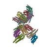
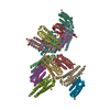
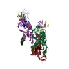
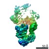
| Title | X-ray structure of Desulfovibrio desulfuricans bacterioferritin: the diiron centre in different catalytic states (as-isolated structure) | ||||||
 Components Components | bacterioferritin | ||||||
 Keywords Keywords | IRON STORAGE/ELECTRON TRANSPORT / bacterioferritin / 24 subunits in the active molecule / diiron centre / haem Fe-coproporphyrin III cofactor / IRON STORAGE-ELECTRON TRANSPORT COMPLEX | ||||||
| Function / homology |  Function and homology information Function and homology informationferroxidase / ferroxidase activity / ferric iron binding / iron ion transport / intracellular iron ion homeostasis / heme binding / cytosol Similarity search - Function | ||||||
| Biological species |  Desulfovibrio desulfuricans (bacteria) Desulfovibrio desulfuricans (bacteria) | ||||||
| Method |  X-RAY DIFFRACTION / SYNCHROTRON / MAD / Resolution: 1.95 Å X-RAY DIFFRACTION / SYNCHROTRON / MAD / Resolution: 1.95 Å | ||||||
 Authors Authors | Macedo, S. / Romao, C.V. / Mitchell, E. / Matias, P.M. / Liu, M.Y. / Xavier, A.V. / LeGall, J. / Teixeira, M. / Lindley, P. / Carrondo, M.A. | ||||||
 Citation Citation | Journal: NAT.STRUCT.BIOL. / Year: 2003 Title: The nature of the di-iron site in the bacterioferritin from Desulfovibrio desulfuricans Authors: Macedo, S. / Romao, C.V. / Mitchell, E. / Matias, P.M. / Liu, M.Y. / Xavier, A.V. / LeGall, J. / Teixeira, M. / Lindley, P. / Carrondo, M.A. #1: Journal: Acta Crystallogr.,Sect.D / Year: 2001Title: Structure determination of bacterioferritin from Desulfovibrio desulfuricans by the MAD method at the Fe K-edge Authors: Coelho, A.V. / Macedo, S. / Matias, P.M. / Thompson, A.W. / LeGall, J. / Carrondo, M.A. | ||||||
| History |
| ||||||
| Remark 350 | GENERATING THE BIOMOLECULE THE ACTIVE BIOLOGICAL UNIT IS A 24-MER. SINCE THE ASYMMETRIC UNIT ...GENERATING THE BIOMOLECULE THE ACTIVE BIOLOGICAL UNIT IS A 24-MER. SINCE THE ASYMMETRIC UNIT CONTAINS PARTS OF TWO DIFFERENT SPHERES, APPLY THE SYMMETRY OPERATIONS (-Z, 1/2+X, 1/2-Y) AND (Y-1/2, 1/2-Z, -X) TO MONOMERS A, B, G, H, I, J, M AND N OR THE SYMMETRY OPERATIONS (Z+1/2, 1/2-X, -Y) AND (1/2-Y, -Z, X-1/2) TO THE REMAINING MONOMERS IN THE ASYMMETRIC UNIT TO GENERATE THE 24-MER. |
- Structure visualization
| Structure viewer | Molecule: MolmilJmol/JSmol |
|---|
- Downloads & links
-Download
| PDBx/mmCIF format | 1nfv.cif.gz | 627.7 KB | Display | PDBx/mmCIF format |
|---|---|---|---|---|
| PDB format | pdb1nfv.ent.gz | 520.6 KB | Display | PDB format |
| PDBx/mmJSON format | 1nfv.json.gz | Tree view | PDBx/mmJSON format | |
| Others |  Other downloads Other downloads |
-Validation report
| Arichive directory | https://data.pdbj.org/pub/pdb/validation_reports/nf/1nfvftp://data.pdbj.org/pub/pdb/validation_reports/nf/1nfv | HTTPS FTP |
|---|
-Related structure data


-Links
 PDBj
PDBj


- Assembly
| Deposited unit | 
| |||||||||||||||||||||
|---|---|---|---|---|---|---|---|---|---|---|---|---|---|---|---|---|---|---|---|---|---|---|
| 1 |
| |||||||||||||||||||||
| 2 | 
| |||||||||||||||||||||
| 3 | 
| |||||||||||||||||||||
| Unit cell |
| |||||||||||||||||||||
| Components on special symmetry positions |
| |||||||||||||||||||||
| Details | the biologically relevant molecule is formed applying the following symmetry operations to chains A, B, G, H, I, J, M and N: -z, 1/2+x, 1/2-y and the combination of the three operations 1/2-z, -x, 1/2+y; z, x, y; x-1, y, z; |
-Components
-Protein , 1 types, 16 molecules ABCDEFGHIJKLMNOP
| #1: Protein | Mass: 19906.281 Da / Num. of mol.: 16 / Source method: isolated from a natural source / Source: (natural) Desulfovibrio desulfuricans (bacteria) / Strain: ATCC 27774 / References: GenBank: 14326006, UniProt: Q93PP9*PLUS |
|---|
-Non-polymers , 6 types, 2006 molecules 


| #2: Chemical | ChemComp-FE /  Mass: 55.845 Da / Num. of mol.: 32 / Source method: obtained synthetically / Formula: Fe Mass: 55.845 Da / Num. of mol.: 32 / Source method: obtained synthetically / Formula: Fe#3: Chemical | ChemComp-SO4 /  Mass: 96.063 Da / Num. of mol.: 51 / Source method: obtained synthetically / Formula: SO4 Mass: 96.063 Da / Num. of mol.: 51 / Source method: obtained synthetically / Formula: SO4#4: Chemical | ChemComp-GOL /  Mass: 92.094 Da / Num. of mol.: 24 / Source method: obtained synthetically / Formula: C3H8O3 Mass: 92.094 Da / Num. of mol.: 24 / Source method: obtained synthetically / Formula: C3H8O3#5: Chemical | ChemComp-FEC /  Mass: 708.538 Da / Num. of mol.: 8 / Source method: obtained synthetically / Formula: C36H36FeN4O8 Mass: 708.538 Da / Num. of mol.: 8 / Source method: obtained synthetically / Formula: C36H36FeN4O8#6: Chemical | ChemComp-3PY / |  Mass: 104.061 Da / Num. of mol.: 1 / Source method: obtained synthetically / Formula: C3H4O4 Mass: 104.061 Da / Num. of mol.: 1 / Source method: obtained synthetically / Formula: C3H4O4#7: Water | ChemComp-HOH / | Mass: 18.015 Da / Num. of mol.: 1890 / Source method: isolated from a natural source / Formula: H2O |
|---|
-Details
| Has protein modification | N |
|---|
-Experimental details
-Experiment
| Experiment | Method: X-RAY DIFFRACTION / Number of used crystals: 2 |
|---|
- Sample preparation
| Crystal | Density Matthews: 2.29 Å3/Da / Density % sol: 54 % | ||||||||||||||||||||||||
|---|---|---|---|---|---|---|---|---|---|---|---|---|---|---|---|---|---|---|---|---|---|---|---|---|---|
| Crystal grow | Temperature: 293 K / Method: vapor diffusion, sitting drop / pH: 5.5 Details: ammonium sulfate, sodium acetate, pH 5.5, VAPOR DIFFUSION, SITTING DROP, temperature 293K | ||||||||||||||||||||||||
| Crystal grow | *PLUS pH: 3.6 Details: Coelho, A.V., (2001) Acta Crystallogr., Sect.D, 57, 326. | ||||||||||||||||||||||||
| Components of the solutions | *PLUS
|
-Data collection
| Diffraction |
| ||||||||||||||||||
|---|---|---|---|---|---|---|---|---|---|---|---|---|---|---|---|---|---|---|---|
| Diffraction source |
| ||||||||||||||||||
| Detector |
| ||||||||||||||||||
| Radiation |
| ||||||||||||||||||
| Radiation wavelength |
| ||||||||||||||||||
| Reflection | Resolution: 1.95→30 Å / Num. all: 271259 / Num. obs: 271259 / % possible obs: 98.9 % / Observed criterion σ(F): 0 / Observed criterion σ(I): 0 / Redundancy: 3.3 % / Biso Wilson estimate: 28 Å2 / Rmerge(I) obs: 0.066 / Net I/σ(I): 6.4 | ||||||||||||||||||
| Reflection shell | Resolution: 1.95→2.02 Å / Redundancy: 3.1 % / Rmerge(I) obs: 0.343 / Mean I/σ(I) obs: 2.1 / % possible all: 97.6 | ||||||||||||||||||
| Reflection | *PLUS Lowest resolution: 30 Å / Num. measured all: 905598 | ||||||||||||||||||
| Reflection shell | *PLUS % possible obs: 97.6 % |

- Processing
| Software |
| |||||||||||||||||||||||||||
|---|---|---|---|---|---|---|---|---|---|---|---|---|---|---|---|---|---|---|---|---|---|---|---|---|---|---|---|---|
| Refinement | Method to determine structure: MAD Starting model: polyalanine model, built from scratch Resolution: 1.95→30 Å / Num. restraintsaints: 327899 / Isotropic thermal model: anisotropic / Cross valid method: THROUGHOUT / σ(F): 0 / σ(I): 0 / Stereochemistry target values: Engh & Huber Details: electron density above diiron site not modelled, seems to be a mixture of states
| |||||||||||||||||||||||||||
| Displacement parameters | Biso mean: 31.6 Å2 | |||||||||||||||||||||||||||
| Refine analyze | Occupancy sum non hydrogen: 24247.95 | |||||||||||||||||||||||||||
| Refinement step | Cycle: LAST / Resolution: 1.95→30 Å
| |||||||||||||||||||||||||||
| Refine LS restraints |
| |||||||||||||||||||||||||||
| LS refinement shell | Resolution: 1.95→2.02 Å /
| |||||||||||||||||||||||||||
| Software | *PLUS Name: SHELXL / Version: 97 / Classification: refinement | |||||||||||||||||||||||||||
| Refinement | *PLUS Lowest resolution: 30 Å / % reflection Rfree: 1.9 % | |||||||||||||||||||||||||||
| Solvent computation | *PLUS | |||||||||||||||||||||||||||
| Displacement parameters | *PLUS | |||||||||||||||||||||||||||
| LS refinement shell | *PLUS Rfactor Rfree: 0.015 |
